|
|
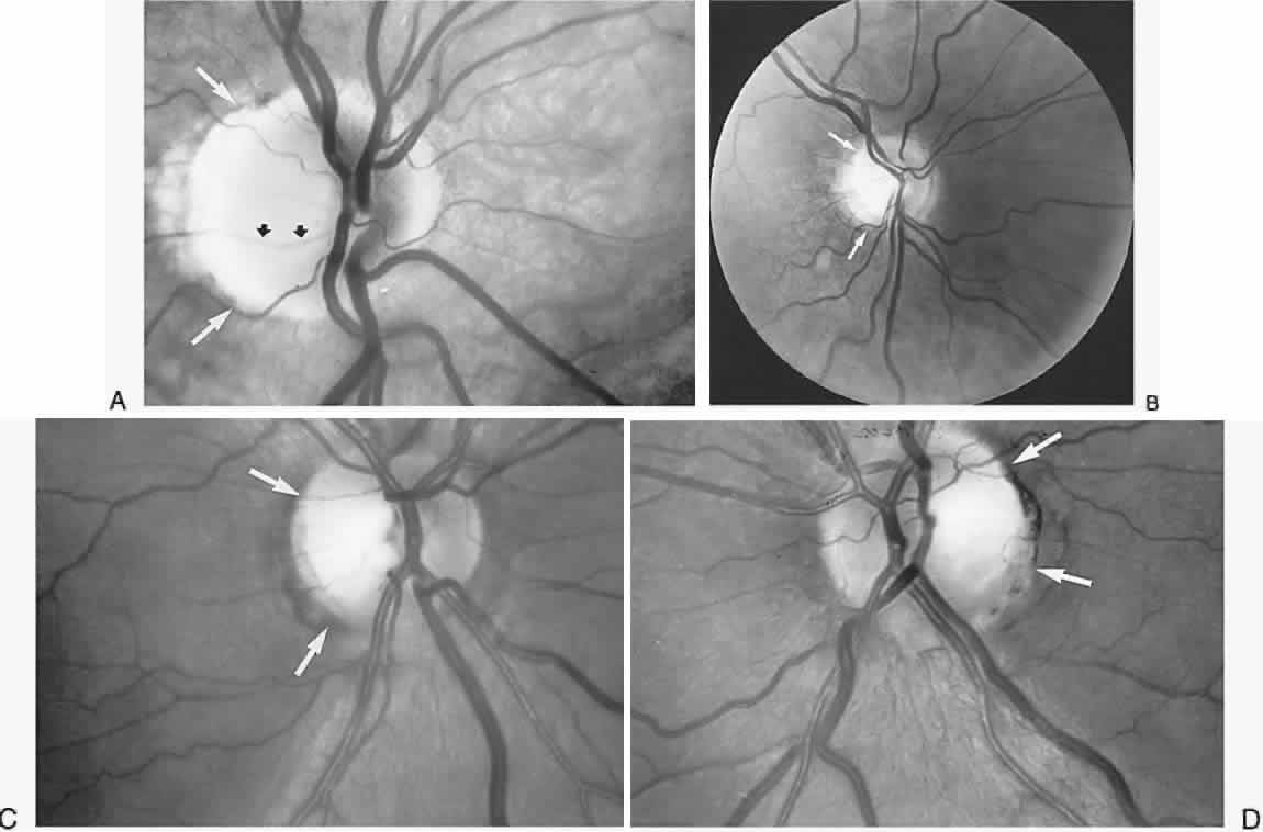
| Fig. 20. Dominant optic atrophy. White arrows indicate circumscribed temporal atrophy. A. Right eye with focal temporal atrophy and excavation (see vessel depression, black arrows). B. Right eye of the son of the patient in (A). C and D. Patient with right eye acuity of 20/80 and left eye acuity of 20/100; see D-15 color test and Humphrey visual fields (see Fig. 21). |