|
|
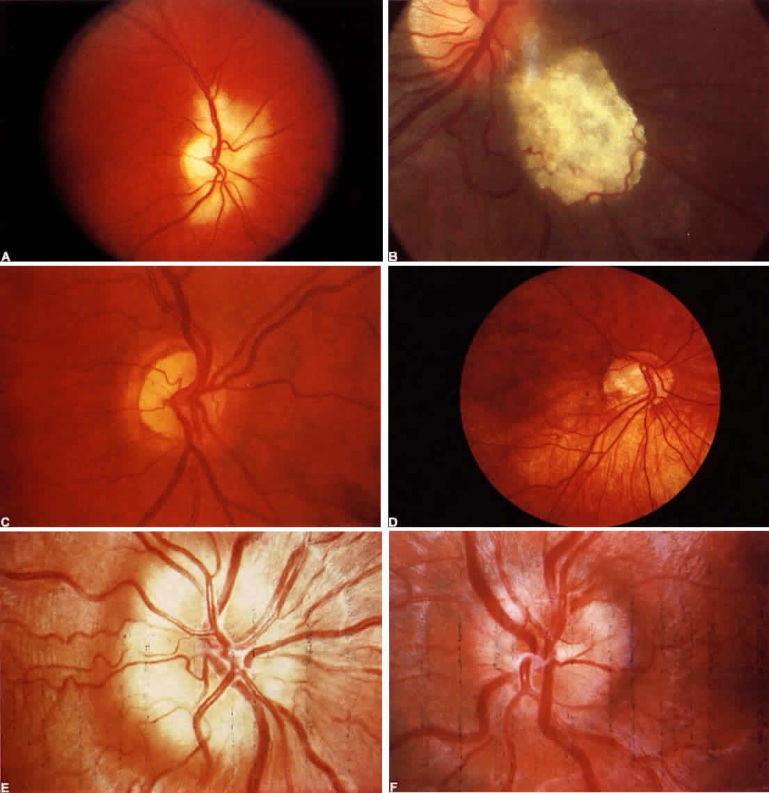
| Color Plate 5-1. A. Myelinated nerve fibers. Retina is white, opaque, with feathered edges. B. Calcified astrocytic hamartoma of retinal nerve fiber layer in tuberous sclerosis. C. Hypoplasia of the optic nerve. Disk is small and with pigment rim and surrounding paler ring. Disk vessels appear disproportionately large. D. Inferior crescent. Disk is small and horizontally oval with scleral crescent at lower border. Contiguous inferior fundus sector is hypopigmented and appears albinotic; foveal reflext is indistinct. E. Pseudopapilledema; congenital elevated disk (compare with true papilledema, F). Note absence of central cup, vessels arise at disk apex. Vascular anomalies include excessive number of major disk vessels and multiple bifurcations. Nerve fiber layer does not obscure vessels at disk margins. F. Chronic moderate papilledema (compare with pseudopapilledema in E.) Note retention of central cup, flame hemorrhage at superior border, absence of anomalous vessel pattern, small arterioles are obscured in nerve fiber layer. |