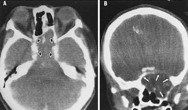
Fig. 44.
Sphenoethmoidal mucocele with visual loss. Computed tomography (CT) axial section (
A
) and CT coronal section (
B
) demonstrate expansion and deformation of sinus walls (
arrows
).