|
|
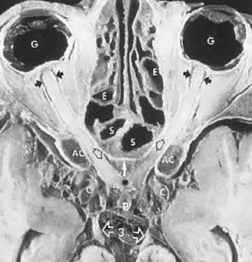
| Fig. 11. Axial anatomic section of the course of the optic nerves. G, globes; small black arrows just behind globes show the subarachnoid space within the optic nerve sheaths; S, sphenoid and E, ethmoid air cells; AC, anterior clinoids with dark bone marrow; open black arrows on the canalicular segments of the optic nerves indicate the thin bony medial optic canal wall shared with the sphenoid sinus; white arrow indicates the tuberculum of the planum sphenoidale; asterisk indicates chiasm bordered by carotid arteries, C; P, pituitary stalk; oculomotor nerves, 3 with open arrows. (Courtesy of Dr. Renate Unsold) |