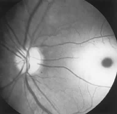
Fig. 4.
“Cherry-red spot” of advanced Tay-Sachs disease (gangliosidosis). Note the central foveal window surrounded by a ring of densely opaque retinal ganglion cell layer; also, optic atrophy.