|
|
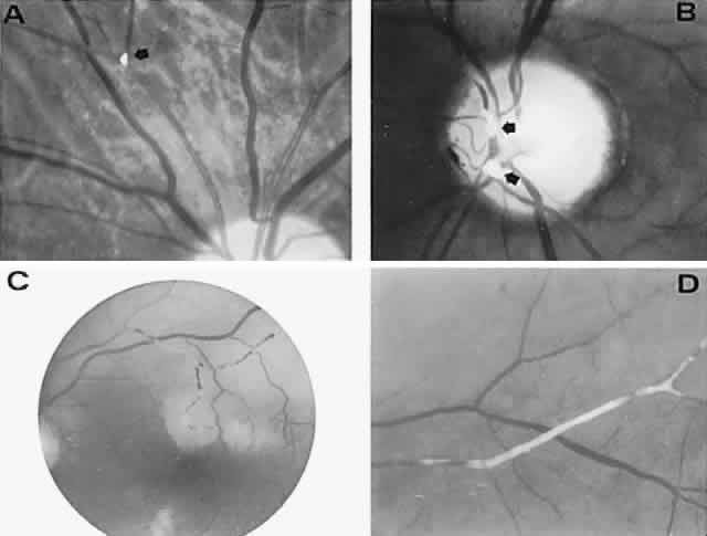
| Fig. 5. Retinal microembolic phenomena. A. Bright cholesterol plaque (arrow) impacted at an arterial bifurcation. Thin crystal does not obstruct flow. B. Cholesterol crystals in disc vessels (arrows). Often, the plaque appears larger than the vessel diameter. C. Infarcted opaque retina. The artery contains emboli (? fibrin platelets) that have obstructed flow. D. Reactive opacification of the arterial wall. Fluorescein angiography demonstrated flow through this formerly occluded vessel. |