|
|
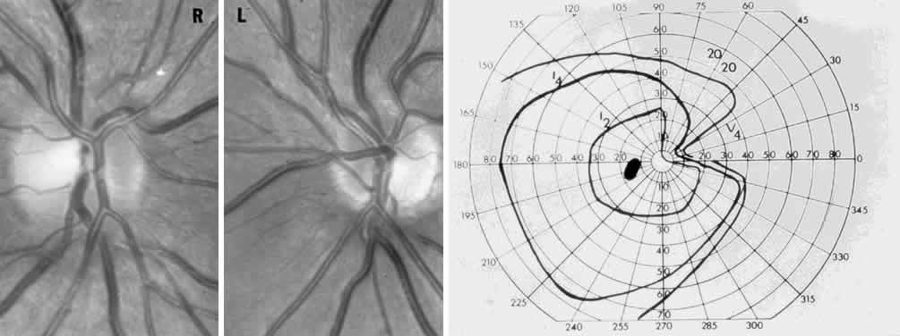
| Fig. 12. Relative optic nerve hypoplasia. Top. R, right optic disc; L, left optic disc. Note smaller left disc, with otherwise normal morphology and no peripapillary pigment disturbance. Bottom. Left visual field shows a dense nasal wedge defect. A left afferent pupil defect was present, and the case misdiagnosed as optic neuritis due to multiple sclerosis. |