|
|
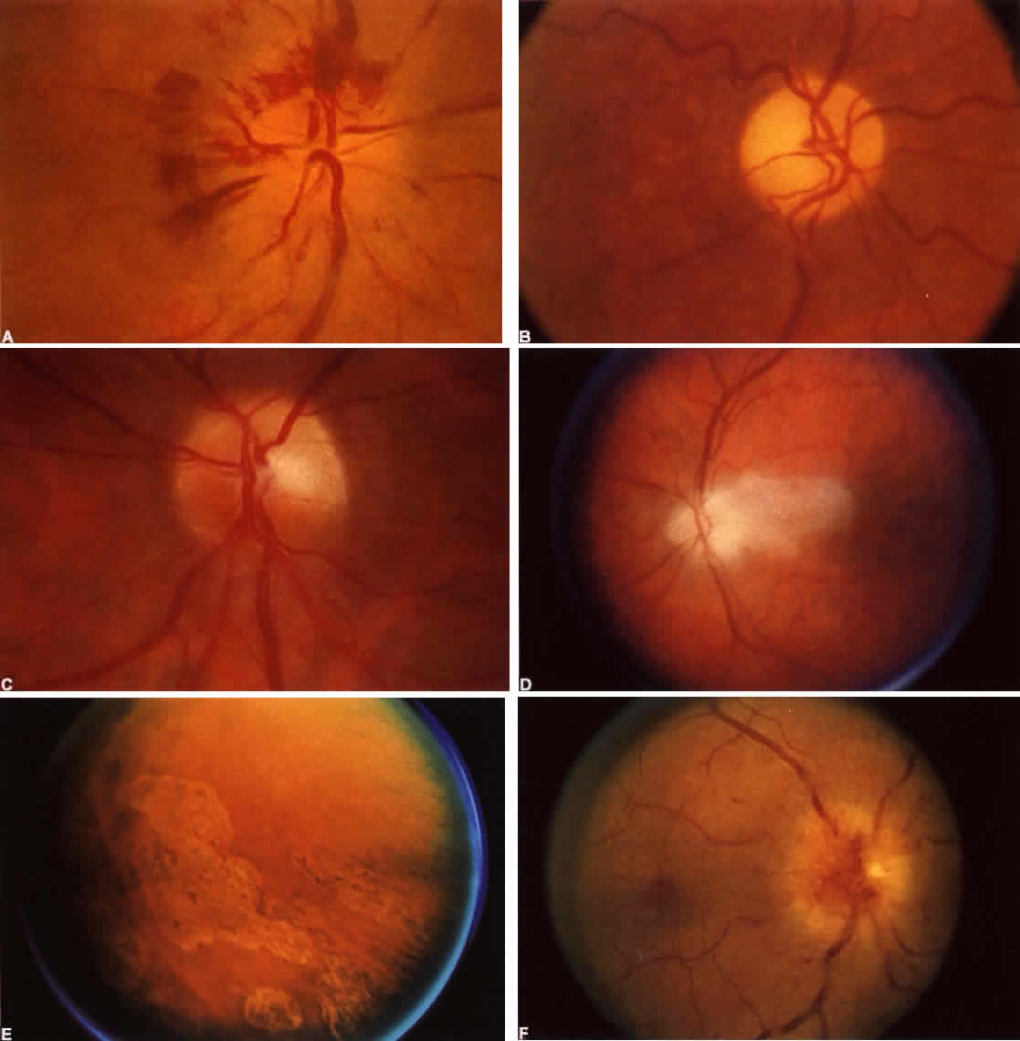
| Color Plate 5-3. A. Ishcemic optic neuropathy with disk edema and “flame” hemorrhages in nerve fiber layer. B. Disk atrophy after ischemic optic neuropathy. Note arteriolar narrowing. C. Superior segmental atrophy after disk infarct, with inferior field defect. Inferior half of disk appears hyperemic. D. Cranial arteritis. Milk pale edema of disk extending into macula. E. Cranial arteritis. Pigmentary changes 3 months after choroidal infarcts. Diabetic papillopathy. Note florid telangiectasia of disk capillaries and cyst at fovea. |