Chapter 1
Topographic Anatomy of the Eye: An Overview
D. J. JOHN PARK and JAMES KARESH
Main Menu   Table Of Contents

Search

GENERAL DIMENSIONS AND TOPOGRAPHY OF THE GLOBE
RELATIONSHIP OF THE GLOBE TO THE BONY ORBIT
SURFACE ANATOMY OF THE GLOBE AND THE SCLERA
THE CORNEA
THE IRIS
THE ANTERIOR CHAMBER AND AQUEOUS HUMOR
ANTERIOR CHAMBER ANGLE AND AQUEOUS OUTFLOW STRUCTURES
THE CILIARY BODY AND POSTERIOR CHAMBER
THE POSTERIOR CHAMBER
THE LENS
THE CHOROID
THE RETINA
THE INTRAOCULAR OPTIC NERVE
THE VITREOUS
REFERENCES

Topography is the study of the gross surface relationships between different aspects of the surface of a structure or between various objects related to that structure. The globe is a highly complex object in terms of its own three-dimensional shape and because of the number of other anatomic elements contained within it and surrounding it. In addition, a number of vascular and neural structures pass into and out of the globe, further complicating its topography. However, to adequately understand the globe as an organ of vision, it is first essential to understand its structure and the complex interrelationships that exist between its internal organization and the external elements surrounding it. A number of excellent texts have been written covering the various aspects of ocular anatomy and histology, and the reader is referred to them for more detailed descriptions of these subjects.1–3 Much of what is discussed in this overview can be found in these texts.
Back to Top
GENERAL DIMENSIONS AND TOPOGRAPHY OF THE GLOBE

As a three-dimensional object, the globe approximates an irregular oblong spheroid that can be divided topographically into segments of two modified spheres of different radii of curvature. (Figs. 1, 2, and 3) The cornea constitutes the anterior segment, covers one-sixth of the surface area, and has a radius of curvature of approximately 8 mm. The larger posterior segment is composed of the scleral shell and has a radius of curvature of approximately 12 mm. This segment is not perfectly spherical because of the three following features: (1) the anterior portion is flatter and more weakly curved than the posterior portion;1 (2) the entire globe is slightly flattened from above so that its greatest vertical diameter is slightly less than its greatest horizontal diameter; and (3) the scleral bulges outward more temporally than nasally.

Fig. 1 Normal human eye, anterior aspect. N, nasal; T, temporal. The cornea (a) is anterior to the iris (b) and pupil (c). The elliptic shape of the anterior corneal margin (arrows) is compared to the round shape of its posterior one (dotted line). The collarette of the iris is evident at b1. The pupil is displaced slightly to the nasal side of the eye. The limbal area and external scleral sulcus surround the margin of the cornea (d). The sclera (e) is peripheral to the limbus. The sclera has been made artificially transparent to show the ora serrata (f), which is farther posterior on the temporal than on the nasal side. The superior and inferior rectus tendons (g) are curved and are inserted obliquely to the axis of the eye. The tendons of the medial and lateral recti (h) also have curved insertions that are not oblique to the horizontal meridian. Each rectus muscle has two anterior ciliary arteries (aca) except the lateral rectus, which has one. The measurements of all these structures are given in millimeters. (Hogan M, Alvarado J, Weddell J: Histology of the Human Eye—An Atlas and Textbook. Philadelphia: WB Saunders, 1971:46)

Fig. 2 Drawing of the posterior eye. T, temporal; N, nasal. The optic nerve (a) with its central vessels and surrounding meningeal sheaths is seen. Its center is located 3 mm nasal and 1 mm inferior to the posterior pole of the eye. Surrounding it are the short posterior ciliary arteries and nerves. The approximate position of the macula is at x. Along the horizontal meridian, which bisects the eye, are the long posterior ciliary arteries and nerves (b). The exits of four vortex veins are shown, one for each quadrant (c). The curved, oblique insertions of the superior oblique (d) and inferior oblique (e) muscles are seen. The cut ends of the four rectus muscles are at f. (Hogan M, Alvarado J, Weddell J: Histology of the Human Eye—An Atlas and Textbook. Philadelphia: WB Saunders, 1971:51)

Fig. 3 Drawing of the upper half of the eye. T, temporal; N, nasal. The contrasting degrees of curvature of the cornea (a) and sclera (b) are evident. At the limbus (c), where they join, is the external scleral sulcus. The relation of the ora serrata (d) to the surface is shown. The nasal displacement of the optic nerve (e) with respect to the posterior pole of the eye makes the three layers of the temporal eye longer than those on the nasal side. The slightly curved oblique insertion of the superior rectus muscle is at f, and the tendinous oblique insertion of the superior oblique muscle is at g. Two vortex veins are seen (h), and the long posterior ciliary arteries and nerves are at i. (Hogan M, Alvarado J, Weddell J: Histology of the Human Eye—An Atlas and Textbook. Philadelphia: WB Saunders, 1971:53)

Anteriorly, the center of the external surface of the cornea is called the anterior pole. Because the vertical dimension of the cornea is less than the horizontal dimension, this pole is closer to the inferior and superior border of the cornea and farther away from its nasal and temporal border (Fig. 4). Posteriorly, the center of the external surface of the sclera is called the posterior pole. For practical purposes, this point is equidistant from any point along the limbus. The geometric (anatomic), visual, and optic axes describe the globe in the antero-posterior dimension. The geometric axis connects the anterior pole and the posterior pole and is the basis for topographical reference for the gross elements of the globe. When this axis is measured from the external surface of the cornea to the external surface of the sclera, it is referred to as the external axis, and when measured from the internal surface of the cornea to the anterior surface of the retina, it is called the internal axis. The visual axis represents a line drawn from the fovea centralis to the point of fixation passing through the nodal point. It passes slightly nasal to the corneal summit. The optic axis represents a line passing through the anterior pole, the center of the lens, and the nodal point. Whereas the visual and optic axes relate to the fovea, nodal point, and other aspects of visual function, the geometric axis is purely a contrived dimension that is quite variable between individuals.

Fig. 4 A. Anterior and posterior diagram of the corneal edges showing the elliptic shape anteriorly and the round shape posteriorly. Also noted are the vertical and horizontal diameters of the anterior and the posterior cornea; the radius of curvature of the cornea and of the sclera (B); the corneal height and the central 4 mm of the cornea, which is optically important; and the comparative thickness of the central and peripheral cornea (C). (Hogan M, Alvarado J, Weddell J: Histology of the Human Eye—An Atlas and Textbook. Philadelphia: WB Saunders, 1971:61)

The anatomic equator and the geometric equator are different.1,2 The former represents a circumferential line on which every point is equidistant from the anterior and posterior poles. The latter is a simple circle whose diameter is perpendicular to the geometric axis and whose center is equidistant from the anterior and posterior poles. The anatomic equator divides the globe into two unequal halves, termed anterior and posterior hemispheres. If the globe were an ideal sphere, the anatomic equator would be a perfect circle in the coronal plane, perpendicular to the geometric axis, and thus coincident with the geometric equator. However, because the sclera bulges more temporally, the anatomic axis3 is farther posterior on its temporal side than on its nasal side. On average, the anatomic equator is approximately 13 to 14 mm behind the limbus. This is in contrast to what may be considered the functional equator for purposes of muscle surgery. This latter concept is based on the arc of contact the rectus muscles have with the globe, which is approximately 4 mm posterior to the anatomic equator on the temporal side and 4 mm anterior to it on the nasal side. A meridian is defined as a line encircling the globe in the antero-posterior direction, which passes the anterior and posterior pole and is perpendicular to the anatomic equator. There are two cardinal meridians for purposes of measuring the globe. The sagittal meridian divides the globe into nasal and temporal hemispheres and arbitrarily is based on the 6 o'clock and 12 o'clock positions on the limbus. The horizontal or transverse meridian is perpendicular to the sagittal meridian, being based on the 3 o'clock and 9 o'clock positions on the limbus and divides the globe into superior and inferior hemispheres.

The various dimensions of the globe have been extensively measured.1 These are generally fairly constant in the adult population and are relatively unrelated to either sex or race, usually varying by no more than 1 mm in various studies. However, in extreme degrees of axial hyperopia and myopia, the anteroposterior diameter of the globe may vary by as much as 3 mm from the normal measurement.4 The outer anteroposterior diameter of the globe averages 24.15 mm (range, 21.7–28.75 mm), whereas the internal anteroposterior diameter averages 22.12 mm.1 In high hypermetropia and myopia, the anteroposterior diameter can be as low as 20 mm and as high as 29 mm, respectively.4 The transverse diameter (diameter of the globe at the anatomic equator measured from the nasal to the temporal side) averages 23.48 mm, and the vertical diameter (diameter of the globe at the anatomic equator measured superiorly to inferiorly) averages 23.48 mm.1 The circumference of the globe at the anatomic equator averages 74.91 mm.1 At birth, the globe is more spherical than the adult globe and has an anteroposterior diameter approximately two-thirds that of the adult globe (16–17mm).5 By 3 years of age, this increases to within 1 mm of the average adult size (22.5–23 mm), reaching the normal adult size by approximately 13 years of age.5 When fully developed, the globe weighs approximately 7.14 to 7.5 g,2,6,7 has a volume of approximately 6.5 to 7.2 mL,7 and has a surface area of approximately 22.86 cm2.8

As already noted, the globe is asymmetric. This is demonstrated by the tilt of the anatomic equator, the more anterior placement of the nasal vortex veins compared with the temporal ones, and the more anterior placement of the medial rectus muscle insertion compared with the lateral rectus muscle insertion. Likewise, the optic nerve is positioned 3 mm nasal and 1 mm inferior to the posterior pole. In addition, the lens and pupil are slightly displaced nasally, with the anterior chamber angle being narrower nasally than it is temporally. The ciliary body is 1 mm in the anteroposterior dimension nasally than temporally. In contrast, the temporal retina is positioned approximately 1 mm farther anteriorly than the nasal retina.

Back to Top
RELATIONSHIP OF THE GLOBE TO THE BONY ORBIT
The globe is positioned in the anterior portion of the orbit and constitutes approximately 20% of the entire volume of the orbit. It is slightly closer to the lateral orbital wall than the medial wall and is nearer to the roof than the floor of the orbit. The posterior pole lies approximately 18 mm (14–24 mm)7 from the apex of the orbit. At its closest distance to the bony orbit the globe is approximately 4 mm from the roof, 4.5 mm from the lateral wall, 6.5 mm from the medial wall, and 6.8 mm from the floor.3 The lateral orbital rim is considerably recessed posteriorly compared to the medial orbit, which continues anteriorly to end at the nasal bridge. This leaves approximately one half of the globe exposed laterally, such that a line connecting the lateral orbital rim to the anterior lacrimal crest would pass well behind the ora seratta temporally and near the junction of the ciliary body and iris nasally. Thus the globe is most vulnerable to trauma on its lateral side, and surgical approach is easier here. In contrast, the anterior pole of the globe lies just posterior to a line drawn from the superior orbital rim to the inferior orbital rim and can vary from 12 mm anterior to 10 mm posterior to this line in healthy individuals. The prominence of the globes is dependent on orbital volume, globe size, orbital fat volume, and structure of the eyelids and conjunctiva, all of which vary between individuals and shows significant differences between ethnicities. Based on a study of 681 individuals with no history of orbital or endocrine disease, high myopia, or buphthalmos, Migliori and Gladstone found that white men had a mean protrusion value measured from the lateral orbital rim of 16.5 mm compared to 18.5 mm in black men, and 15.4 mm in white women compared to 17.8 mm in black women.9

The geometric axis of the globe is parallel to the medial orbital wall and is at a 45-degree angle to the lateral orbital wall. Thus divergent axis of each orbit is approximately 23 degrees. The two lateral walls of both orbits form a 90-degree angle.10 The distance between the geometric axes of the globes, or the interpupillary distance, is 58 to 60 mm.

Back to Top
SURFACE ANATOMY OF THE GLOBE AND THE SCLERA
The sclera forms five-sixths of the outer tunic of the eye, consists of dense fibrous tissue, and contains openings and canals for the various vessels and nerves entering and exiting the globe (Figs. 1, 2, and 3). Its mechanical properties and limited distensibility help contain the intraocular pressure and prevent deformations of the globe during extraocular movement. It is relatively avascular and principally composed of collagen with scant fibrocytes and ground substance. Externally it is white, whereas internally it is slightly brown because of the presence of pigmented melanocytes. This most interior layer of the sclera, adjacent to the uvea, is called the lamina fusca, whereas the most exterior layer of the sclera, consisting of delicate fibrous and elastic tissue connecting the bulbar fascia and the sclera, is called the episcleral tissue. If the sclera is thin, as in childhood, or its hydration content decreases to less than 40% or increases to more than 80%, the sclera becomes more lucent, such that it appears blue or slate grey. As the sclera thins, the underlying uvea absorbs more light and the scattering of shorter wavelength light by the scleral collagen imparts a blue hue.

The scleral thickness varies considerably in the anteroposterior dimension. It is thickest near the optic nerve, measuring 1 to 1.35 mm, and gradually thins anteriorly, where it measures 0.4 to 0.6 mm in thickness at the equator. The sclera is thinnest beneath the tendonous insertions of the extraocular muscles, where it measures approximately 0.3 mm in thickness.11,12 At the limbus (the transitional area between cornea and sclera), the sclera thickness is approximately 0.825 mm. In older individuals, it is unclear whether the apparently thinner sclera is caused by age-related atrophy13 or changes in scleral hydration.14

The anterior scleral foramen is defined by a zone in which the scleral fibers merge with the collagen fibrils of the corneal stroma. Usually the sclera is beveled internally to overlap the cornea anteriorly in the sagittal meridian, making the cornea appear as an ellipse. For this reason, the external diameter of the cornea averages 10.6 mm vertically and 11.6 mm horizontally.12 Internally, the anterior scleral foramen is more nearly circular, with a diameter of 11.6 mm. Both the external and internal margins of the sclera at the sclerocorneal junction extend slightly anteriorly, creating an external and internal annular groove, called the external and internal scleral sulcus, respectively. The external scleral sulcus is approximately 1 mm in depth. However, most of this is filled in by the attachments of the conjunctiva and Tenon's fascia as they blend into the limbus, and thus is less apparent. The external scleral sulcus is more prominent nasally than temporally. The internal scleral sulcus is circular with an internal groove formed by the slight anterior projection of the sclera near the limbus. It is bound anteriorly by Schwalbe's ring, posteriorly by the scleral spur, and contains Schlemm's canal, externally, and the corneoscleral meshwork, internally. Along its inner surface, inserts the anterior tendons of the longitudinal ciliary muscles.15 The posterior margin of internal scleral sulcus extends anteriorly as the scleral spur and is the attachment for the ciliary muscle. At the base of the sulcus is Schlemm's canal. A so-called pectinate ligament can occasionally be found to bridge the internal scleral sulcus.

The posterior scleral foramen allows the ganglion cell axons to exit the globe and enter the orbital segment of the optic nerve. It is a short canal located 3 mm nasal to the vertical meridian and 1 mm inferior to the horizontal meridian in the posterior hemisphere. It has an internal diameter of 1.5 to 2.0 mm and an external diameter of 3.0 to 3.5 mm. It is approximately 0.7 mm long and exits the globe in a nasal direction at an oblique angle. The network of collagenous and elastic scleral fibers that traverse the foramen in the innermost one-third of the foramen is referred to as the lamina cribosa. It provides mechanical strength and anchorage points for the neural and vascular elements as they traverse the canal. The inner surface of the lamina cribosa is concave16 and is displaced posteriorly in the presence of sustained ocular hypertension. Fibroelastic fibers from the outer two-thirds of the sclera turn outward to merge with the dural sheath of the optic nerve.

Emissary channels allow the passage of nerves and vessels through the scleral tunic. These can be divided into anterior, middle, and posterior emissary channels (Figs. 5, 6).

Fig. 5 Schematic drawing of the distribution of the ciliary arteries. IMC, intramuscular ciliary arterial circle; MAC, major arterial circle of the iris; PCA, posterior ciliary artery; ACA, anterior ciliary arteries; lpca, long posterior ciliary artery; a, recurrent branch of the long posterior ciliary artery; b, long posterior ciliary artery; c, recurrent branch of the long posterior ciliary artery; d, anterior choroidal branch of the ciliary intramuscular circle; e, branch of the IMC; f, iris artery arising from a ciliary process branch of the MAC; g, branch to the anterior choroid from the anterior ciliary artery; h, branch to the anterior choroid from the MAC; i, iris artery arising from the MAC branch to the choroid; j, iris artery arising from the anterior ciliary artery. (Reprinted with permission from Bron AJ, Tripathi RC, Tripathi BJ. Wolff's Anatomy of the Eye and Orbit, 8th Edition. London: Chapman & Hall Medical, 1997)

Fig. 6 The blood supply of the eye. K, branch of short posterior ciliary to the optic nerve; I, anastomoses between choroidal and central vessels. In the case of the artery this is capillary only; s, vein from ciliary muscle to vena vorticosa; t, branch of anterior ciliary vein from ciliary muscle; o, recurrent artery. (From Meyer PAR. Eye 1989;3:121, with permission)

The anterior emissary channels are located near the limbus, through which the anterior ciliary arteries and veins and the ciliary nerves traverse. The anterior ciliary arteries travel with the rectus muscles, two in each muscle with the exception of the lateral rectus, which has one artery. Each artery enters the sclera just anterior to the insertions and travels obliquely to supply the ciliary body and feed the major arterial circle of the iris. Smaller branches turn externally toward the limbus, where they anastamose with subconjunctival vessels to form the episceral plexus. These recurrent branches of the limbal vessels supply the perilimbal conjunctiva within 3 to 6 mm of the limbus. The anterior ciliary veins leave the ciliary body and travel in a channel that is frequently shared with a branch of the posterior ciliary nerve, which passes close to the surface before looping into the ciliary body. This neurovascular loop is called the nerve loop of Axenfeld and is present in approximately 12% of individuals.17 Clinically, it appears as 1- to 2-mm dome-shaped gray area, that is often pigmented.

The middle emissary channels are located several millimeters posterior to the geometric equator and transmit the vortex veins. Their position can be somewhat variable. Temporally, these veins are closer to the sagittal meridian, and nasally they are farther away from this meridian. Internally, their position relative to the sclera varies from the equator to 6.36 mm posterior to the equator, with the most common position being 3 mm posterior to the equator.18,19 Externally, their exit position from the sclera varies from 13.75 to 25 mm posterior to the limbus. The superotemporal and superonasal veins exit more posteriorly (20.2 mm and 19.3 mm from the limbus, respectively) than the inferotemporal and inferonasal veins (17.4 mm and 18 mm from the limbus, respectively). The superotemporal vein exits the sclera approximately 8 mm, the superonasal vein 7 mm, the inferotemporal vein 5.5 mm, and the inferonasal vein 6 mm posterior to the equator. The distance each vein travels in the sclera is variable and ranges from 1.25 to 8.5 mm. When they exit the sclera, the vortex veins travel loosely between it and the bulbar fascia for 5 to 10 mm, with the superior temporal vein having the longest episcleral course.

On average, the choroid of each eye contains seven vortex veins (range, 5 to 8). They are more frequent nasally then temporally. In more than 50% of cases, the main trunks of these veins are incomplete, and they leave their scleral canals as branches. Usually only one vein is seen to exit the sclera in the superior temporal quadrant in contrast to the other quadrants where one or more veins may be found exiting. Generally, the veins tend to exit between the extraocular muscles and only rarely exit under the belly of a muscle.

The posterior emissary channels transmit up to 20 short posterior ciliary arteries and 10 short posterior ciliary nerves and two long posterior arteries and nerves. The short posterior ciliary arteries and nerves enter the sclera in an annular fashion around and within 1 to 2 mm of the optic nerve. These nerves and arteries are more closely approximated to the nerve medially than temporally. The two long posterior ciliary arteries and nerves pierce the sclera on either side of the optic nerve approximately along the horizontal meridian. The medial nerve and artery are approximately 3.6 mm from the medial aspect of the nerve sheath, whereas the temporal nerve and artery are approximately 3.9 mm from the temporal aspect of the nerve sheath and in close approximation to the posterior insertion of the inferior oblique muscle. They pass obliquely through the sclera for a distance of 3 to 7 mm. There may be additional long posterior ciliary arteries and nerves that insert into the posterior sclera, most commonly in the inferior aspect of the globe. The entrance of these additional vessels is usually more anterior than that of the vessels along the horizontal meridian.

Both the insertions of the rectus and oblique muscles have specific topographical reference points that deserve considerable attention (Figs. 1, 2, 3, and 7). The tendons of the rectus muscles insert into the sclera and its fascial coverings at various distances from the limbus. The tendonous insertions of the medial and lateral rectus muscles are relatively linear, whereas those of the inferior and superior rectus muscles are somewhat curvilinear and obliquely placed so that their nasal insertion is closer to the limbus than their temporal insertion. The distance from the midpoint of the rectus muscle tendons to the limbus varies from study to study but generally is approximately 5.7 mm, 6.7 mm, 7.5 mm, and 7.9 mm for the medial, inferior, lateral, and superior rectus muscles, respectively (Table 1).2,18,20–22 This configuration describes the spiral of Tillaux. The distance between the insertions is approximately 7.4 mm between the medial and inferior rectus muscles, 8.5 mm between the inferior rectus and lateral rectus muscles, 7.2 mm between the lateral rectus and superior rectus muscles, and 8 mm between the superior rectus and medial rectus muscles.

Fig. 7 Schematic diagram to illustrate the distances of various structures entering or leaving the globe from the limbus. The right globe is viewed from behind. (Reprinted with permission from Bron AJ, Tripathi RC, Tripathi BJ. Wolff's Anatomy of the Eye and Orbit, 8th Edition. London: Chapman & Hall Medical, 1997)


TABLE 1. Measurements of the Tendinous Insertions of the Rectus Muscles


 Tendon Width (mm)Distance From Tendon to Limbus at Its Nearest Insertion (mm)Distance From Tendon to Limbus at Its Furthest Insertion (mm)Distance From Tendon to Limbus at the Midpoint of Its Insertion (mm)
Medial rectus10.4 (8–13)5.565.7 (4.5–7.5)
Inferior rectus8.6 (7–12)5.586.7 (5.5–8)
Lateral rectus9.6 (8–13)6.777.5 (6–9)
Superior rectus10.4 (7–12)6.5–6.79–117.9 (6.5–10)

Compiled from multiple sources.2,7–10


The insertion of the superior oblique muscle is beneath the anterior portion of the superior rectus muscle. It has a wide curvilinear tendinous insertion that faces posterolaterally and is convex in that direction. The width of the insertion varies between 7 and 18 mm. The anterior and lateral edge of the tendon is just anterior to the geometric equator of the globe, approximately 12 to 14 mm (average, 13.8 mm)23 posterior to the limbus, only a few millimeters (3–4.5 mm)23 posterior to the lateral insertion of the superior rectus tendon, and close to the lateral edge of the superior rectus muscle. The majority of the tendon is posterior to the geometric equator. The medial and posterior edge of the tendon approximately 17 to 19 mm (average, 18.8 mm) posterior to the limbus and as much as 14 mm (average, 13.6 mm) posterior to the medial insertion of the superior rectus tendon. This edge of the tendon is approximately 8 mm superior to the posterior pole of the globe. The superolateral vortex vein is only a few millimeters posterior to the medial insertion of the superior oblique tendon and very close to the lateral edge of the superior rectus muscle.

The inferior oblique muscle has a very short or absent tendon at its insertion into the sclera. This insertion is inferior to the horizontal meridian and lateral to the sagittal meridian, placing it in the inferior lateral quadrant of the posterior aspect of the globe. The insertion is convex in the superior direction and measures 5 to 14 mm in length. At its most posterior point, the insertion is 3 to 6 mm lateral to the lateral aspect of the optic nerve and approximately 1 mm below and 1 to 3 mm (average, 2.2 mm) temporal to the macula.24 The most anterior aspect of its insertion is approximately 10 mm posterior to the midpoint of the lateral rectus muscle insertion. The posterior edge of the insertion and the muscle are only a few millimeters anterior to the inferolateral vortex vein.

Back to Top
THE CORNEA
The cornea is a tough transparent and avascular tissue that, along with the precorneal tear film, forms the major refracting surface for the eye and serves as a barrier between the environment and the inside of the eye (Fig. 4). The precorneal tear film ranges in thickness from 6 to 20 μm but averages approximately 7 μm.25 This tear film consists of an outer layer of lipid produced by the sebaceous glands of the eyelid and caruncle, a middle layer of aqueous fluid produced by the lacrimal and accessory lacrimal glands, and an inner layer of mucoprotein produced by conjunctival goblet cells. Anteriorly, the conjunctival surface of the eyelids is adjacent to and in contact with the tear film layer. During the course of blinking, this layer is distributed over the surface of the corneal epithelium continually rewetting and renewing the entire surface.

The external dimensions of the cornea are 11.6 to 12.6 mm horizontally and 10.6 to 11.7 mm vertically.12 These measurements are approximately 0.1 mm less in females. When viewed from the anterior surface, the cornea is oval because of a more prominent limbus superiorly and inferiorly.26 When viewed posteriorly, the cornea is circular and has a diameter of 11.6 mm. The thickness of the cornea varies from 0.51 to 0.56 mm centrally to 0.63 to 0.67 mm peripherally.27,28 An increasing number of studies have found that long-term use of contact lenses (hard and soft) is associated with decreased central corneal thickness.29–34 However, peripherally the thickness has been measured as high as 0.74 mm in normal individuals.1,12,35,36 Posteriorly, the corneal endothelium is bathed by the aqueous humor. The surface area of the cornea is approximately 1.3 cm3 or approximately one-fourteenth the area of the globe.12 The height of the cornea (i.e., the distance from a plane through the peripheral visible border of the cornea to the apex of the cornea externally) is 2.68 mm.1 The fresh cornea weighs 180 mg and has a specific gravity of approximately 1.05. Because the curvature of the cornea is greater than that of the scleral shell, a slight sulcus, the external scleral sulcus, marks the junction between the cornea and sclera. The radius of curvature of the anterior surface of the cornea ranges from 7.2 to 8.4 mm, and the radius of curvature of the posterior surface ranges from 6.2 to 6.8 mm in white men. The cornea is more curved in Asians than in whites and in women than in men. Although a wide variety of different corneal shapes can be seen between individuals, it is frequently more curved at the temporal cornea than the nasal cornea, and in the vertical plane than in the horizontal plane.37

The optical zone of the cornea is the central one-third, approximately 4 mm in diameter. The anterior and posterior surfaces of the cornea are relatively spherical in this region. However, they tend to diverge as they extend toward the periphery, where the cornea is slightly flattened. However, in individuals with some degree of corneal astigmatism the central optical zone may be somewhat ellipsoidal.

The size and shape of the cornea change throughout life. The size of the cornea in a newborn relative to the adult eye is fairly large, attaining adult size (1.3 cm2) between the ages of 1 and 2 years (Table 2).1 The most rapid period of corneal growth is in the first 6 months after birth. The newborn cornea is flatter than the adult cornea and is usually more curved in its periphery than centrally. On reaching adulthood, the average corneal radius decreases with age and the cornea becomes more spherical, thus contributing to the reduction of retinal image quality through the lifespan.38


TABLE 2. Comparison of the Newborn and Adult Cornea


 Newborn (mm)Adult (mm)
External diameter of horizontal base1011.8
External diameter of corneal arc1418.2
Internal diameter of corneal arc1116.4
Mean thickness0.80.9
Oblique thickness1.11.6
External height33.4
Internal height1.12.7

(Adapted from Duke Elder S. Wybar K. The eye. In Duke-Elder S (ed): The Anatomy of the Visual System, Vol II, pp 75–386. St. Louis, CV Mosby, 1961)


The cornea consists of five layers: (1) the epithelium; (2) Bowman's layer; (3) stroma; (4) Descemet's membrane; and (5) endothelium. The epithelium, which makes up approximately 10% of the total corneal thickness and measures approximately 50 to 60 μm, is a stratified, nonkeratinized, nonsecretory squamous epithelium that is 5 to 7 cells in thickness centrally, increasing at the limbus to 15 cells and to more than 20 cells in the conjunctiva. The epithelial layer consists of three distinct cellular types: a single layer of cuboidal basal cells, an intermediate layer of polygonal cells (also referred to as wing cells) that is 2 to 3 cells in thickness, and a superficial layer of flattened epithelial cells that is 3 to 4 cells in thickness. The basal layer consists of columnar cells that are quite regular in shape and size, measuring 18 μm in height by 10 μm in width. As basal cells divide, daughter cells move toward the surface of the cornea and differentiate into polygonal cells, forming the intermediate layer of the epithelium. The cells of the superficial layer are terminally differentiated squamous cells that measure 45 μm in length by 4 μm in height. Basal cells are adherent to its basement membrane, which is 48-nm-thick and is separated from these cells by a layer of granular material that is 23-nm-thick. At the limbus it becomes considerably thicker, forming the basal layer of the conjunctival epithelium. The basement membrane is strongly attached to the underlying Bowman's layer.39

Bowman's layer, which consists of interwoven collagen fibrils, is 8 to 14 μm in thickness, with the peripheral one-third being slightly thicker than the central two-thirds. Peripherally, the collagen fibrils of Bowman's layer become more loosely arranged and end abruptly at the limbus. The deep surface of Bowman's layer merges into the corneal stroma.

The corneal stroma measures 500 μm in thickness, constituting approximately 90% of the corneal thickness. It consists of approximately 200 to 250 collagen lamellae, each having a thickness of 2 μm and a width ranging from 9 to 260 μm.2 The lamellae span the entire length of the cornea. These lamellae, as well as the collagen fibrils composing them, are parallel, regularly arranged, and layered. The individual collagen fibrils measure 34 to 40 nm in diameter and are separated by a space measuring 20 to 50 nm.2 Peripherally, as the stroma blends into the sclera, the fibrils measure 60 to 70 nm and are less regularly spaced. In contrast, the scleral collagen fibrils measure 28 to 280 nm in diameter, and the collagen bundles measure 30- to 50-μm-wide and 10-μm-thick at the scleral spur and 100- to 140-μm-wide and 10- to 16-μm-thick elsewhere. The periodicity of the corneal stromal collagen fibrils is 62 to 64 nm, whereas that of the scleral stromal fibrils is 80 nm.

Descemet's membrane consists of regularly arranged layers of very fine collagen filaments and includes at its most posterior limit the basement membrane of the corneal endothelium. In adults this membrane is 10- to 12-μm-thick, whereas at birth it is only 3- to 4-μm-thick. At the edge of the cornea, Descemet's membrane branches out to form the sheets of trabeculae forming part of the aqueous outflow apparatus. In adults older than age 20 years, Descemet's membrane demonstrates thickenings (Hassall-Henle warts) that bulge into the anterior chamber.

A single layer of endothelial cells measuring 5 μm in height and 18 to 20 μm in width make up the posterior layer of the cornea. Approximately 500,000 hexagonal cells are present in this layer, and this number decreases with age. Each cell contains 20 to 30 microvilli that project into the anterior chamber and measures 0.1 to 0.2 μm in width by 0.5 to 0.6 μm in height. Corneal endothelial cells are continuous with the endothelial cells that line the space of the iridocorneal angle and the anterior surface of the iris.39

The cornea is richly supplied with sensory nerves. The corneal nerve fibers are derived from the ophthalmic division of the trigeminal nerve. A few superficial sensory nerve endings enter the cornea from the subconjunctival and episcleral areas, but most of the nerve supply is provided transsclerally from the long posterior ciliary nerves. After passing through the choroid and ciliary body, these nerves enter the anterior sclera a short distance posterior to the limbus, where they divide to form the annular plexus. Seventy to 80 large corneal nerves composed of fibers from the branching ciliary nerves enter the cornea radially. Most pass into the middle one-third of the peripheral cornea as myelinated nerve bundles that lose their myelin sheath in the peripheral 1 to 2 mm of cornea. These sensory nerves are found in all corneal layers but are more commonly found in the anterior third of the corneal stroma. The nerves subdivide into smaller side branches and eventually turn abruptly 90 degrees to proceed towards the corneal surface. A well-developed neural net arborizes in the surface corneal epithelium after penetrating Bowman's layer.

The blood supply and the lymphatics to the cornea are provided by subconjunctival, episcleral, and scleral vessels. These vessels supply the limbus as well and arise from the anterior ciliary arteries.40 Two sets of vessels originate from these arteries: terminal vessels that supply the peripheral corneal arcade before becoming a venous plexus and recurrent vessels that supply a portion of the peripheral cornea measuring approximately 0.5 mm before traveling posteriorly to supply 3 to 6 mm of perilimbal conjunctival and episcleral tissue. These latter vessels anastomose with recurrent conjunctival vessels from the fornix.

Back to Top
THE IRIS

The iris is the anterior extension of the uvea that forms a mobile diaphragm between the posterior chamber and anterior chamber of the eye (Fig. 8).1,2 The central aperture of the iris forms the pupil, through which light enters the eye. The diameter of the pupil, which varies from 1.5 to 8 mm, is regulated by movement of the iris, thus impacting the amount of light that enters the eye. The iris extends transversely in a radial direction from the center of the eye to approximately 1 mm posterior to the corneoscleral junction where it joins the ciliary body.41 The anterior surface of the iris is visible through the cornea and is separated from the cornea by the anterior chamber of the eye. Its posterior surface faces the posterior chamber, and its central portion is in contact with the anterior surface of the lens. The diameter of the iris is approximately 12 mm. The cornea magnifies the image of the iris by approximately 10%.

Fig. 8 Composite drawing of the surfaces and layers of the iris. Beginning at the upper left and proceeding clockwise, the iris cross section shows the pupillary (a) and ciliary portions (b), and the surface view shows a brown iris with its dense, matted anterior border layer. Circular contraction furrows are shown (arrows) in the ciliary portion of the iris. Fuchs' crypts (c) are seen at either side of the collarette in the pupillary and ciliary portion and peripherally near the iris root. The pigment ruff is seen at the pupillary edge (d). A blue iris surface shows a less dense anterior border layer and more prominent trabeculae. The iris vessels are shown beginning at the major arterial circle in the ciliary body (e). Radial branches of the arteries and veins extend toward the pupillary region. The arteries form the incomplete minor arterial circle (f), from which branches extend toward the pupil, forming capillary arcades. The sector below it demonstrates the circular arrangement of the sphincter muscle (g) and the radial processes of the dialator muscle (h). The posterior surface of the iris shows the radial contraction furrows (i) and the structural folds (j) of Schwalbe. Circular contraction folds are also present in the ciliary portion. The pars plicata of the ciliary body is at k. (Hogan M, Alvarado J, Weddell J: Histology of the Human Eye—An Atlas and Textbook. Philadelphia: WB Saunders, 1971:207)

The shape of the iris resembles the cut base of a cone, with the apical opening defining the pupillary border surrounding the pupillary aperture (Fig. 9) and the basal aperture defining the peripheral or ciliary border. Thus, in accordance with the convexity of the underlying lens, the pupillary plane of the iris is anterior to its periphery. When compared to the center of the cornea, the center of the pupil is slightly displaced inferiorly and nasally.

Fig. 9 Pupillary portion of the iris. The dense, cellular anterior border layer (a) terminates at the pigment ruff (b) in the pupillary margin. The sphincter muscle is at (c). The arcades (d) from the minor circle extend toward the pupil and through the sphincter muscle. The sphincter muscle and the iris epithelium are close to each other at the pupillary margin. Capillaries, nerves, melanocytes, and clump cells (e) are found within and around the muscle. The three to five layers of dilator muscle (f) gradually diminish in number until they terminate behind the midportion of the sphincter muscle (arrow), leaving low, cuboidal epithelial cells (g) to form the anterior epithelium to the pupillary margin. Spur-like extensions from the dilator muscle form Michel's spur (h) and Fuch's spur (i), which extend anteriorly to blend with the sphincter muscle. The posterior epithelium (j) is formed by tall columnar cells with basally located nuclei. Its apical surface is contiguous with the apical surface of the anterior epithelium. (Reprinted with permission from Hogan MJ, Alvarado JA, Weddell JE. Histology of the Human Eye—An Atlas and Textbook. Philadelphia: WB Saunders, 1971)

The collarette of the iris is a wavy circular line approximately 1 to 1.5 mm from the pupillary border, between the inner one-third and outer two-thirds on the anterior surface of the iris. It marks the zone that separates two concentric rings of the iris: the inner pupillary zone (1–2 mm in width), which contains the sphincter muscle, and the outer ciliary zone (3–4 mm in width). The collarette is the thickest portion of the iris, measuring 0.6 to 1.0 mm; the iris becomes thinner toward the pupil and even thinner in the periphery (0.3–0.5 mm), where it joins the ciliary body.41

The anterior surface of the iris (Fig. 10) is also characterized by small connecting crests in the pupillary zone, deep radial slits known as Fuchs crypts or stomates at the collarette, and deep round ruts, which are mostly incomplete and concentric to the pupillary border in the ciliary zone. The radial ridges are the result of the radially oriented blood vessels and the connective tissue surrounding them in the iris stroma. There is an incomplete circle of arteries (minor circle of the iris) at the collarette. The blood supply to the iris is from the long posterior ciliary arteries that form the major arterial circle of the ciliary body located just peripheral to the iris root.42,43 The anterior surface of a darkly pigmented iris is dense and thickly matted with few visible fibrils, compared with a blue or less darkly pigmented iris. The posterior surface of the iris is black or dark brown, except in albino subjects, and is also characterized by various types of folds or plicae. Schwalbe's contraction plicae are radial folds in the pupillary region. Schwalbe's structural plicae are wider than the contraction plicae and are located in the ciliary zone. Circular plicae are fine circular folds present near the papillary margin caused by variations in epithelial thickness.

Fig. 10 Surface anatomy of the front of the iris. (Reprinted with permission from Bron AJ, Tripathi RC, Tripathi BJ. Wolff's Anatomy of the Eye and Orbit, 8th Edition. London: Chapman & Hall Medical, 1997)

The iris consists of five layers. The anterior border layer consists of densely packed melanocytes and fibroblasts with little collagen. This layer is thickest at the collarette and thin or absent near iris crypts. It is thinner in nonpigmented irides than in pigmented ones. No endothelium is present over this layer in the adult, although endothelium may cover the iris at birth.1,2 The number of melanocytes present in this layer determines the iris color. Accumulation of Schwann cells is partially responsible for the formation of iris nodules in the anterior border layer, whereas accumulations of melanocytes form “iris freckles.” Capillaries are normally seen in this layer very close to its anterior surface. Peripherally, this layer of cells becomes continuous with cells on the surface of the ciliary body.

The iris stroma consists of loosely arranged collagen as well as melanocytes and fibroblasts. Mucopolysaccharide ground substance and fluid are also present within this iris meshwork. The stroma is loosely connected to the anterior border layer as well as to the sphincter and dilator muscles. The collagen fibrils in the iris stroma are 60 nm in diameter and have an axial periodicity of 50 to 60 nm.44,45

The sphincter muscle is 0.75 to 0.8 mm in width and 0.1 to 0.17 mm in thickness. At the pupillary border, it is separated from the pigment epithelium of the posterior iris surface by a thin collagen band. Posteriorly and peripherally, it is firmly attached to a dense collagen layer that is also attached to the dilator muscle. The sphincter muscle cells are spindle-shaped and oriented circumferentially to the pupillary margin. These cells are wrapped in bundles by a collagen layer. These cells are supplied by parasympathetic nerve fibers from the long ciliary nerves with synapses in the ciliary ganglion. Sympathetic innervation is also supplied to this muscle and its surrounding connective tissue.46

The anterior epithelial layer of the iris and the dilator muscle are approximately 12.5-μm-thick. The cells in this layer consist of an epithelial apical portion and a muscular basal portion. The muscle layer is approximately 4-μm thick and consists of spindle-shaped cells measuring 7 μm in width and 60 μm in length. The muscular portion of these epithelial cells contains numerous myofilaments, whereas the apical portion contains pigment. The dilator muscle portion of the anterior epithelial cell layer extends from the iris root to the stroma posterior to the sphincter muscle. The epithelial cell layer extends onto the ciliary body as a pigmented layer of cells. The dilator muscle is supplied by sympathetic fibers from the superior cervical ganglion. The apical portion of the anterior epithelial layer (i.e., the surface facing posteriorly) is joined to the apical portion of the posterior epithelial layer (i.e., the surface facing anteriorly) by tight junctions and desmosomes. Cilia from both the anterior and posterior epithelial layers often present in areas of separation between the two cell layers.

The posterior epithelial layer of the iris is very heavily pigmented. The cells constituting this layer measure 36 to 55 μm in height and 16 to 25 μm in width. As this layer of cells extends onto the ciliary body, it becomes depigmented. A thin basement membrane is present over the posterior surface of these cells.

Back to Top
THE ANTERIOR CHAMBER AND AQUEOUS HUMOR

The anterior chamber represents an area bordered anteriorly by the posterior surface of the cornea, posteriorly by the anterior surface of the iris and the pupillary portion of the lens, and peripherally by the trabecular meshwork, scleral spur, ciliary body, and iris root, which comprise the anterior chamber angle (Fig. 11). The cellular lining of this space includes the corneal and trabecular meshwork endothelium and the melanocytes and fibroblasts of the anterior border layer of the iris and ciliary body. On cross-section, the anterior chamber appears to be ellipsoidal (shaped like a kidney bean). The greatest diameter of the anterior chamber ranges from 11.3 to 12.4 mm, or approximately the diameter of the cornea. In the horizontal meridian, the anterior chamber angle is 1 mm posterior to the corneal periphery, and in the sagittal meridian it is 0.75 to 1 mm posterior to it.47 The narrowest portion of the anterior chamber is at the angle. However, as the iris changes direction when it inserts into the ciliary body, there is a slight widening of the angle in its furthest recess.

Fig. 11 Drawing of a meridional section of the limbal area. The histologic limit of the corneolimbal junction is outlined by the dotted plane commencing at the termination of Bowman's layer and curving posteriorly toward Schlemm's canal, then extending forward to end at Descemet's membrane. Another definition of the limbus is that used by the pathologist; the anterior limit (corneolimbal junction) is formed by a plane joining the ends of Bowman's layer and Descemet's membrane; the posterior limit by a plane constructed 1.5 mm posterior to the corneolimbal junction at a right angle to the scleral surface in the superior and inferior limbus, and 2 mm in the horizontal meridian. The limbus has the following gross anatomic parts: conjunctival epithelium (a); conjunctival stroma (b); Tenon's capsule and episclera (c); and the limbal or corneoscleral stroma at (d). The longitudinal portion of the ciliary muscle is indicated at (e) and circular and radial bundles of the muscle at (f). (Reprinted with permission from Hogan MJ, Alvarado JA, Weddell JE. Histology of the Human Eye—An Atlas and Textbook. Philadelphia: WB Saunders, 1971)

The shortest distance between a coronal plane drawn through the root of the iris and the corneal apex is approximately 4.2 mm, whereas this distance from a coronal plane drawn through the pupil to the corneal apex (anterior chamber depth) is approximately 3.5 mm (1.99–4.75 mm).48 There is considerable variability in anterior chamber depth based on refractive error, age, sex, ethnicity, genetics, and the amplitude of accommodation.4,49–53 The anterior chamber is deeper in myopic eyes relative to hyperopic ones.54 Generally, in hypermetropia, the anterior chamber depth ranges from 3 to 3.5 mm, in emmetropia it ranges from 3.1 to 3.6 mm, and in myopia it ranges from 3.3 to 3.8 mm. The anterior chamber depth also decreases with age, probably because of thickening of the lens.55,56 At age younger than 15 years, the anterior chamber depth is approximately 3.6 to 3.65 mm. Between 15 and 35 years, it has been measured at 3 to 3.7 mm, and between the ages of 35 and 55 years it ranges from 2.8 to 3.3 mm. Finally, at older than age 55, this distance has been variously measured between 2.7 and 3.2 mm. Orbscan topographical evaluations of the anterior chamber depth have shown that men and those with darker irides have deeper chambers than women and those with lighter irides.48 In a study of ocular refraction and its biometric determinants in 114 twin pairs, Lyhne et al. found that the anterior chamber depth had a high degree of concordance between twins and that a dominant genetic effect was the most likely explanation.57 With maximal accommodation, the anterior chamber depth decreases by an average of 0.24 mm.58

The anterior chamber contains aqueous humor. This is a crystal-clear fluid with a specific gravity between 1.0034 and 1.0036. The index of refraction of this fluid is 1.3336 and is less than that of the lens, which is 1.39.59 The volume of the anterior chamber and the aqueous humor it contains is approximately 250 μL. It decreases by 0.11 μL per year of life and is approximately 0.69 μL larger per diopter of myopia.60 In comparison, the posterior chamber is much smaller, with a volume of 60 μL.59 The balance between the rate of aqueous production and outflow determines the intraocular pressure. The aqueous humor provides nutrients for the avascular lens and cornea and the egress for waste products from these structures. It has 0.1% to 0.2% of the concentration of plasma protein (20 mg/100 mL) and a higher concentrations of amino acids than plasma. When the protein concentration increases, the resultant scattering of light (Tyndall effect) is visible as flare biomicroscopically.61 Ascorbate is present at approximately 20 times that of normal plasma concentrations and may be important in protecting the anterior segment structures from the oxidative effects of ultraviolet light.62 Lactate concentrations are relatively high due to the glycolytic activity of the lens.63 The rate of aqueous humor production is approximately 2 to 2.5 μL/min. Approximately 1% of the anterior chamber and 1.5% of the posterior chamber volume of aqueous humor are replaced each minute.61,64

Back to Top
ANTERIOR CHAMBER ANGLE AND AQUEOUS OUTFLOW STRUCTURES
The corneoscleral limbus is the circumferential zone that contains transition of corneal to sclera65,66 (Fig. 11). It is approximately 1 to 1.5 mm in width in the horizontal meridian and 1.5 to 2 mm in width in the vertical meridian. The posterior edge is referred to as the scleral limbus and the anterior edge as the corneal limbus. The corneal limbus is defined by a line that connects the termination of Bowman's layer (biomicroscopically apparent as the terminations of the marginal corneal vascular arcades) to the termination of Descemet's membrane (apparent as Schwalbe's line on gonioscopy). The scleral limbus is clinically less apparent and defined by a line passing through the scleral spur and perpendicular to the external surface. The limbus can be further divided into three zones: (1) the superficial limbus, which is contains the episclera, Tenon's capsule, conjunctival stroma, and limbal conjunctival epithelium; (2) the middle limbus, which contains the termination of the corneal stroma as it projects posteriorly and transitions into the scleral stroma; and (3) the deep limbus, which consists of the trabecular meshwork and Schlemm's canal.11

The specialized structures that comprise the anterior chamber angle accommodate aqueous fluid outflow. Internally, it is bounded posteriorly by the iris root and anteriorly by the internal edge of the corneal limbus. The iris root lies approximately 1.5 mm posterior to the corneoscleral margin (Figs. 12, 13). This distance ranges from 1.75 mm superiorly to 1.45 mm inferiorly in the vertical meridian and 1 mm nasally and temporally in the horizontal meridian. Therefore, the iris curves posteriorly at the periphery forming the angle recess. Gonioscopically, the anterior chamber angle can be visualized as the ciliary band, scleral spur, trabecular meshwork, Schlemm's canal, and Schwalbe's ring.67

Fig. 12 Drawing of the aqueous outflow apparatus and adjacent tissues. Schlemm's canal (a) is divided into two portions. An internal collector channel (Sondermann) (b) opens into the posterior part of the canal. The sheets of the corneoscleral meshwork (c) extend from the corneolimbus (e) anteriorly to the scleral spur (d). The rope-like components of the uveal meshwork (f) occupy the inner portion of the trabecular meshwork; they arise in the ciliary body (CB) near the angle recess and end just posterior to the termination of Descemet's membrane (g). An iris process (h) extends from the root of the iris to merge with the uveal meshwork at approximately the level of the anterior part of the scleral spur. The longitudinal ciliary muscle (i) is attached to the scleral spur but has a portion that joins the corneoscleral meshwork (arrows). Descemet's membrane terminates within the deep corneolimbus. The corneal endothelium becomes continuous with the trabecular endothelium at j. A broad transition zone (double-headed arrows) begins near the termination of Descemet's membrane and ends where the uveal meshwork joins the deep corneolimbus. (Hogan M, Alvarado J, Weddell J: Histology of the Human Eye—An Atlas and Textbook. Philadelphia: WB Saunders, 1971:137)

Fig. 13 Meridional section of the eye showing the blood supply of the limbal area. An anterior ciliary artery (ACA) divides to form an episcleral (E) and a major perforating (MP) branch. The episclearl branches produce episcleral, conjunctival (C), and intrascleral (IS) nutrient vessels. The conjunctival vessels form the superficial marginal plexus of the cornea (SMP). Two sets of vessels arise from the superficial marginal plexus: one (1) extends forward to form the peripheral corneal arcades; the other forms recurrent vessels (2) that run posteriorly to supply 3 to 6 mm of the perilimbal conjunctiva. The latter eventually anastomose with the recurrent conjunctival vessels from the fornices. The major perforating artery passes through the sclera to join the major arterial circle (MAC) of the iris (note long ciliary arfery, LCA). At 3, a branch from the major perforating artery passes forward to form the intrascleral arterial channels of the limbus. This region often is supplied by a vessel that arises directly from the anterior ciliary artery as an episcleral vessel, such as the one indicated at 4. The major venous drainage from the limbus is into the episcleral veins, which then unite with the ophthalmic veins. The deep scleral venous plexus (5) is close to Schlemm's canal (SC). An aqueous vein (arrows) arises from the deep scleral plexus and joins the episcleral veins. The intrascleral venous plexus (6) forms an extensive network in the limbal stroma. An important part of the drainage from the ciliary plexus (CP) is into the deep and intrascleral venous plexuses. One of these channels is seen at 7. (Hogan M, Alvarado J, Weddell J: Histology of the Human Eye—An Atlas and Textbook. Philadelphia: WB Saunders, 1971:120)

The ciliary band lies at the apex of the angle recess and is the posterior-most landmark of the anterior chamber angle. It represents the anterior most tip of the ciliary body, including the insertion of the ciliary muscles to the scleral spur. It is visible as a dark band just anterior to the iris root on gonioscopy and may only be apparent in individuals with wide-open angles.

Gonioscopically, the scleral spur appears as a pale translucent line separating the ciliary band from the trabecular meshwork. It is a triangular ridge that marks the internal boundary of the scleral limbus and forms the posterior boundary of the internal scleral sulcus. The anterior tendons of the long ciliary muscle inserts on its inner surface, and the corneoscleral meshwork attaches to its anteromedial base. A population of loosely aggregated, circularly oriented contractile fibroblasts is present within the scleral spur.68

The trabecular meshwork is a complex meshwork composed of sheets of connective tissue beams that extend from the termination of Descemet's membrane to the scleral spur and the junction of the iris and ciliary body. The meshwork is triangular in shape, with its base at the iris root and scleral spur and its apex at the termination of Descemet's membrane. The anterior portion of the meshwork contains 3 to 5 perforated layers, whereas the posterior portion contains 15 to 20 layers. Ultrasound biomicroscopy and A-scan biometry of the trabecular meshwork demonstrate that this structure has a mean width of 0.58 mm (0.40–0.80 mm).69 Myopic eyes tend to have a taller trabecular meshwork than eyes that are hyperopic. The trabecular meshwork consists of a larger, bulkier outer corneoscleral meshwork, which is connected to the scleral spur and adjacent to Schlemm's canal, and a thinner, looser uveal meshwork, which lies internal to the corneoscleral meshwork and extends from Schwalbe's line to the ciliary muscle.67–73 The corneoscleral meshwork and Schlemm's canal lie in the internal scleral sulcus, which is bordered posteriorly by the scleral spur and the groove formed between the scleral spur and the sclera as it extends anteriorly toward the limbus. This main body of the sclera also forms the external border of the internal scleral sulcus. The scleral spur forms the main site of attachment for the corneoscleral meshwork and the longitudinal muscle of the ciliary body.74,75 The collagen fibrils of the internal portion of the scleral spur resemble the trabecular meshwork and are approximately 34 nm in diameter. In the external portion of the spur they are thicker, measuring 80 nm in diameter.

The corneoscleral meshwork consists of flattened bands of trabeculae forming layers perforated by numerous oval openings.72,76–79 The layers converge anteriorly, where they merge with the inner corneal lamellae while the trabecular cells interface with keratocytes. The thickness of this meshwork posteriorly at the scleral spur is 120 to 150 μm and is composed of 8 to 15 layers. There is a considerable amount of branching and interlacing of these trabeculae. Each trabecular layer is approximately 5- to 12-μm thick and separated from each other by 5 to 20 μm. The intratrabecular space varies from 2 to 20 μm in width. These apertures are irregular in size and progressively decrease in size as they extend toward Schlemm's canal.11 The trabecular layers are composed of circularly oriented collagen fibrils having a periodicity of 64 nm and are surrounded by a layer of material with a periodicity of 100 nm. The collagen fibrils have a diameter ranging from 10 to 40 nm. Tropocollagen bundles measuring up to 0.4 μm in cross-section are also present. There is also a matrix of ground substance and a covering of a single layer of endothelial cells. These cells are very thin, measuring 4 to 5 μm in thickness in their periphery and 12 μm in thickness in their nuclear region. The cells may be up to 40 μm in length. With age, the number of trabecular endothelial cells decreases, and the amount of fibrillar material and plaques increases, corresponding to the age-related decrease in aqueous outflow facility.80

The uveal meshwork resembles the scleral meshwork but is more delicate.67,70–73 These lamellae are usually two or five layers in thickness posteriorly and extend from Schwalbe's ring and pass over the corneoscleral meshwork and insert into the ciliary body and scleral spur. The trabecular cord may be contiguous with the circular, radial, or meridional muscle fibers of the ciliary body.72 The cords of the uveal meshwork measure 4 to 6 μm in diameter and enclose openings that are rhomboidal and large, measuring 25 to 27 μm in size.

Iris processes, sometimes called pectinate fibers, arise from the iris root and bridge the anterior chamber angle. They often insert into the scleral spur but may also insert into the trabecular meshwork. These processes resemble the anterior iris stroma and number no more than 100 for the entire anterior chamber circumference. These processes may measure 100 μm in diameter near the iris root and thin as they insert onto the scleral spur or trabecular meshwork.

Schlemm's canal conducts aqueous fluid through collector channels to the episcleral venous network. The canal lies external to the trabecular meshwork in the outermost portion of the internal scleral sulcus. Posteriorly, it is bounded by the sclera externally and the scleral spur internally. Anteriorly, it is bounded by the sclera externally and the corneoscleral meshwork internally. The canal itself usually consists of a single lumen but may break into two or three lumens and then recombine into a single lumen as it circles the anterior chamber.71 It is approximately 36 mm in circumferential length.81 Its cross-section is elliptical in appearance and ranges from 200 to 400 μm along its anteroposterior length and 10 to 25 μm in its shorter axis.82 It is slightly narrower in children than adults. The cross-sectional area and the length of the inner wall of Schlemm's canal are significantly smaller in those with primary open angle glaucoma.83 Multiple varicosities or collecting vessels may appear at various points in the canal.84 The shape of the canal itself may vary from oval to slit-like.

Schlemm's canal is basically a venous channel lined with a single layer of endothelial cells measuring 0.1 to 0.2 μm in thickness and 10 μm in diameter71 (Fig. 14). Larger-diameter endothelial cells measuring 20 to 50 μm are present on the internal wall of the canal. The connective tissue external wall of the canal measures 5 to 10 μm, whereas it appears to be thicker in the internal canal wall, measuring 10 to 20 μm between the endothelial cell basement membrane and the nearest trabecular space. The junction between a trabecular space and Schlemm's canal consists of a layer of trabecular endothelium, followed by the connective tissue of the canal wall and finally the endothelium and basement layer of the canal.74,79,85 Multiple giant cytoplasmic vacuoles measuring up to 14 μm in length by 5 μm in width are present in the cells of the inner wall of Schlemm's canal.71 The number of these vacuoles in serial section ranges from 0.3 to 0.9 for each 10 μm. The giant vacuoles are concentrated in the region underlying the collector channels and are more numerous on the inner wall of the canal.86

Fig. 14 Schematic drawing showing the circular course and related vessels of the canal of Schlemm. The canal divides into two or more portions intermittently. The drawing is divided into four portions by the dotted lines. The internal collector channels of Sondermann are labeled in the upper right sector as they extend into the trabecular meshwork. The external collector channels are seen in the upper and lower right sectors, arising from the canal and uniting with the deep intrascleral plexus or extending directly to the episcleral veins. The deep and intrascleral venous plexuses are external to the canal. In the upper left sector an aqueous vein (1) arises from the deep scleral plexus and another (2) arises from Schlemm's canal and runs directly to the episcleral venous plexus. External collector veins are seen to arise from the canal and join the deep scleral plexus. In the lower left sector the arteries of the deep sclera are seen to be in close relation to the canal of Schlemm. (Reprinted with permission from Hogan MJ, Alvarado JA, Weddell JE. Histology of the Human Eye—An Atlas and Textbook. Philadelphia: WB Saunders, 1971)

The external wall of Schlemm's canal is characterized by the presence of 25 to 35 endothelial-lined external collector veins that either join the deep scleral venous plexus or continue directly through the sclera to become aqueous veins and join with the episcleral plexus of veins.73,87–89 These veins are more numerous nasally than temporally. Occasionally with the aid of a slit lamp, an aqueous vein can be seen externally positioned in the limbal region between 1 and 5 o'clock and between 7 and 11 o'clock. There is no arterial connection to the collector veins.84 However, an incomplete arterial circle originating from the anterior ciliary arteries is in close approximation to the collector veins and even embedded into the wall of the canal. Endothelial-lined internal collector channels may also exist as direct connections between the canal and the anterior chamber.70,87,90–94 Such channels exist between the internal wall of Schlemm's canal and the internal aspect of the trabecular meshwork. The width of these internal collector channels may be as large as 12 to 15 μm. The anastomoses and webs of vessels associated with the canal and the various venous plexuses into which theses vessels drain are extremely extensive and complicated.

Back to Top
THE CILIARY BODY AND POSTERIOR CHAMBER

The ciliary body has three basic functions: aqueous production and removal, accommodation, and the formation of vitreous mucopolysaccharide. The ciliary body is somewhat triangular in meridional sections and present circumferentially around the internal surface of the globe (Figs. 15, 16). It is narrower nasally (4.5–5.2 mm) than temporally (5.6–6.3 mm) and is composed of the pars plicata and the pars plana. The pars plicata constitutes the anterior 2 mm and has an estimated surface area of 600 mm2, whereas the pars plana comprises the posterior 3.5 to 4.5 mm and has a surface area of 245mm2.7 The anterior margin of the ciliary body is at the scleral spur, and thus 1.5 mm posterior to the corneal limbus in the horizontal meridian and 2 mm posterior in the vertical meridian.95 It terminates posteriorly at the ora serrata, which is approximately 7.5 to 8 mm posterior to the corneal limbus temporally, 6.5 to 7 mm nasally, and 7 mm inferiorly and superiorly. This somewhat corresponds to the tendinous insertion sites of the rectus muscles. Anteriorly and externally, the ciliary body forms a part of the posterior portion of the anterior chamber angle. The iris is attached to its anterior and internal surface. Internally, it lies free and juts internally slightly anterior to the equator of the lens. Externally, it is adjacent to the sclera with the perichoroidal space intervening between the two. The internal surface of the ciliary body is adjacent to the vitreous. The space formed by the posterior surface of the iris and the internal and slightly anterior projection of the anterior-most ciliary processes is called the ciliary sulcus, which is angulated slightly anteriorly.96

Fig. 15 The ciliary body showing the ciliary muscle and its components. The cornea and sclera have been dissected away, but the trabecular meshwork (a), Schlemm's canal (b), and two external collectors (c), as well as the scleral spur (d), have been left undisturbed. The three components of the ciliary muscle are shown separately, viewed from the outside and sectioned meridionally. Section 1 shows the longitudinal ciliary muscle; in section 2, the longitudinal ciliary muscle has been dissected away to show the radial ciliary muscle; in section 3, only the innermost circular ciliary muscle is shown. According to Calasans (1953), the ciliary muscle originates in the ciliary tendon, which includes the scleral spur (d) and the adjacent connective tissue. The cells originate as paired V-shaped bundles. The longitudinal muscle forms long V-shaped trellises (e) that terminate in the epichoroidal stars (f). The arms of the V-shaped bundles formed by the radial muscle meet at wide angles (g) and terminate in the ciliary processes. The V-shaped bundles of the circular muscle originate at such distant points in the ciliary tendon that their arms meet at a very wide angle (h). The iridic portion is shown at i joining the circular muscle cells. (Hogan M, Alvarado J, Weddell J: Histology of the Human Eye—An Atlas and Textbook. Philadelphia: WB Saunders, 1971:305)

Fig. 16a A. The inner aspect of the ciliary body showing the pars plicata (a) and the pars plana (b). The ora serrata is at c, and posterior to it the retina shows cystoid degeneration (d). The bays (e) and dentate processes (f) of the ora are shown; linear ridges or striae (g) project forward from the dentate processes across the pars plana to enter the valleys between the ciliary processes. The zonular fibers arise from the pars plana beginning 1.5 mm from the ora serrata. They curve forward from the sides of the dentate ridges into the ciliary valleys, then from the valleys to the lens capsule. Zonules coming from the valleys on either side of a ciliary process have a common point of attachment on the lens. The zonules attach up to 1 mm from the equator posteriorly and up to 1.5 mm from the equator anteriorly. At the equatorial border, the attaching zonules give a crenated appearance to the lens. The ciliary processes vary in size and shape and often are separated from each other by lesser processes. The radial (h) and circular furrows (i) of the peripheral iris are shown.

Fig. 16b B. Anterior view of the ciliary processes shows the zonules attaching to the lens. Zonules form columns (a) on either side of the ciliary processes (b), which meet on a single site (c) as they attach to the lens. These two columns form a triangle having its base on the ciliary body and its apex on the lens. The zonules form a tentlike structure (d) as they become attached to the lens capsule. The equatorial surface of the lens is crenated (e) by the attachment of the zonule. The iris is pulled upward, showing its posterior surface with the radial (f) folds and the circular furrows (g). (Hogan M, Alvarado J, Weddell J: Histology of the Human Eye—An Atlas and Textbook, pp 272, 273. Philadelphia, WB Saunders, 1971)

The posterior 3.5- to 4.5-mm width of the ciliary body adjacent to the ora serrata is darkly pigmented and constitutes the pars plana (orbiculus ciliaris). The ora serrata contains thin anterior extensions, known as dentate processes. The intervening scalloped spaces are called bays. This surface feature is more prominent nasally than temporally. The surface of the pars plana is relatively smooth except for the presence of ciliary striae of Schultze, which are subtle dark ridges that extend from dentate processes to the valleys between ciliary processes. Occasionally, dentate processes extend and merge with the posterior end of a ciliary process. This meridional fold of retinal and ciliary epithelium is called a meridional complex.97 The vitreous base also attaches to the posterior ciliary epithelium over the most posterior 1.5 to 2 mm of the pars plana anterior to the ora serrata, in addition to its attachment to the peripheral retina just posterior to the ora serrata. The anterior edge of the vitreous base is often visible as a grey line on dilated gonioscopy.

The pars plicata (corona ciliaris) constitutes the anterior 2 mm and is characterized by the ciliary processes, which consist of 70 to 80 prominent ridges and are arranged radially.98 These ridges are continuous with the posterior iris and end as blunt-shaped knobs extending out behind the posterior surface of the iris. The wider anterior head of the processes may be fused with that of adjacent processes. Deep valleys are present between each of these processes and contain smaller accessory processes. In general, the folds measure 2 mm in length and 0.5 mm in width. Their height is approximately 0.8 to 1.0 mm. Small intermediate processes are present in the valleys between each of the major processes.

The zonules arise posteriorly from the inner surface of the ciliary body in the area of the pars plana approximately 1.5 mm anterior to the ora serrata.99–102 They curve forward from the bays of the pars plana and in the valleys between the ciliary processes to the lens capsule. Zonules also arise from the ciliary processes to attach onto the lens capsule. They attach onto the lens capsule up to 1 mm posterior to and 1.5 mm anterior to the lens equator. The zonules show considerable variability in size and shape.99,100 They may taper or widen and branch. Generally they consist of fine bundles measuring 50 to 80 μm in diameter. More often they form large bundles measuring 130 to 150 μm in diameter. Individual zonular fibers are composed of collagen and measure 5 to 7.5 nm in cross-section. The zonules attach to the nonpigmented epithelium by merging with the internal limiting membrane on the surface of and within the folds present in these cells.103

The arterial supply to the ciliary body is via the long posterior ciliary and anterior ciliary arteries.40,43,104–108 These anastomose to form the major arterial circle of the iris, which is just posterior to the anterior chamber angle. Venous drainage is into the choroidal vasculature and vortex veins. The extensive vascular network within the ciliary body combined with its extensive surface area provides a diffusing surface area of approximately 6.7 cm2. The long posterior ciliary nerves travel in the external portion of the choroid and terminate at ganglia within the ciliary body. An extensive parasympathetic network is present around the ciliary muscle. Sympathetic fibers to the iris and ciliary muscle, as well as the ciliary body vasculature, are also present.

The ciliary body can be divided into four layers: the epithelial layer, the stroma, the ciliary muscle, and the supraciliary space. The epithelial layer is responsible for aqueous secretion and is composed of the internal limiting membrane, nonpigmented epithelium, pigmented epithelium, and the external limiting membrane.

The basement membrane of the nonpigmented ciliary epithelium is the innermost layer and forms the internal limiting membrane of the ciliary body. It is contiguous with the internal limiting membranes of the retina and iris. This membrane thickens considerably with aging. It can vary in thickness from 0.5 to 2 μm. The nonpigmented epithelium is continuous with the epithelium covering the posterior surface of the iris and continues posteriorly as a single-cell layer to become continuous with the sensory retina at the ora serrata.68 In the area of the pars plicata, these cells are short and cuboidal, measuring 12 to 15 μm in width by 10 to 15 μm in height. In the pars plana, these cells are more nearly columnar, measuring 30 μm in height by 6 to 9 μm in width.

The pigmented epithelium is also a single-cell layer continuous with the retinal pigment epithelium posteriorly and dilator muscle of the iris anteriorly. Its apical aspect is firmly attached to the apical aspect of the nonpigmented epithelium. This apposition is maintained by the intercellular junctions called puntum adhaerens and is vital to the secretory function of the epithelium. In the area of the pars plana, these pigmented cells measure 10 to 15 μm in height by 10 μm in width. In the area of the ciliary processes of the pars plicata, they measure 8 to 12 μm in height by 8 μm in width. Externally, the basement membrane of this epithelium is attached to the ciliary body stroma and forms the external limiting membrane of the ciliary body. It is contiguous anteriorly with the basement membrane of the dilator epithelium and posteriorly with that of the retinal pigment epithelium (cuticular layer of Bruch's membrane).11

The ciliary body stroma is a connective tissue matrix containing collagen, vessels, nerves, and cells. It can be divided into the outer ciliary muscle stroma, which separates bundles of the ciliary muscle fibers, from the inner connective tissue layer, which separates the pigment epithelium from the ciliary muscle in the area of the pars plicata. Posteriorly, the stroma is continuous with the stroma of the choroid. The inner connective tissue layer consists of connective tissue, a mixed cellular population including melanocytes, fibroblasts, and lymphocytes, and blood vessels. It becomes somewhat hyalinized with age. It is thicker over the pars plicata and thinner over the pars plana. The major arterial circle of the ciliary body runs within it anteriorly. Elsewhere it contains large fenestrated (15–30 μm in diameter) and smaller nonfenestrated capillaries and veins. Anteriorly, it is continuous with the iris stroma.

The ciliary muscle has longitudinal, radial, and circular portions. It consists of bundles of nonstriated muscle fibers, with the radial (oblique) fibers running between the more external longitudinal (meridional) fibers and the more internal circular (sphincteric) fibers.109 Ciliary muscle fibers arise as V-shaped pairs, with the narrow angle meeting posteriorly. Most of the fibers run externally in the meridional direction and form the longitudinal muscle. Their corresponding pair runs at progressively broader, oblique angles to form the radial muscle. Those fibers that diverge at the extreme wide angles run in a circular orientation and become the circular muscle. Posteriorly, the ciliary muscle is attached by tendinous insertions to the elastic fibers of Bruch's membrane in the pars plana. Anteriorly, the longitudinal fibers attach to the scleral spur, whereas the radial and circular fibers attach to the posterior portion of the uveal meshwork and the iris wall.

The supraciliaris represents a portion of the ciliary body extending from the longitudinal muscle fibers. This tissue consists of ribbons of pigmented connective tissue that extend outwardly from the ciliary muscle to become continuous with the sclera.

Back to Top
THE POSTERIOR CHAMBER
The posterior chamber is an irregular-shaped space containing aqueous fluid. It is bounded anteriorly by the posterior surface of the iris, centrally by the lens capsule and the junction of the lens and iris, and peripherally by the ciliary body and iris. The posterior border of this chamber is the anterior vitreous face. However, it may also be considered the interface of the posterior zonules with the anterior vitreous. The size of the posterior chamber is somewhat dependent on the size of the pupillary aperture and is approximately 65 μL.7 The posterior chamber can be divided into three portions: the posterior chamber proper (pre-zonular compartment) containing aqueous fluid and lying between the posterior surface of the iris and a plane defined by the anterior-most zonules, the zonular portion (canal of Hannover) lying between the anterior and posterior zonules and containing collagen fibrils as well as zonules that obliquely traverse this space, and the retrozonular space of Petit lying between the posterior zonules and the anterior vitreous.
Back to Top
THE LENS
The lens is a transparent biconvex structure located posterior to the iris and the pupil and anterior to the vitreous body. Its anterior curved surface is a slightly flattened ellipsoid whose apex is called the anterior pole. Its posterior surface is more curved than its anterior surface. Its apex is called the posterior pole. A line joining the two poles forms the axis of the lens, and the marginal circumference of the lens is its equator. The lens measures approximately 10 mm in diameter and 4 mm in thickness in adults. Encircling the equator of the lens at a distance of 0.5 mm are the ciliary processes of the ciliary body. The temporal border of the lens is slightly behind its nasal border, and the superior border of the lens is slightly in front of the inferior border. The direction of the axis of the lens differs from the visual axis by 4 degrees.

The posterior surface of the lens fits into a depression in the anterior vitreous face (the patellar fossa) and is in close approximation with this surface over a circular area. The vitreous face in this area may be slightly thickened. This has been termed the ligamentum hyaloideocapsulare. There is a slight space (Berger's space) between the posterior pole of the lens and the area surrounding it and the anterior vitreous face.

The dimensions of the lens vary considerably with age and accommodation.68 Measured anatomically, the lens axis is between 3.5 and 4 mm in individuals younger than age 20 years. This increases to approximately 4 and 4.14 mm between the ages of 20 and 50 years, 4.77 mm between the ages of 50 and 60 years, and 5 mm between the ages of 80 and 90 years. The equatorial diameter of the lens is between 6 and 6.5 mm at birth. This increases to 7.5 mm at age 1 yeare, 8.2 mm between ages 2 and 3 years, 8.8 mm at age 12 years, and 9 mm in the adult. The radius of curvature of the lens is extremely variable. At birth it is more nearly spherical. However, it begins to flatten before the age of 2 years. Optical measurements of the radius of curvature of the anterior lens surface range from 8.4 to 13.8 mm, with an average of 10 mm. Similar measurements of the posterior lens surface range from 4.6 to 7.5 mm, with an average of 6 mm. The radius of curvature of the anterior and posterior lens surfaces decreases considerably during accommodation from an average of 10 mm anteriorly and 6 mm posteriorly to 5.33 mm.110 The lens weight is 65 mg at birth and doubles by age 1 year. It progressively increases to 174 mg between ages 20 and 30 years, 204 mg between ages 40 and 50 years, and 266 mg between ages 80 and 90 years111–113 (Table 3). As the specific gravity of the lens increases with age, its volume decreases in proportion to its weight, being 163 mL between the ages of 20 and 30 and 244 mL between ages 80 and 90 years. The lens has a refractive index of approximately 1.36 in the periphery and 1.4 in the inner zone.39


TABLE 3. Measurements of Lens Capsule Thickness


Age (yrs)Anterior Pole (μm)Anterior Max (μm)Equator (μm)Posterior Max (μm)Posterior Pole (μm)
2.5812–15718–222
35142117234
711421992.3

(Adapted from Hogan M, Alvarado J, Weddell J: Histology of the Human Eye—An Atlas and Textbook. Philadelphia, WB Saunders, 1971)


The lens is composed of three parts: (1) an elastic capsule; (2) a lens epithelium; and (3) the lens fibers (Fig. 17). The capsule is a thick elastic basement membrane that envelops the entire lens. The anterior surface is thicker than the posterior surface and increases in thickness throughout life. Thickness increases toward the equator on both anterior and posterior surfaces, measuring 20 μm, but not at the equator itself. The capsule is thinnest at the posterior pole, measuring approximately 3 μm.

Fig. 17 Embryonal and adult lens to show the sutures and arrangement of the lens cells. A. Drawing of the embryonal nucleus. The anterior Y suture is at (a) and the posterior at (b). The lens cells are depicted as wide, colored bands. Those cells that attach to the tips of the Y sutures at one pole of the lens attach to the fork of the Y at the opposite pole. It can be seen if the lens cells attaches to the tip of a Y suture anteriorly or its distance from the equator is shorter at that pole of the lens. B. Adult lens cortex. The anterior and posterior organization of the sutures is more complex. Those lens cells that arise from the tip of a branch of the suture insert farther anteriorly or posteriorly into a fork at the posterior pole. This arrangement conserves the shape of the lens. This drawing shows the suture to lie in a single plane for pictorial reasons, but it should be remembered that it extends throughout the thickness of the cortex and nucleus to the level of the Y sutures in the embryonal nucleus. C. Schematic representation of the adult lens, showing the nuclear zones, epithelium and capsule. The thickness of the lens capsule in various zones is shown. (Reprinted with permission from Hogan MJ, Alvarado JA, Weddell JE. Histology of the Human Eye—An Atlas and Textbook. Philadelphia: WB Saunders, 1971)

On either side of the lens equator, suspensory ligaments, or zonules, arising from the ciliary body support the lens laterally by attaching to the lens capsule over an area 2.5 mm in width. The zonules extend in two sheet-like layers, one posteriorly from the bays of the pars plana and the other anteriorly from the valleys between the ciliary processes and from the sides of these processes to the lens.99–102,114 As these fibers attach onto the lens capsule, they spread out in a “brush-like” expansion. Relaxation of accommodation places the zonules under tension. This tents up the otherwise smooth lens capsule in those areas where the zonules are attached. The fibers themselves are composed of minute fibrillae measuring 8 to 12 nm in diameter. There is also a granular component consisting of mucopolysaccharide and protein associated with these zonules. The zonules are mucin-covered and birefringent, forming a sheet-like membrane because of the associated mucus. Approximately 140 large zonular fiber bundles are present circumferentially to support the lens.

The epithelium of the lens is present beneath the anterior capsule and equatorial region of the lens. There is no epithelium under the posterior capsule. The apices of the lens epithelium face the lens capsule. The epithelial cells under the anterior lens capsule measure approximately 5 to 8 μm in height and 11 to 17 μm in width. They are almost cuboidal in shape and have round nuclei displaced slightly toward the lens cortex. The epithelial cells between the anterior portion and equatorial portions of the lens are more cylindrical. Mitoses are present in this area, and the nuclei are more centrally placed. At the equator, they become transformed into lens fibers.9

The lens fibers comprise the main mass of the lens. At the equator, the epithelial cells are long, ribbon-like, and extend backward along the inner surface of the lens capsule. Anteriorly, these cells extend under their adjacent neighbor. The nuclei of these cells are flattened and eventually disappear as cells are pushed in toward the cortex and incorporated into the lens cortex as lens fibers. The fibers are U-shaped and run meridionally from the anterior to the posterior lens surface. The total number of lens cells in the adult has been estimated to be between 2100 and 2300. The cortical cells measure 8 to 12 mm in length, 7 μm in width, and 4.6 μm in thickness. Cells as thin as 2 μm have been measured in the lens cortex.

A cross-section of the lens demonstrates the presence of what appears to be a bow-shaped arrangement of cell nuclei extending from the equator anteriorly. Anteriorly and posteriorly, the lens cells terminate at the lens sutures. These sutures form an erect Y shape anteriorly and an inverted Y shape posteriorly. The anterior suture is formed by the interdigitation of the apical processes of the lens cells. The posterior suture is formed by the interdigitation of the basal processes of the lens cells.

The lens continues to accumulate cells throughout life, such that the central portion of the lens, or nucleus, becomes less pliable, more compact, sclerosed, and yellow. The increased density eventually reduces visual acuity. In addition to this nuclear sclerosis, the lens can become opaque because of a variety of other factors, including disorganization of fiber membranes or of lens proteins and accumulation of colored, insoluble proteins in the lens.115

Back to Top
THE CHOROID

The choroid is a highly vascularized and pigmented tissue that is the posterior extension of the ciliary body (Fig. 18). It extends from the ora serrata anteriorly to the optic nerve posteriorly. The choroid is light to dark brown in color and sponge-like in appearance.

Fig. 18 The uveal blood vessels. The blood supply of the eye is derived from the ophthalmic artery. Except for the central retinal artery that supplies the inner retina, almost the entire blood supply of the eye comes from the uveal vessels. There are two long posterior ciliary arteries, one entering the uvea nasally and one temporally along the horizontal meridian of the eye near the optic nerve (a). These two arteries give off three to five branches (b) at the ora serrata that pass directly back to form the anterior choriocapillaries. These capillaries nourish the retina from the equator forward. The short posterior ciliary arteries enter the choroid around the optic nerve (c). They divide rather rapidly to form the posterior choriocapillaris that nourishes the retina as far anteriorly as the equator (the choriocapillaris is not shown in this drawing). This system of capillaries is continuous with those derived from the long posterior ciliary arteries. The anterior ciliary arteries (d) pass forward with the rectus muscles, and then pierce the sclera to enter the ciliary body. Before joining the major circle of the iris, they give off 8 to 12 branches (e) that pass back through the ciliary muscle to join the anterior choriocapillaris. The major circle of the iris (f) lies in the corona ciliaris and sends branches posteriorly into the ciliary body as well as forward into the iris (g) and limbus (h). Some branches join the episcleral system of vessels (i). The circle of Zinn (j) is formed by pial branches (k) as well as branches from the short posterior ciliary arteries. The circle lies in the sclera and furnishes part of the blood supply to the optic nerve and disc. The vortex veins exit from the eye through the posterior sclera (l) after forming an ampulla (m) near the internal sclera. Venous branches that join the anterior and posterior part of the vortex system are meridionally oriented and are fairly straight (n), whereas those joining the vortices on their medial and lateral sides are oriented circularly about the eye (o). The venous return from the iris and ciliary body (p) is mainly posterior into the vortex system, but some veins cross the anterior sclera and limbus (q) to enter the episcleral system of veins. (Hogan M, Alvarado J, Weddell J: Histology of the Human Eye—An Atlas and Textbook. Philadelphia: WB Saunders, 1971:326)

The venous channels within this structure occupy an extensive amount of space in a fashion similar to that of erectile tissue. The thickness of the choroid is difficult to estimate postmortem because of the extreme vascular nature of this tissue. Estimates range from 0.2 to 0.3 mm at the posterior pole to 0.1 to 0.15 mm at the ora serrata.68 The thickness of the choroid diminishes with age because of a decrease in the size and number of the venous channels in its stroma.

External to the choroid lies the sclera. There is a potential space between these two structures. Posteriorly, this is called the suprachoroidal space, whereas anteriorly it is termed the supraciliary space. The vessels and nerves supplying the choroid traverse this space after penetrating the sclera. This potential space extends from the scleral spur anteriorly to the optic nerve head posteriorly. Delicate collagen lamellae stretch from the sclera into the external surface of the choroid. Internally, the choroid is closely adherent to the retinal pigment epithelium and the intervening Bruch's membrane. It is for this reason that retinal detachments occur between the neural retinal layers and the pigment epithelium instead of between the pigment epithelium and the choroid.

The blood supply to the choroid is extensive, being provided by the long posterior ciliary arteries, the short posterior ciliary arteries, and the anterior ciliary arteries. As previously noted, there is an anterior ciliary arterial circle. Posteriorly, the short posterior ciliary arteries anastomose to form the circle of Zinn in the sclera at the edge of the optic disc. The posterior ciliary arteries have short arterioles that form the choriocapillaris.114 The venous drainage of the choroid is via the vortex veins. Where the various venous channels converge to form the vortex veins just internal to the sclera, there is a venous dilatation or ampulla measuring 1.5 to 2 mm in diameter.

The choroid consists of four layers: (1) suprachoroid; (2) stroma; (3) choriocapillaris; and (4) Bruch's membrane. The outermost layer of the choroid, the suprachoroid, is situated between the inner layer of the pigmented sclera (lamina fusca) and the large vessel layer of the choroidal stroma. The 30-μm-thick layer contains long, ribbon-like fibers of collagen that form a loose branching network that connects the sclera to the choroids. Numerous melanocytes are present in this layer in addition to ganglion cells, nerve plexuses, elastic fibers, and fibroblasts. Similar cells extend throughout the choroidal stroma. The extensive number of melanocytes in the choroid and suprachoroid gives these structures their dark brown coloration.

The stroma of the choroid contains a large meshwork of branching small arteries and arterioles as well as venous channels. This stromal layer can be divided into two portions. Closest to the suprachoroidal layer is a layer (of Haller) of large arteries and veins. The arteries are unfenestrated, and the veins in this layer coalesce to form the vortex veins. Internal to Haller's layer at the center of the choroid is a layer (of Sattler) of highly intertwined, unfenestrated, medium-sized vessels in a loose collagen stroma. The small arteries are between 40 and 90 μm in diameter, and the arterioles are approximately 20 to 40 μm in diameter. The venous channels in the stroma measure from 100 to 300 μm in diameter for the larger veins to 10 to 40 μm in diameter for the smaller veins.116 Short posterior ciliary nerves supply most of the vascular structures within the choroid.

The choriocapillaris forms the capillary bed of the choroid. Posteriorly, the meshwork of vessels in this layer measures 3 to 18 μm in cross-section, whereas at the equator these vessels measure 6 to 36 μm in cross-section by 36 to 400 μm in length. In the macular region, the diameter of these vessels is approximately 20 μm, and elsewhere it ranges from 18 to 50 μm. These capillaries are considerably wider in cross-section than capillaries elsewhere in the body. They have fenestrations measuring 80 nm in diameter.

The most interior portion of the choroid is Bruch's membrane, which was formed in part by the basement membrane of the pigmented epithelium and by the thicker basement membrane of the choriocapillaris. This membrane extends from the optic disc margin to the ora serrata, becoming continuous with a similar structure in the ciliary body. The membrane is 2 to 4 μm in thickness near the optic disc and thins to 1 to 2 μm peripherally. Bruch's membrane thickens with aging.117

Bruch's membrane consists of five layers: the basement membrane of the pigment epithelium, the inner collagenous zone, the elastic layer, the outer collagenous zone, and the basement membrane of the choriocapillaris.118–120 The basement membrane of the pigment epithelium is 0.3 μm in thickness and consists of fine filaments that join the cell membrane of the pigment epithelium to the adjacent collagen. A space measuring 100 nm separates this membrane from the cell membrane of the pigment epithelium. The inner collagenous zone is 1.5-μm thick. It consists of interwoven collagen fibrils that are oriented in all directions and are firmly adherent to the basement membrane of the pigment epithelium. These collagen fibrils are 60 nm in diameter and have a periodicity of 64 nm. The elastic layer is 0.8-μm thick and is composed of multiple rod-like elastic fibers and some collagen fibers. Irregular spaces exist between the elastic fibers that form a woven pattern that is two to four fibers in thickness. Peripherally, the elastic layer is less continuous. The outer collagenous zone is 0.7 μm in thickness and is similar in structure to the inner collagenous layer. The basement membrane of the choriocapillaris forms the most external portion of Bruch's membrane. It is 0.14 μm in thickness.

Back to Top
THE RETINA

The retina is a very thin, delicate, and transparent membrane, with a surface area of approximately 266 mm2.1–3,97,121,122 It is loosely attached to the choroid via the pigment epithelium. The retina consists of two distinct layers: the neurosensory retina and the retinal pigment epithelium. These two layers extend over the inner surface of the eye from the optic disc posteriorly to the ora serrata anteriorly, where the retinal pigment epithelium becomes continuous with the pigmented epithelium of the ciliary body and the neurosensory retina becomes continuous with the nonpigmented epithelium of the ciliary body. The retinal layers are thickest at the optic disc, measuring 0.56 mm in thickness.123 Peripherally, the retina thins so that at the equator it is 0.18 mm in thickness and at the ora serrata it is 0.1 mm in thickness. The major topographical landmarks of the retina include the optic nerve head, the area centralis, the peripheral retina, the retinal vasculature, and the ora serrata (Fig. 19).

Fig. 19 A. Fundus photograph of left human eye showing topographic demarcation of the area centralis that measures 5.5 to 6 mm in diameter (outermost circle) and its subdivisions (inner circles). From an anatomic standpoint, the zones demarcated are in fact horizontally elliptical rather than circular as depicted here. The central area of the macular region is represented by the fovea centralis (2), approximately 1.85 mm in diameter, which has a central pit, the foveola (1), 0.35 mm in diameter. The anatomically distinguishable retinal belts that surround the fovea centralis are the parafovea (3), 0.5 mm wide, and perifovea (4), 1.5 mm wide. B. Transverse section of the fovea retina matched to the fundus photograph show in (A). Photomicrograph, original magnification ×70. (From Tripathi RC, Tripathi BJ. In: Davson, H, ed. The Eye. Academic Press, 1984)

The optic nerve head is just nasal to the posterior pole of the eye. Its edge is located approximately 3.42 ± 0.34 mm from the foveola. The optic disc has an average vertical diameter of 1.86 ± 0.21 mm and an average horizontal diameter of 1.75 ± 0.19 mm. Here, all the layers of the retina terminate, with the exception of the nerve fiber layer. The inner layer of the retina terminates before the outer layers, both of which are separated from the optic disc by a thin layer of glial cells called the intermediary tissue of Kuhnt (Fig. 20).

Fig. 20 Schematic diagram of the blood–brain barrier at the optic nerve head. Plasma proteins may leak readily from the choroid via the highly fenestrated choriocapillaris and into the nerve head across the sclera, through the glial mantle, and directly, at the level of the choroid. Entry of proteins into the retina is blocked by the presence of a series of tight junctions between the lining glial cells and the adjacent pigment epithelium. (From Tso MOM, Shih C-Y, McLean IW. Arch Ophthalmol 93:815, 1975, with permission)

The area centralis (macula) is subdivided into the fovea with the central foveola and the surrounding parafovea and perifovea. It is located in the posterior fundus, temporal to the optic disc, and bounded by the superotemporal and inferotemporal retinal vascular arcades. It is elliptical in shape, with its long axis oriented horizontally, and has an average diameter of 5.5 mm, which corresponds to approximately 15 degrees of the central visual field. The macula lutea represents a yellow zone around the fovea. It is horizontally oval, with a diameter of approximately 3 to 5 mm, depending on the method of observation. The yellow color of this zone may be caused by the presence of xanthophyll pigment in the bipolar and ganglion cells in this area.

The foveola represents a focal concave depression, where the retina is thinner, and marks the center of the area centralis. It is located approximately 4 mm temporal and 0.8 mm inferior to the center of the optic disc. The average thickness of the foveola as measured by ocular coherence tomography and scanning retinal thickness analyzer ranges from 108 to 178 μm.124,125 It is 0.35 mm in diameter and 0.25 mm in depth. This area subtends 1 degree of the centermost visual field and represents the area of the sharpest visual acuity. This is because of the high density of cone cells, absence of rod cells, avascularity, and peripheral displacement of the inner layers of the retina. This displacement of the inner retina creates the downward slope called the clivus, which meets the floor of the foveola. The optical reflectance is formed by the inner limiting membrane by the concavity of the foveola and the radius of curvature is formed by the clivus decreases significantly with age.126 The inner nuclear layer, inner plexiform layer, ganglion cell layer, and nerve fiber layer are absent in the foveola.

The fovea surrounds the foveola and has a diameter of 1.85 mm, which corresponds to the central 5 degrees of the visual field. The average thickness of the retina in this area is 0.25 mm. The foveal avascular zone measures approximately 0.4 mm in diameter. The entire vascular supply to the fovea is via the choriocapillaris.

The parafoveal central retina is an annular zone 0.5 mm in width, and along with the fovea, measures 2.5 mm in diameter. It contains the largest number of nerve cells in the entire retina. The thickness of the photoreceptor layer in this portion of the retina is 40 to 45 μm. There are 100 cones per 100 μm2 in this area.

The perifoveal central retina or near peripheral retina measures 1.5 mm in width beyond the parafoveal retina. The outer boundary of this area is 2.75 mm from the foveal center. There are nine to 12 cones per each 100 μm2 in the perifoveal central retina. These cones are surrounded by rods. Henle's fiber is absent in this area.

The midperiphery consists of an annular area 3-mm wide surrounding the near peripheral retina. The cones in this area number approximately eight to 10 per 100 μm2. These cones are separated from each other by at least three rods.

The far peripheral retina extends in width 9 to 10 mm beyond the midperipheral retina temporally and 16 mm nasally. Ganglion cells in this area are quite large and widely spaced. In this area, there are only six to seven cones per 100 μm2, and they have shortened outer segments.

Beyond the far peripheral retina is an anterior fringe of the retina that constitutes the ora serrata. It consists of dentate processes with intervening scalloped-shaped bays and marks the zone where the retinal pigment epithelium and the attenuated neurosensory retina tran-sition into the pigmented ciliary epithelium and the nonpigmented ciliary epithelium of the pars plana, respectively. In this area, cones are poorly developed and replace rods. The rod and cone layers ultimately fuse and the plexiform layer disappears as the area of the ora serrata is approached. Approximately 0.5 mm before reaching the ora serrata, ganglion cells, rods, and the nerve fiber layer cease to be present. The ora serrata is 2-mm wide on its temporal side and 0.7- to 0.8-mm wide on its nasal side, and located approximately 6 to 7 mm posterior to the corneoscleral junction. Cystoid degeneration is often present in the peripheral retina near its termination at the ora serrata in the outer plexiform layer, is more prominent nasally, and becomes more pronounced with age.127

The outer one-third of the retina, which is avascular, receives nourishment by diffusion from the choriocapillaris. The central retinal artery supplies the remainder of the inner two-thirds of the retina.42,128–131 These vessels travel in the nerve fiber layer and send fine branches to the inner nuclear layer. One branch of the central retinal artery and vein is supplied to each of the four quadrants of the retina. An additional small cilioretinal artery may on occasion separately supply the macular area.128,132 The veins and arteries frequently cross, with the vein lying deeper than the artery.133 The two will often share a common adventitial sheath.

The retina consists of 10 layers: the retinal pigment epithelium, rod and cone layer, outer limiting membrane, outer nuclear layer, outer plexiform layer, inner nuclear layer, inner plexiform layer, ganglion cell layer, nerve fiber layer, and internal limiting membrane (Figs. 21, 22).

Fig. 21 Morphological organization of the retina. A. Transverse section of the retina showing pigmented epithelium (1) attached to the sensory retina that consists of photoreceptor layer (2); external limiting membrane (3); outer nuclear layer (4); outer plexiform layer (5); inner nuclear layer (6); inner plexiform layer (7); ganglion cell layer (8); nerve fiber layer (9); and internal limiting membrane (10). Ch, Choroid. Photomicrograph, original magnification ×245. B. Diagrammatic representation of the elements that comprise the retina. 1, Pigment epithelium; 2, photoreceptor layer consisting of rods (R) and cones (C); 3, external limiting membrane; 4, outer nuclear layer; 5, outer plexiform layer; 6, inner nuclear layer; 7, inner plexiform layer; 8, ganglion cell layer; 9, nerve fiber layer; 10, internal limiting membrane. (From Tripathi RC, Tripathi BJ. In: Davson H, ed. The Eye. Academic Press, 1984, with permission)

Fig. 22 Magnification of the retina, showing the slope or clivus (CL) meeting the floor of the foveola (F). The junction (arrows) marks the termination of the inner nuclear layer (IN) and of the retinal capillaries (astericks). The ganglion cells (G) terminate approximately 30 μm before the inner layer. The internal limiting membrane continues uninterrupted. HL, Henle's fibre layer; P, photoreceptors of retina. Photomicrograph, original magnification ×485. (From Tripathi RC, Tripathi BJ. In: Davson H, ed. The Eye. Academic Press, 1984, with permission)

The retinal pigment epithelium is the most external layer of the retina.68,134–137 It plays an important role in the transport of nutrients and metabolites through the blood–retinal barrier, metabolism of photoreceptor segments, the production of extracellular matrix material, the absorption of scattered light, and the turnover of vitamin A.138 It consists of a uniform layer of single cells and extends from the optic disc to the ora serrata. The cells are hexagonal in shape. There are 4.2 to 6.1 million such cells in each eye. In the macula, these cells are more pigmented and measure 14 μm in diameter and 10 to 14 μm in height. At the ora serrata they are flatter, with a diameter of up to 60 μm. These cells contain pigment granules measuring up to 1 μm in diameter and 2 to 3 μm in length. They also have a significant number of microvilli on the internal apical surface, which interface with the outer segments of the photoreceptors. At its basal surface, these cells are firmly adherent to their basement membrane, which forms the inner most layer of Bruch's membrane. Between cells, laterally, there is a complex series of intercellular junctions, of which the zonula occludentes form the external boundary of the blood–retinal barrier.139

The rod and cone layer lies internal to the pigment epithelium.140,141 There are approximately 110 to 125 million rods and 6.3 to 6.8 million cones in the retina. The fovea contains the highest density of cones, with estimates ranging from 147,300/mm2 to as high as 300,000/mm2.11,142 The number of cones decreases rapidly over an area extending to approximately 10 degrees from the fovea and then gradually decreases to a level of approximately 5000/mm2 near the peripheral retina. Cones are the only photoreceptors in the central fovea. Studies using special preparations that limit artefactual cell shrinkage have demonstrated that the distance between the centers of each cone is approximately 3.8 μm.143 The central rod-free area within the fovea measures 0.57 mm in diameter and contains 35,000 cones. There are 100,000 cones within the central 1.75-mm2 surface area of the fovea, 2500 of which are closely packed in the foveola.144 The cones in the fovea measure 80 μm in length. Their outer segments are 45 μm in length and 2 μm in thickness, and their inner segments measure 20 to 30 μm in length and 2 to 3 μm in thickness.144 This more closely approximates the shape and size of rods, which can measure up to 120 μm in length. In contrast, cones in the area of the equator measure 37 to 40 μm in length, and at the ora serrata they measure 6 μm in length. The foveola is free of rods, which first appear 130 μm from the center of the fovea. The density of rods increases to approximately 160,000/mm2 at a distance of 5 to 6 mm from the fovea. Their numbers decrease toward the retinal periphery until they reach a density ranging from 23,000 to 50,000/mm2.

The external limiting membrane is a thin fenestrated membrane extending to the edge of the optic disc, where it terminates along with the outer segments of the photoreceptors.68 This structure is not a true membrane. Rather, it is formed by the terminal bars (zonulae occludentes) connecting the cell membranes of the rods and cones to those of the Müller cells.145

The outer nuclear layer contains the cell bodies of the rods and cones. Near the nasal edge of the disc, this layer measures 45 μm in thickness and contains eight to nine rows of nuclei. At the temporal disc, this layer is thinner, measuring only 22 μm in thickness with only four rows of nuclei. At the fovea it increases in thickness to 50 μm and contains 10 rows of nuclei, all of which represent cones. The number of nuclei and the thickness of this layer decrease peripherally. For the remainder of the retina, with the exception of the ora serrata, this layer is 27-μm-thick. Here, a single row of cone cell nuclei lie adjacent to the external limiting membrane, with four rows of rod cell nuclei internal to it.

The outer plexiform layer represents the zone in which the first-order neurons of the retina synapse with the second-order ones. The outer portion is composed of the inner fibers of the photoreceptors and the inner portion consists of dendrites of bipolar and horizontal cells. The processes of Müller cells surround their fibers and dendrites. There is an extensive net of neural processes in this area. In the macular area, where this layer thickens considerably to 51 μm because of the oblique and elongated course of the rod and cone axons in this area, it is also called the fiber layer of Henle.

The inner nuclear layer contains horizontal cells, Müller cells, bipolar cells, and amacrine cells. The cell nuclei of the horizontal cells form the outermost layer, followed by the bipolar cell nuclei, then the Müller cell nuclei, with the amacrine cell nuclei forming the innermost layer. The inner nuclear layer is only two cell layers in thickness at the edge of the fovea and is absent within the fovea.

The inner plexiform layer contains the synaptic connections between the second-order (bipolar cell) and third-order neurons (ganglion cell). It also harbors fibers from amacrine cells and interplexiform cells, processes of Müller cells, and a rich microvasculature. Its thickness ranges from 18 to 36 μm, and it contains a higher density of synapses than the outer plexiform layer. Like the inner nuclear layer, the inner plexiform layer is displaced peripherally and is absent at the foveola.

The ganglion cell layer primarily contains the soma of the third-order neurons, although neuroglial elements, Müller cell processes, and tributaries of the retinal vasculature may be present. It is approximately 10- to 20-μm thick for most of the peripheral retina and consists of one layer of nuclei. However, in the macula it increases in thickness to between 60 and 80 μm, with 6 to 8 layers of nuclei at the edge of the foveola. These cells diminish in number as they approach the foveola, where they disappear entirely. The ganglion cells are generally rounded and have a diameter of 25 to 30 μm except in the macular area, where their diameter is approximately 12 μm. In general, the ganglion cells are multipolar, with axons projecting into the nerve fiber layer.

The nerve fiber layer consists of the axons of the ganglion cells as they proceed from all areas of the retina toward the optic disc to form the optic nerve. These fibers are accompanied by the processes of glial and Müller cells. There is a well-developed branching system of arteries and veins in this layer. The nerve fiber layer is thickest around the optic nerve, where it measures approximately 80 to 100 μm temporally and nasally and 140 to 160 μm superiorly and inferiorly by optical coherence tomography.146,147 The average thickness of the retinal nerve fiber layer is 45 μm.146 This thickness is reduced in the peripheral retina. The retinal nerve fiber layer progressively gets thinner with age.148,149 In general, the fibers from the nasal retina traveling in this layer are radially arranged. The fibers from the retina temporal to the macula arch above and below this area to enter the optic disc at its upper and lower edges. The fibers from the macula form the papillomacular bundle and travel directly toward the temporal disc.

The most internal layer of the retina is the inner limiting membrane.68,114,150,151 It is 0.4-μm thick at the periphery of the fovea and 10- to 20-μm thick within the fovea144,152 and composed of processes from the Müller cells as well as some glial cell processes. The basement membrane of these processes is approximately 0.5 μm in thickness. The remaining portion of the membrane is formed by vitreous fibrils and mucopolysaccharides.

Back to Top
THE INTRAOCULAR OPTIC NERVE
The optic nerve is formed by axons of the retinal ganglion cell layer as they proceed posteriorly through the sclera toward the lateral geniculate body. Each optic nerve contains approximately 1.2 million axons.150,153,154 This number represents approximately 40% of all afferent nerve fibers from all the cranial nerves combined. The diameter of these axons ranges from 0.7 to 8 μm and averages approximately 1 μm. The intraocular portion of the optic nerve is composed of the optic disc and the prelaminar, laminar, and retrolaminar portions of the nerve.

The intraocular portion lies within the limits of the posterior sclera and measures approximately 1 mm in length (Fig. 23). The internal surface is in contact with the vitreous. When viewed on the retina, it is termed the optic disc. It is somewhat larger in males than in females and in blacks than in whites.155 The surface area of the disc averages 2.34 ± 0.47 mm2.156,157 In one study, age and gender had no significant effect on disc size; however, myopic discs were found to be larger than hypermetropic discs.156 The disc is pale pink and may be round or slightly oval. Its horizontal diameter averages 1.75 ± 0.19 mm, whereas its vertical diameter averages 1.8 ± 0.21 mm.1 The center of the optic disc is approximately 4 mm medial and 0.8 to 1 mm superior to the foveola.158 It is 27 mm from the nasal limbus and 31 mm from the temporal limbus. The optic nerve pierces the sclera approximately 3 mm medial and 1 mm inferior to the posterior pole of the globe.

Fig. 23 Three-dimensional drawing of the intraocular portion of the optic nerve and part of the orbital optic nerve. Where the retina terminates at the optic disc edge, the Müller cells (1a) are in continuity with the astrocytes, forming the internal limiting membrane of Elschnig (1b). In some specimens, Elschnig's membrane is thickened in the central portion of the disc to form the central meniscus of Kuhnt (2). At the posterior termination of the choroid on the temporal side, the border tissue of Elschnig (3) lies between the astrocytes surrounding the optic nerve canal (4) and the stroma of the choroid. On the nasal side, the choroidal stroma is directly adjacent to the astrocytes surrounding the nerve. This collection of astrocytes (4) surrounding the canal is known as the border tissue of Jacoby. This is continuous with a similar glial lining called the intermediary tissue of Kuhnt (5) at the termination of the retina. The nerve fibers of the retina are segregated into approximately 1000 bundles or fascicles by astrocytes (6). On reaching the lamina cribrosa (upper dotted line), the nerve fascicles (7) and their surrounding astrocytes are separated from each other by connective tissue. This connective tissue is the cribriform plate, which is an extension of scleral collagen and elastic fibers through the nerve. The external choroid also sends some connective tissue to the anterior part of the lamina. At the external part of the lamina cribrosa (lower dotted line), the nerve fibers become myelinated, and columns of oligodendrocytes and a few astrocytes are present within the nerve fascicles. The astrocytes surrounding the fascicles form a thinner layer here than in the laminar and prelaminar portion. The bundles continue to be separated by connective tissue all the way to the chiasm (Sep). This connective tissue is derived from the pia mater and is known as the septal tissue. A mantle of astrocytes (GI.M), continuous anteriorly with the border tissue of Jacoby, surrounds the nerve along its orbital course. The dura (Du), arachnoid (Ar), and pia mater (Pia) are shown. The central retinal vessels are surrounded by a perivascular connective tissue throughout its course in the nerve; this connective tissue blends with the connective tissue of the cribriform plate in the lamina cribrosa; it is called the central supporting connective tissue strand here. (Anderson D, Hoyt W: Ultrastructure of the intraorbital portion of human and monkey optic nerve. Arch Ophthalmol 82:506, 1969)

Ophthalmoscopically, the appearance of the optic disc depends on several factors, including the termination of the surrounding retinal and choroidal layers, the degree at which the nerve enters the scleral canal and leaves the globe, the size of the globe, the shape, size, and orientation of the scleral canal, the branching of the vessels at its surface, the glial tissue within the nerve head, the size of the optic cup, and the number of nerve fibers present in the nerve head.159 Generally, the termination of the retinal layers occurs in an oblique fashion so that the inner layers end before the outer layers. This shelving of the retinal layers is more prominent nasally than temporally, where the termination of the retinal layers may be vertical. When the retinal pigment epithelium and retina terminate before reaching the optic disc, a choroid crescent is visible. If the retinal and choroidal layers terminate before reaching the optic disc, a scleral crescent is visible. When the pigment epithelium is folded or prominent, a pigment crescent may be visible. The optic disc may appear tilted if the optic nerve exits at a more oblique angle, as is often the case in axial myopia.

The central portion of the optic disc contains the central retinal artery and vein.160–162 The central retinal artery is a branch of the ophthalmic artery. The central retinal vein drains into both the cavernous sinus and the superior ophthalmic vein. In general, these vessels are present on the nasal side of the optic cup and disc and divide on the surface of the disc into superior and inferior branches, which further divide into nasal and temporal branches. Rarely, the branching of these vessels may appear to occur within the substance of the nerve so that only their branches and not their main trunks are visible within the optic cup.

The nerve head is surrounded by a mound of unmyelinated nerve axons as they converge into the optic disc. This convergence of the nerve fiber layer is called the papilla and varies in height depending on the amount of glial tissue present, the branching of the retinal vessels, and the size of the optic disc and cup.163 Although nerves in the intraocular portion are unmyelinated, a thin layer of astrocytes associated peripherally with processes from Müller cells is present (the inner limiting membrane of Elschnig). Centrally, this membrane may be quite thickened, forming what is called the central meniscus of Kuhnt. At the disc margin, the retinal axons bend sharply over the edge of the disc. The axons in this area become segregated as bundles by neuroglial cells oriented at right angles to the fascicles. The astrocytes present in the disc form a three-dimensional basket-like network.163 Surrounding the nerve and separating it from the retina and choroid is the circular intermediate glial tissue of Kuhnt.

The laminar portion of the optic nerve head consists of retinal ganglion cell axons and central retinal vessels that pass through a porous connective tissue region of the posterior sclera, called the lamina cribrosa.164 The lamina cribrosa forms a scaffolding for the optic nerve axons and serves to anchor the axons to each other and to the sides of the optic nerve canal.165–168 The lamina is formed by collagen and elastic tissue from the inner one-third of the sclera. This tissue extends across the optic nerve canal to form a sieve-like membrane through which the optic nerve fascicles pass. Pores are significantly larger in the superior and inferior quadrants than in the nasal and temporal quadrants, and the differences are more pronounced for peripheral regions compared to central regions.168 The anterior portion of the lamina is concave with the concavity facing anteriorly.16,163 The lamina cribrosa terminates posteriorly at the point where the initial septae of the optic nerve are formed by pial tissue. As in the prelaminar portion of the optic nerve, astrocytes surround the axonal bundles, separating them from the connective tissue of the lamina cribrosa and the vessels within the nerve. Glial tissue surrounds the nerve, and outside this glial tissue is a connective tissue layer of Elschnig.

Posterior to the lamina cribosa lies the retrolaminal optic nerve, which represents a transition zone from the optic nerve head to the orbital portion of the nerve. The nerve axons in this area become myelinated. Pial septae originating from the posterior surface of the lamina cribrosa surround the nerve fascicles as they leave the lamina. As the nerve leaves the sclera, it becomes surrounded by a connective tissue sheath that externally is continuous with the sclera and represents the dura overlying the arachnoid and pial tissues. The subarachnoid and subdural spaces end blindly at the point where the optic nerve exits the sclera.

The scleral canal is usually conical in shape, being narrower internally than externally. However, it may also be cylindrical and, rarely, narrow in its midportion, and wide both internally and externally. Although the optic disc is approximately 1.5 mm in diameter, the diameter of the nerve as it exits the globe is approximately 3 to 4 mm because of the presence of the optic nerve sheath. At this most anterior aspect of the orbital portion of the optic nerve, the incomplete arterial circle of Zinn is present surrounding the optic nerve sheath.169 These vessels anastomose with the short and long posterior ciliary arteries as well as the pial vessels and provide nourishment to the laminar and prelaminar portions of the nerve head.

Back to Top
THE VITREOUS

The vitreous body is a clear, transparent, gel-like substance that fills the eye between the lens and retina (Figs. 24, 25). It is 16.5 mm in axial length with a volume of approximately 4 mL, comprising approximately two-thirds the volume of the globe. Because the vitreous is approximately 99% water, it has a specific gravity of 1.0053 to 1.0089 and weighs approximately 4 g.170–171 The refractive index of the vitreous is 1.334.172–174 Its viscosity is 1.8 to 2 times that of water in mL.175,176 Its gel-like consistency is caused by a matrix composed of small-diameter collagen fibrils (12–25 nm) in hyaluronic acid. Soluble proteins and small molecules are also present in small amounts.177

Fig. 24 Vitreous relations in the anterior eye. The ora serrata (1) is the termination of the retina. The vitreous base (2) extends forward about 2 mm over the ciliary body and posteriorly approximately 4 mm over the peripheral retina. The collagen in this region is oriented at a right angle to the surface of the retina and ciliary body, but anteriorly over the pars plana it is more parallel to the inner surface of the ciliary body. The posterior hyaloid (4) is continuous with the retina, and the anterior hyaloid (3) with the zonules and lens. The ligamentum pectinatum is at 5, and the space of Berger is at 6. (Hogan M, Alvarado J, Weddell J: Histology of the Human Eye—An Atlas and Textbook. Philadelphia, WB Saunders, 1971:612)

Fig. 25 Developmental landmarks of the normal youthful vitreous gel and surrounding structures. The anterior surface of the vitreous is attached to the posterior lens capsule in a circular zone approximately 8 mm in diameter (Wieger's ligament). Central to this is a potential space (Berger's space) lying anterior to the residual structures of the primary vitreous. The remaining primary vitreous constitutes Cloquet's canal with funnel-shaped openings anteriorly behind the lens (space of Erggelet) and posteriorly over the optic nerve head (space of Martegiani). The vitreous is adherent to the eyewall in the region of the vitreous base, along the margins of the optic nerve head and surrounding the fovea. (Reprinted with permission from Michels RG, Wilkenson CP, Rice TA. Retinal Detachment. St. Louis: Mosby, 1990)

The shape of the vitreous is somewhat spherical, conforming to the space in which it is present. Anteriorly, the patellar fossa represents a depression in the vitreous face adjacent to the posterior lens capsule. The vitreous is loosely attached to the ciliary processes and zonular fibers of the lens at an annular region 1 to 2 mm in width and 8 to 9 mm in diameter called the hyaloideocapsular ligament (of Wieger),178,178a and this attachment becomes looser with age. At the center of the hyaloideocapsular annulus is “Erggelet's”179 or “Berger's”180 space. Arising from this space is the canal of Cloquet, which runs posteriorly through the central vitreous to a funnel-shaped region anterior to the optic disc, the area of Martegiani.181 The lumen of Coquet's canal, which represents the remnant of the hyaloid artery,178,182,183 is devoid of vitreous collagen filaments and is surrounded by multifenestrated sheaths that were the basal laminae of the hyaloid artery wall.184,185 The canal lies mainly below the horizontal meridian and has an S-shaped course with the downward dip of the canal in its central portion. Anteriorly, the canal is about 1 to 2 mm wide. In the area of the patellar fossa it measures 4 to 5 mm.

The vitreous base refers to a three-dimensional zone extending from 1.5 to 2 mm anterior to the ora serrata, to 1 to 3 mm posterior to the ora serrata, and including several millimeters into the vitreous body itself.149,186 The posterior portion of the vitreous base contains thicker collagen fibers that are more densely packed.187 Just posterior to the ora serrata, heavy bundles of vitreous fibrils attach to the basal laminae of retinal glial cells.149

The cortex of the vitreous, which circumscribes the entire vitreous, is approximately 100 μm in thickness and consists of a condensation of collagen fibrils, cells, proteins, and mucopolysaccharides.112,149,188,189 The cortical vitreous is more dense than the central vitreous. The anterior vitreous cortex, also referred to as the anterior hyaloid face, runs anteriorly from the anterior vitreous base.112 Fibrils in the vitreous cortex at the ora serrata range in diameter from 10.8 to 12.4 nm.187 The posterior vitreous cortex, or posterior hyaloid face, is adjacent to the retina and runs posterior from the ora serrata. The posterior vitreous cortex consists of densely packed collagen fibrils. At the meeting of the retina with the vitreous cortex, an electron-lucent space of approximately 40 nm is present separating the cortex from the internal limiting membrane.150 Delicate fibrils extend across this lucent space.152

The vitreous is strongly adherent in two places.68,190 Anteriorly, it is firmly attached to the nonpigmented epithelium of the ciliary body. The attachment is approximately 2 to 3 mm in width, encompassing the posterior aspect of the pars plana and the most anterior aspect of the peripheral retina. Posteriorly, it is attached around the optic disc but not to the disc itself. There also may be strong attachments around the macula in some individuals.191,192 The vitreous may also be somewhat adherent to the retinal vessels.193,194

Back to Top
REFERENCES

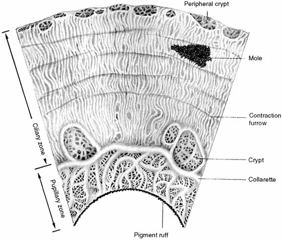
1. Duke-Elder S, Wybar K: The eye. In Duke-Elder S (ed): The Anatomy of the Visual System, Vol II. St. Louis: CV Mosby, 1961:75–386

2. Hogan M, Alvarado J, Weddell J: Histology of the Human Eye—An Atlas and Textbook. Philadelphia: WB Saunders, 1971

3. Whitnall S: The Anatomy of the Human Orbit and Accessory Organs of Vision. London: Oxford University Press, 1932

4. Stenström S: Untersuchungen über die Variation und Kovariation der optischen Elemente des menshlichen Auges. Acta Ophthalmol (Copenh) 26:1, 1946

5. Sorsby A, Sheridan M: The eye at birth: measurement of the principal diameters in forty-eight cadavers. J Anat 94:192, 1960

6. Testut L: Traité d'anatomie humaine, 5th ed. Paris, 1905

7. Weiss L: Uber das wachstum des menschlichen auges und uber die veranderung der muskelinsertionen am wachsenden auge (anatomische hefte). Arb Anat Inst Weisbaden 8:191, 1897

8. Brubaker RF, Pederon JE: Ciliochoroidal detachment. Surv Ophthalmol 27:281, 1983

9. Migliori ME, Gladstone GJ: Determination of the normal range of exophthalmometric values for black and white adults. Am J Ophthalmol Oct 15;98(4):438–442, 1984

10. Lemke BN: Anatomy of the ocular adnexa and orbit. In Smith BC (ed): Ophthalmic Plastic and Reconstructive Surgery, Vol. 1, Part 1. St. Louis: C.V. Mosby, 1987

11. Bron AJ, Tripathi RC, Tripathi BJ: Wolff's Anatomy of the Eye and Orbit, 8th ed. London: Chapman & Hall, 1997

12. Maurice D: Cornea and sclera. In Davson H (ed): Physiology of the Eye, 2nd ed. Boston: Little, Brown and Co, 1969:6,14,15

13. Vannas S, Teir H: Observations on struture and age changes in the human sclera. Acta Ophthalmol 38:268, 1960

14. Weale RA: A Biography of the Eye: Development, Growth Age. London: H.K. Lewis, 1982

15. Tripathi RC, Tripathi BJ: Functional anatomy of the anterior chamber angle. In Duane TD, Jaeger EA (eds): Biomedical Foundations of Ophthalmology. Philadelphia: Lippincott, 1982

16. Hayreh S, Vrabec F: The structure of the head of the optic nerve in the rhesus monkey. Am J Ophthalmol 61:136, 1966

17. Stevenson TC: Intrascleral nerve loops: a clinical study of frequency and treatment. Am J Ophthalmol 55:935, 1963

18. Fuchs E: Beitrage Beiträge zur normalen Anatomie des Augapfels. Albrecht Von Graefes Arch Klin Exp Ophthalmol 30:1, 1894

19. Rutin U: Fundus appearance in the normal eye. I. The choroid. Am J Ophthalmol 64:821, 1967

20. Von Noorden G: Burian-Von Noorden's Binocular Vision and Ocular Motility. St. Louis: CV Mosby, 1985

21. Apt L: An anatomical evaluation of rectus muscle insertions. Trans Am Ophthalmol Soc 78:365, 1980

22. Lang J, Horn T, von den Eichen U: Über die äusseren Augenmuskeln und ihre Ansatzzonen. Gegenbaurs Morphol Jahrb 126:817, 1980

23. Fink W: Surgery of the Vertical Muscles of the Eyes. Springfield, IL:. Charles C Thomas, 1962

24. Stallard H: Eye Surgery. Baltimore: Williams & Wilkins, 1950

25. Mishima S: Some physiologic aspects of the precorneal tear film. Arch Ophthalmol 73:233, 1965

26. Klyce SD, Beuerman RW: Structure and function of the cornea. In Kaufman HE, Barron BA, McDonald MB, Waltman SR (eds): The Cornea. New York: Churchill Livingston, 1988

27. Rapuano CJ, Fishbaugh JA, Strike DJ: Nine point corneal thickness measurements and keratometry readings in normal corneas using ultrasound pachymetry. Insight 18:16, 1993

28. Eysteinsson T, Jonasson F, Sasaki H, Arnarsson A, Scerrisson T, Sasaki K, Stefansson E: Reykjavik Eye Study Group: Central corneal thickness, radius of the corneal curvature and intraocular pressure in normal subjects using non-contact techniques: Reykjavik Eye Study. Acta Ophthalmol Scand 80:11, 2002

29. Liu Z, Pflugfelder SC: The effects of long-term contact lens wear on corneal thickness, curvature, and surface regularity. Ophthalmology 107:105, 2000

30. Myrowitz EH, Melia M, O'Brien TP: The relationship between long-term contact lens wear and corneal thickness. CLAO J 28:217, 2002

31. Pflugfelder SC, Liu Z, Feuer W, Verm A: Corneal thickness indices discriminate between keratoconous and contact lens-induced corneal thinning. Ophthalmology 109:2336, 2002

32. Sanchis-Gimeno JA, Lleo A, Alonso L, Rahhal MS, Martinez-Soriano F: Differences in corneal anatomy in a pair of monozygotic twins due to continuous contact lens wear. Cornea 22:243, 2003

33. Perez JG, Meijome JM, Jalbert I, Sweeney DF, Erickson P: Corneal epithelial thinning profile induced by long-term wear of hydrogel lenses. Cornea 22:304, 2003

34. Gonzalez-Meijome JM, Gonzales-Perez J, Cervino A, Yebra-Pimentel E, Parafita MA: Changes in corneal structure with continuous wear of high-Dk soft contact lenses: a pilot study. Optom Vis Sci. 80:440, 2003

35. von Bahr G: Corneal thickness: Its measurement and changes. Am J Ophthalmol 42:251, 1956

36. Martola E, Baum J: Central and peripheral corneal thickness. Arch Ophthalmol 79:28, 1968

37. Koch DD, Haft EA: Introduction to corneal topography. In Sanders DR, Koch DD (eds): An Atlas of Corneal Topography. Thorofare, NJ:. SLACK Inc., 1993

38. Guirao A, Redondo M, Artal P: Optical aberrations of the human cornea as a function of age. J Opt Soc Am A Opt Image Sci Vis 17:1697, 2000

39. Snell RS, Lemp MA: Clinical Anatomy of the Eye. Cambridge, MA:. Blackwell Scientific Publications, Inc., 1989

40. Morrison J, van Buskirk EM: Anterior collateral circulation in the primate eye. Ophthalmology 90:707, 1983

41. Brancato R, Bandello F, Lattanzio :. Atlas of Iris Fluorescein Angiography. Milano: Ghedini Editore, 1995

42. Hayreh S: The ophthalmic artery. III. Branches. Br J Ophthalmol 46:212, 1962

43. Virchow H: Graefe-Saemisch handbuch der gesammten Augenheilkunde. Leipzig: Engelmann, 1910

44. Tousimis A, Fine B: Ultrastructure of the iris: The intercellular components. Am J Ophthalmol 48:397, 1959

45. Tousimis A, Fine B: Ultrastructure of the iris: The intracellular stromal components. AMA Arch Ophthalmol 62:974, 1959

46. Ehinger B: Ocular and orbital vegetative nerves. Acta Physiol Scand (suppl 268) 67:1, 1966

47. LaGrange F: Un moyen de relevement rélévement de la tension oculaire. Ann Ocul (Paris) 157:447, 1920

48. Cosar CB, Senar AB: Orbscan corneal topography system in evaluating the anterior structures of the human eye. Cornea 22:118, 2003

49. Aizawa K: The depth of the anterior chamber. Nippon Ganka Gakkai Zasshi 62:2283, 1958

50. Tornquist R: Shallow anterior chamber in acute glaucoma. Acta Ophthalmol (Copenh) (suppl 39) 31:1, 1953

51. Weekers R, Grieten J, Lavergne G: Study of the dimensions of the human anterior chamber. Ophthalmologica 142:361, 1961

52. Weekers R, Grieten J: The measurement of the depth of the anterior chamber in clinical practice. Bull Soc Belge Ophtalmol 129:361, 1961

53. Weekers R, Grieten J, Lekeux M: Etude des dimension de la chambre anterieure antérieure de l'oeil humain. IV. L'intumescence cristallinienne et ses conséquences chirurgicales. Ophthalmologica 146:57, 1963

54. Touzeau O, Allouch C, Borderie V, Kopito R, Laroche L: Correlation between refraction and ocular biometry. J Fr Ophtalmol 26:355, 2003

55. Wong TY, Foster PJ, Ng TP, Tielsch JM, Johnson GJ, Seah SK: Variations in ocular biometry in an adult Chinese population in Singapore: the Tanjong Pagar Survey. Invest Ophthalmol Vis Sci 42:73, 2001

56. Fontana ST, Brubaker RF: Volume and depth of the anterior chamber in the normal aging human eye. Arch Ophthalmol 98:1803, 1980

57. Lyhne N, Sjolie AK, Kyvik KO, Green A: The importance of genes and environment for ocular refraction and its determiners: a population based study among 20–45 year old twins. Br J Ophthalmol 85:1470, 2001

58. Garner LF, Yap MK: Changes in ocular dimensions and refraction with accommodation. Ophthalmic Physiol Opt 17:12, 1997

59. Stamper R: Aqueous humor: Secretion and dynamics. In Tasman W, Jaeger E (eds): Duane's Foundations of Clinical Ophthalmology, Vol 2., Philadelphia: JB Lippincott, 1992

60. Brubaker RF, Nagataki S, Townsend DJ, Burns RR, Higgins RG, Wentworth W: The effect of age on aqueous humor formation in man. Ophthalmology 88:283, 1981

61. Caprioli J: The ciliary epithelia and aqueous humor. In Hart WM (ed): Adler's Physiology of the Eye: Clinical Application, 9th ed. St. Louis: Mosby, 1992

62. DiMatteo J: Active transport of ascorbic acid into lens epithelium of the rat. Exp Eye Res 66:731, 1998

63. Hoskins HD, Kass MA: Aqueous humor outflow. In Hoskins HD, Kass MA (eds): Becker-Schaffer's Diagnosis and Therapy of the Glaucomas. St. Louis: Mosby, 1989

64. Johnson F, Maurice D: A simple method of measuring aqueous humor flow with intravitreal fluoresceinated dextrans. Exp Eye Res 39:791, 1984

65. Van Buskirk EM: The anatomy of the limbus. Eye 3:101, 1989

66. Luo YR, Luo WB: A study of the surgical anatomy of the corneo-scleral limbus. Zhonghua Yan Ke Za Zhi 29:362, 1993

67. Burian H, Braley A, Allen L: External and gonioscopic visibility of the ring of Schwalbe and the trabecular zone. Trans Am Ophthalmol Soc 52:389, 1955

68. Tamm E, Flugel C, Stefani FH, Rohen JW: Contractile cells in the human scleral spur. Exp Eye Res 54:531, 1992

69. Stegman Z, Sokol J, Liebmann JM, Cohen H, Tello C, Ritch R: Reduced trabecular meshwork height in juvenile primary open-angle glaucoma. Arch Ophthalmol 114:660, 1996

70. Salzmann M: The Anatomy and Histology of the Human Eyeball. Brown EVL (trans): Chicago: University of Chicago Press, 1912

71. Allen L, Burian H, Braley A: The anterior border ring of Schwalbe and the pectinate ligament. Arch Ophthalmol 53:799, 1955

72. Ashton N, Brini A, Smith R: Anatomical studies of trabecular meshwork of normal human eye. Br J Ophthalmol 40:257, 1956

73. Holmberg A: Schlemm's canal and the trabecular meshwork. An electron microscopic study of the normal structure in man and monkey (cerecopithecus ethiops). Doc Ophthalmol (Den Haag) 19:339, 1965

74. Moses RA, Grodzki WJ Jr : The scleral spur and scleral roll. Invest Ophthalmol Vis Sci 16:925, 1977

75. Unger H, Jankovsky F: Sehnen und Stützelemente im Trabeculum corneosclerale. Albrecht Von Graefes Arch Klin Exp Ophthalmol 170:355, 1966

76. Flocks M: The anatomy of the trabecular meshwork as seen in tangential section. Arch Ophthalmol 56:708, 1956

77. Garron L, Feeney M, Hogan M, McEwen W: Electron microscopic studies of the human eye. I. Preliminary investigations of the trabeculae. Am J Ophthalmol 46:27, 1958

78. Garron L, Feeney M: Electron microscopic studies of the human eye. II. Study of the trabeculae by light and electron microscopy. Arch Ophthalmol 62:966, 1959

79. Fine B: Structure of the trabecular meshwork and the canal of Schlemm. Trans Am Acad Ophthalmol Otolaryngol 70:777, 1966

80. Gabelt BT, Gottanka J, Lutjen-Drecoll E, Kaufman PL: Aqueous humor dynamics and trabecular meshwork and anterior ciliary muscle morphologic changes with age in rhesus monkeys. Invest Ophthalmol Vis Sci 44:2118, 2003

81. McEwen W: Application of Poiseuille's law to aqueous outflow. Arch Ophthalmol 60:290, 1958

82. Tripathi RC, Tripathi BJ: Human trabecular endothelium, corneal endothelium, keratocytes, and scleral fibroblasts in primary cell culture. A comparative study of growth characteristics, morphology, and phagocytic activity by light and scanning electron microscopy. Exp Eye Res 35:611, 1982

83. Allingham RR, de Kater AW, Ethier CR: Schlemm's canal and primary open angle glaucoma: correlation between Schlemm's canal dimensions and outflow facility. Exp Eye Res 62:101, 1996

84. Ashton N: Anatomical study of Schlemm's canal and aqueous veins by means of neoprene casts, Part 1. Aqueous veins. Br J Ophthalmol 35:291, 1951

85. Rohen J: Das Auge und seine Hilfsorgane. In Bargman W (Kiel) (ed): Handbuch der mikroskopischen anatomie des Menschen, Vol III, Haut und Sinnesorgane, Part IV, Suppl to Vol III/2. Berlin: Springer-Verlag, 1964

86. Parc CE, Johnson DH, Brilakis HS: Giant vacuoles are found preferentially near collector channels. Invest Ophthalmol Vis Sci 41:2984, 2000

87. Theobald G: Schlemm's canal. Its anastomosis and anatomic relations. Trans Am Ophthalmol Soc 32:574, 1934

88. Ascher K:. The Aqueous Veins. Springfield, IL:. Charles C Thomas, 1961

90. Sondermann R: The formation, morphology and function of Schlemm's canal. Acta Ophthalmol (Copenh) 11:280, 1933

91. Francois François J, Neetens A, Collette J: Microradiographic study of the inner wall of Schlemm's canal. Am J Ophthalmol 40:491, 1955

92. Unger H: Anatomy of the angle of the anterior chamber. Klin Monatsbl Augenheilkd 135:161, 1959

93. Iwamoto T: Light and electron microscopy of Sondermann's channels of the human trabecular meshwork. Albrecht Von Graefes Arch Klin Exp Ophthalmol 172:197, 1967

94. Iwamoto T: Further observation on Sondermann's channels of the human trabecular meshwork. Albrecht Von Graefes Arch Klin Exp Ophthalmol 172:213, 1967

95. Prost M: Anatomy of the ciliary body and cyclocryotherapy. Ophthalmologica 188:9, 1984

96. Smith SG, Snowden F, Lamprecht EG: Topographical anatomy of the ciliary sulcus. J Cataract Refract Surg 13:543, 1987

97. Straatsma B, Foos R, Spencer L: The Retina—Topography and Clinical Correlations. Proceedings of The New Orleans Academy of Ophthalmology Symposium on the Retina and Retinal Surgery. St. Louis: CV Mosby, 1969

98. Silverman RH, Lizzi FL, Ursea BG, Rondeau MJ, Eldeen NB, Kaliscz A, Lloyd HO, Coleman DJ: High-resolution ultrasonic imaging and characterization of the ciliary body. Invest Ophthalmol Vis Sci 42:885, 2001

99. McCulloch C: The zonules of Zinn: Its origin, course, and insertion and its relation to neighboring structures. Trans Am Ophthalmol Soc 52:525, 1954

100. Beauviex M: La zonule (e é tude topographique et histologique). Arch Ophthalmol 39:484, 1922

101. Rohen J, Rentsch F: Der Konstructive Bau des Zonulaapparates beim Menschen und dessen funktionelle Bedeutung. Graefes Arch Clin Exp Ophthalmol 178:1, 1969

102. Rohen J: Scanning electron microscopic studies of the zonular apparatus in human and monkey eyes. Invest Ophthalmol Vis Sci 18:133, 1979

103. Holmberg A: Studies of the ultrastructure of the non-pigmented epithelium of the ciliary body. Acta Ophthalmol Scand 33:377, 1955

104. Alm A, Bill A: Ocular and optic nerve blood flow at normal and increased intraocular pressure in monkeys (Macaca iris): A study with radioactively labelled microspheres including flow determination in brain and some other tissues. Exp Eye Res 15:15, 1973

105. Alm A, Bill A: Ocular Circulation. St. Louis: CV Mosby, 1987:183–203

106. Caprioli J, Sears M, Mead A: Ocular blood flow in phakic and aphakic monkey eyes. Exp Eye Res 39:1, 1984

107. Woodlief N: Initial observations on the ocular microcirculation in man. I. The anterior segment and extraocular muscles. Arch Ophthalmol 98:1268, 1980

108. Taniguchi Y: Fine structure of blood vessels in the ciliary body. Jpn J Ophthalmol 6:93, 1962

109. Calasans O: The architecture of the ciliary muscle of man. An Fac Med Univ Sao Paolo 27:3, 1953

110. Gullstrand A: Einführung in die Methoden der Dioptrik des Auges des Menschen. Leipzig, S Hirzel Verlag, 1911

111. Smith P: Diseases of the crystalline lens and capsule. Trans Ophthalmol Soc UK 3:79, 1883

112. Heine L: Physiologisch-anatomische Untersuchungen über die Accomodation des Vogelauges. Albrecht Von Graefes Arch Ophthalmol 45:469, 1898

113. Scammon R, Hesdorffer M: Growth in mass and volume of human lens in postnatal life. Arch Ophthalmol 17:104, 1937

114. Fine B, Tousimis A: The structure of the vitreous body and the suspensory ligaments of the lens. Arch Ophthalmol 65:95, 1961

115. Brown NP, Bron AJ: Lens Disorders: Clinical Manual of Cataract Diagnosis. Oxford: Butterworth-Heinemann Ltd, 1996

116. Hayreh S: Segmental nature of the choroidal vasculature. Br J Ophthalmol 59:631, 1975

117. Guyer DR, Schachat AP, Green WR: The choroids: structural considerations. In Ryan SJ (ed): Retina, 3rd ed., Vol. 1, Part 1. St. Louis: Mosby, Inc., 2001

118. Wolfrum M: Contributions to the anatomy and histology of the choroid in man and higher animals. Albrecht Von Graefes Arch Ophthalmol 67:307, 1908

119. Sumita R: The fine structure of Bruch's membrane in the choroid. Nippon Ganka Gakkai Zasshi 65:1188, 1961

120. Hogan M: Ultrastructure of the choroid. Its role in the pathogenesis of choroidal diseases. Trans Pacific Coast Oto Ophthalmol Soc 42:61, 1961

121. Straatsma B, Hall M, Allen R, Crescitelli F: The Retina—Morphology, Function and Clinical Characteristics. Berkeley: University of California Press, 1969

122. Cohen A: The Retina. St. Louis: CV Mosby, 1987:458–490

123. Thomson A: The Anatomy of the Human Eye. Oxford: Clarendon Press, 1912

124. Landau D, Schneidman EM, Jacobovitz T, Rozenman Y: Quantitative in vivo retinal thickness measurements in healthy subjects. Ophthalmology 104:639, 1997

125. Konno S, Akiba J, Yoshida A: Retinal thickness measurements with optical coherence tomography and the scanning retinal thickness analyzer. Retina 21:57, 2001

126. Gorrand JM, Delori FC: Reflectance and curvature of the inner limiting membrane at the foveola. J Opt Soc Am A Opt Image Sci Vis 16:1229, 1999

127. Sigelman J, Ozanics V: Retina. In Jakobiec F (ed): Ocular Anatomy, Embryology, and Teratology. Philadelphia: Harper & Row, 1982

128. Hayreh S: The central retinal artery of the retina: Its role in the blood supply of the optic nerve. Br J Ophthalmol 47:651, 1963

129. Michaelson I: Retinal circulation in man and animals. Springfield, IL:. Charles C Thomas, 1954

130. Toussaint D, Kuwabara T, Cogan D: Retinal vascular patterns. Arch Ophthalmol 65:575, 1961

131. Bek T, Jensen PK: Three-dimensional structure of human retinal vessels studied by vascular casting. Acta Ophthalmol (Copenh) 71:506, 1993

132. Justice J, Lehmann R: Cilioretinal arteries: A study based on review of stereo fundus photographs and fluorescein angiographic findings. Arch Ophthalmol 94:1355, 1976

133. Weinberg DV, Egan KM, Seddon JM: Asymmetric distribution of arteriovenous crossings in the normal retina. Ophthalmology 100:31, 1993

134. Bairati A Jr, Orzalesi N: The ultrastructure of the pigment epithelium and of the photoreceptor-pigment epithelium junction in the human retina. J Ultrastruct Res 9:484, 1963

135. Tso M, Friedman E: The retinal pigment epithelium. I. Comparative histology. Arch Ophthalmol 78:641, 1967

136. Friedman E, Tso M: The retinal pigment epithelium. II. Histologic changes associated with age. Arch Ophthalmol 79:315, 1968

137. Tso M, Friedman E: The retinal pigment epithelium. III. Growth and development. Arch Ophthalmol 80:214, 1968

138. Zinn KM, Benjamin-Henkind J: Retinal pigment epithelium. In Jakobiec F (ed): Ocular Anatomy, Embryology, and Teratology. Philadelphia: Harper & Row, 1982

139. Hudspeth AJ, Yee AG: The intercellular junctional complexes of the retinal pigment epithelia. Invest Ophthalmol 12:354, 1973

140. Cohen A: Rods and cones. In Fuortes M (ed): Handbook of Sensory Physiology, Vol 7/2. Berlin: Springer-Verlag, 1972

141. Osterberg G: Topography of the layer of rods and cones in the human retina. Acta Ophthalmol (suppl) 6:8, 1936

142. Ahnelt PK: The photoreceptor mosaic. Eye 12:531, 1998

143. Rostgaard J, Qvortrup K: A note about retinal structure and visual acuity. A light microscopic study of the cones in fovea centralis. Acta Ophthalmol Scand 77:45, 1999

144. Yamada E: Some structural features of the fovea centralis in the human retina. Arch Ophthalmol 82:151, 1969

145. Arey L: Retina, Choroid and Sclera. New York: Paul B. Hoeber, 1932:743

146. Jones AL, Sheen NJ, North RV, Morgan JE: The Humphrey optical coherence tomography scanner: quantitative analysis and reproducibility study of the normal human retinal nerve fibre layer. Br J Ophthalmol 85:673, 2001

147. Varma R, Bazzaz S, Lai M: Optical tomography-measured retinal nerve fiber layer thickness in normal Latinos. Invest Ophthalmol Vis Sci 44:3369, 2003

148. Mok KH, Lee VW, So KF: Retinal nerve fiber layer measurement of the Hong Kong Chinese population by optical coherence tomography. J Glaucoma 11:481, 2002

149. Kanamori A, Escano MF, Eno A, Nakamura M, Maeda H, Seya R, Ishibashi K, Negi A: Evaluation of the effect of aging on retinal nerve fiber layer thickness measured by optical coherence tomography. Ophthalmologica 217:273, 2003

150. Polyak S: The Retina. Chicago: University of Chicago Press, 1941

151. Hogan M: The vitreous, its structure and relation to the ciliary body and retina. Invest Ophthalmol 2:418, 1963

152. Foos R: Vitreoretinal juncture: Topographical variations. Invest Ophthalmol 11:801, 1972

153. Oppel O: Microscopic examinations of the number and thickness of the myelinated nerve fibers in the human optic nerve. Albrecht Von Graefes Arch Klin Exp Ophthalmol 166:19, 1963

154. Kupfer C, Chumbley L, Downer J: Quantitative histology of optic nerve, optic tract, and lateral geniculate of man. J Anat 101:393, 1967

155. Quigley HA, Brown AE, Morrison JD, Drance SM: The size and shape of the optic disc in normal human eyes. Arch Ophthalmol 108:51, 1990

156. Agarwal HC, Gulati V, Sihota R: The normal optic nerve head on Heidelberg Retina Tomograph II. Indian J Ophthalmol 51:25, 2003

157. Jonas JB, Thomas R, George R, Berenshtein E, Muliyil J: Optic disc morphology in south India: the Vellore Eye Study. Br J Ophthalmol 87:189, 2003

158. Brown GC, Tasman WS: Congenital Anomalies of the Optic Disc. New York: Grune & Stratton, 1983

159. Digre KB, Corbett JJ: Practical Viewing of the Optic Disc. Burlington: Elsevier Science, Inc., 2003

160. Anderson D, Braverman S: Reevaluation of the optic disc vasculature. Am J Ophthalmol 82:165, 1976

161. Hayreh S: Blood supply of the optic nerve head and its role in optic atrophy, glaucoma, and oedema of the optic disc. Br J Ophthalmol 53:721, 1969

162. Lieberman M, Maumenee A, Green W: Histologic studies of the vasculature of the anterior optic nerve. Am J Ophthalmol 82:405, 1976

163. Wolter J: The human optic papilla. A demonstration of new anatomic and pathologic findings. Am J Ophthalmol 44:48, 1957

164. Dutton JJ: Atlas of Clinical and Surgical Orbital Anatomy. Philadelphia: W.B. Saunders Company, 1994

165. Anderson D: Ultrastructure of the human and monkey lamina cribrosa and optic nerve head. Arch Ophthalmol 82:800, 1969

166. Anderson D: Ultrastructure of the optic nerve head. Arch Ophthalmol 83:63, 1970

167. Anderson D, Hoyt W: Ultrastructure of the intraorbital portion of human and monkey optic nerve. Arch Ophthalmol 82:506, 1969

168. Anderson D, Hoyt W, Hogan M: The fine structure of the astroglia in the human optic nerve and optic nerve head. Trans Am Ophthalmol Soc 65:275, 1967

169. Ducournau D: A new technique for the anatomical study of the choroidal blood vessels. Ophthalmologica 184:190, 1982

170. Visser-Heerema J: Über das spezifische Gewicht der bei der Operation von Netzhautablösungen gewonnenen Flüssigheit. Arch Augenheilkd 109:543, 1936

171. Redslob E: Le corps vitré. Son dévelopment, sa structure, ses propiétes physicochimiques. Paris: Masson et Cie, 1932

172. Richards O, Hague E: Vitreous as removed with large and small needles. Am J Ophthalmol 55:151, 1963

173. Guggenheim I, Franceschetti A: Refraktometrische Untersuchungen des Glaskörpers von Kaninchen un Mensch unter physiologischen und pathologischen Bedingungen. Arch Augenheilkd 98:448, 1928

174. Paufique L, Moreau P: Les greffes de vitre vitré lyophilise lyophilisé. Ann Ocul (Paris) 186:873, 1953

175. Balazs E: Physiology of the vitreous body. In Schepens C (ed): Importance of the Vitreous Body in Retina Surgery with Special Emphasis on Reoperations. St. Louis: CV Mosby, 1960

176. Berman E, Michaelson I: The chemical composition of the vitreous body as related to age and myopia. Exp Eye Res 3:9, 1964

177. Sebag J: The Vitreous: structure, function, and pathobiology. New York: Springer-Verlag, 1989

178. Goldman H: Biomicroscopy of the vitreous body. Arch Ophthalmol 127:334, 1954

178a. Michels RG, Wilkinson CP, Rice TA: Retinal Detachment. St. Louis: Mosby, Inc., 1990

179. Ergglet H: Klinische Befunde bei focaler Beleuchtung mit der Gullstrandischen Nernst-Spaltlampe. Klin Monatsbl Augenheilk 53:449, 1914

180. Busacca A: Observations biomicroscopiques sur le corps ciliaire normal et pathologique. Bull Soc Ophthalmol Fr 68:295, 1955

181. Green WR: Vitreo retinal juncture. In Ryan SJ (ed): Retinal Disease. St. Louis: CV Mosby, 1989

182. Friedenwald J, Stiehler R: Structure of the vitreous. Arch Ophthalmol 14:789, 1935

183. Vogt A: Ueber Berührungspunkte der senilen und der myopichen Bulbusdegeneration. Klin Monatsbl Augenheilkd 72:212, 1924

184. Jack RL: Regression of the hyaloid artery system. An ultrastructural analysis. Am J Ophthalmol 74:261, 1972

185. Bloom GD, Balazs EA, Ozanics V: The fine structure of the hyaloid arteriole in bovine vitreous. Exp Eye Res 31:129, 1980

186. Reeser FH, Aaberg T: Vitreous humor. In Records PE (ed): Physiology of the Human Eye and Visual System. Hagerstown, MD:. Harper & Row, 1979

187. Gartner J: Electronmicroscopic study on the fibrillar network and fibrocyte-collagen interactions in the vitreous cortex at the ora serrata of human eyes with special regard to the role of disintegrating cells. Exp Eye Res 42:21, 1986

188. Brini A, Bronner A, Gerhard J-P, Nordmann J: Biologie et Chirurgie du Corps Vitre. Paris: Masson et Cie, 1968

189. Fine B: Retinal structure: Light and electronmicroscopic observations. In McPherson A (ed): New and Controversial Aspects of Retinal Detachment. New York: Harper & Row, 1968

190. Pau M: Zur Entwicklung der Glaskörperstrukturen und der Zonula. Ophthalmologica 134:320, 1957

191. Gärtner J: Klinische Beobachtungen über Glaskörperadhärenzen am hinteren Augenpol. Klin Monatsbl Augenheilkd 140:161, 1962

192. Schepens C: Clinical aspects of pathologic changes in the vitreous body. Am J Ophthalmol 38:8, 1954

193. Gärtner J: Klinische Beobachtungen über den Zusammenhang der Glaskörpergrenzmembran mit Glaskörpergerüst und Netzhautgefässen in der Ora- Aequatorgegend. Klin Monatsbl Augenheilkd 140:524, 1962

194. Rieger H: Zur Histologie der Glaskörperabhebung, Teir II. Über die Beziehungen des aabgehobenen Glaskörpers zur Netzhaut. Albrecht Von Graefes Arch Klin Exp Ophthalmol 146:447, 1944

Back to Top