|
|
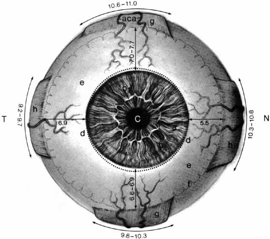
| Fig. 1 Normal human eye, anterior aspect. N, nasal; T, temporal. The cornea (a) is anterior to the iris (b) and pupil (c). The elliptic shape of the anterior corneal margin (arrows) is compared to the round shape of its posterior one (dotted line). The collarette of the iris is evident at b1. The pupil is displaced slightly to the nasal side of the eye. The limbal area and external scleral sulcus surround the margin of the cornea (d). The sclera (e) is peripheral to the limbus. The sclera has been made artificially transparent to show the ora serrata (f), which is farther posterior on the temporal than on the nasal side. The superior and inferior rectus tendons (g) are curved and are inserted obliquely to the axis of the eye. The tendons of the medial and lateral recti (h) also have curved insertions that are not oblique to the horizontal meridian. Each rectus muscle has two anterior ciliary arteries (aca) except the lateral rectus, which has one. The measurements of all these structures are given in millimeters. (Hogan M, Alvarado J, Weddell J: Histology of the Human Eye—An Atlas and Textbook. Philadelphia: WB Saunders, 1971:46) |