|
|
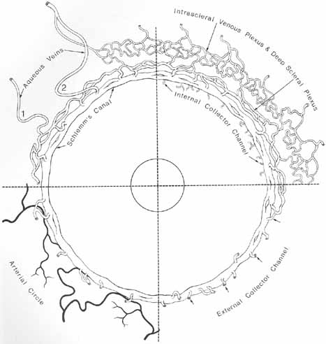
| Fig. 14 Schematic drawing showing the circular course and related vessels of the canal of Schlemm. The canal divides into two or more portions intermittently. The drawing is divided into four portions by the dotted lines. The internal collector channels of Sondermann are labeled in the upper right sector as they extend into the trabecular meshwork. The external collector channels are seen in the upper and lower right sectors, arising from the canal and uniting with the deep intrascleral plexus or extending directly to the episcleral veins. The deep and intrascleral venous plexuses are external to the canal. In the upper left sector an aqueous vein (1) arises from the deep scleral plexus and another (2) arises from Schlemm's canal and runs directly to the episcleral venous plexus. External collector veins are seen to arise from the canal and join the deep scleral plexus. In the lower left sector the arteries of the deep sclera are seen to be in close relation to the canal of Schlemm. (Reprinted with permission from Hogan MJ, Alvarado JA, Weddell JE. Histology of the Human Eye—An Atlas and Textbook. Philadelphia: WB Saunders, 1971) |