|
|
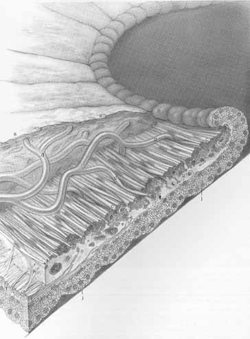
| Fig. 9 Pupillary portion of the iris. The dense, cellular anterior border layer (a) terminates at the pigment ruff (b) in the pupillary margin. The sphincter muscle is at (c). The arcades (d) from the minor circle extend toward the pupil and through the sphincter muscle. The sphincter muscle and the iris epithelium are close to each other at the pupillary margin. Capillaries, nerves, melanocytes, and clump cells (e) are found within and around the muscle. The three to five layers of dilator muscle (f) gradually diminish in number until they terminate behind the midportion of the sphincter muscle (arrow), leaving low, cuboidal epithelial cells (g) to form the anterior epithelium to the pupillary margin. Spur-like extensions from the dilator muscle form Michel's spur (h) and Fuch's spur (i), which extend anteriorly to blend with the sphincter muscle. The posterior epithelium (j) is formed by tall columnar cells with basally located nuclei. Its apical surface is contiguous with the apical surface of the anterior epithelium. (Reprinted with permission from Hogan MJ, Alvarado JA, Weddell JE. Histology of the Human Eye—An Atlas and Textbook. Philadelphia: WB Saunders, 1971) |