|
|
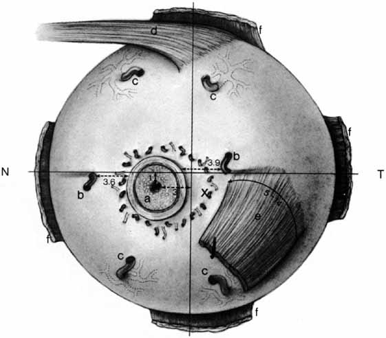
| Fig. 2 Drawing of the posterior eye. T, temporal; N, nasal. The optic nerve (a) with its central vessels and surrounding meningeal sheaths is seen. Its center is located 3 mm nasal and 1 mm inferior to the posterior pole of the eye. Surrounding it are the short posterior ciliary arteries and nerves. The approximate position of the macula is at x. Along the horizontal meridian, which bisects the eye, are the long posterior ciliary arteries and nerves (b). The exits of four vortex veins are shown, one for each quadrant (c). The curved, oblique insertions of the superior oblique (d) and inferior oblique (e) muscles are seen. The cut ends of the four rectus muscles are at f. (Hogan M, Alvarado J, Weddell J: Histology of the Human Eye—An Atlas and Textbook. Philadelphia: WB Saunders, 1971:51) |