|
|
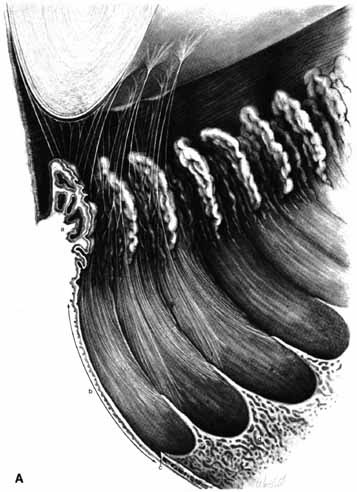
| Fig. 16a A. The inner aspect of the ciliary body showing the pars plicata (a) and the pars plana (b). The ora serrata is at c, and posterior to it the retina shows cystoid degeneration (d). The bays (e) and dentate processes (f) of the ora are shown; linear ridges or striae (g) project forward from the dentate processes across the pars plana to enter the valleys between the ciliary processes. The zonular fibers arise from the pars plana beginning 1.5 mm from the ora serrata. They curve forward from the sides of the dentate ridges into the ciliary valleys, then from the valleys to the lens capsule. Zonules coming from the valleys on either side of a ciliary process have a common point of attachment on the lens. The zonules attach up to 1 mm from the equator posteriorly and up to 1.5 mm from the equator anteriorly. At the equatorial border, the attaching zonules give a crenated appearance to the lens. The ciliary processes vary in size and shape and often are separated from each other by lesser processes. The radial (h) and circular furrows (i) of the peripheral iris are shown. |