|
|
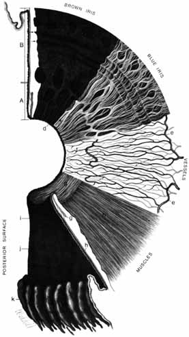
| Fig. 8 Composite drawing of the surfaces and layers of the iris. Beginning at the upper left and proceeding clockwise, the iris cross section shows the pupillary (a) and ciliary portions (b), and the surface view shows a brown iris with its dense, matted anterior border layer. Circular contraction furrows are shown (arrows) in the ciliary portion of the iris. Fuchs' crypts (c) are seen at either side of the collarette in the pupillary and ciliary portion and peripherally near the iris root. The pigment ruff is seen at the pupillary edge (d). A blue iris surface shows a less dense anterior border layer and more prominent trabeculae. The iris vessels are shown beginning at the major arterial circle in the ciliary body (e). Radial branches of the arteries and veins extend toward the pupillary region. The arteries form the incomplete minor arterial circle (f), from which branches extend toward the pupil, forming capillary arcades. The sector below it demonstrates the circular arrangement of the sphincter muscle (g) and the radial processes of the dialator muscle (h). The posterior surface of the iris shows the radial contraction furrows (i) and the structural folds (j) of Schwalbe. Circular contraction folds are also present in the ciliary portion. The pars plicata of the ciliary body is at k. (Hogan M, Alvarado J, Weddell J: Histology of the Human Eye—An Atlas and Textbook. Philadelphia: WB Saunders, 1971:207) |