|
|
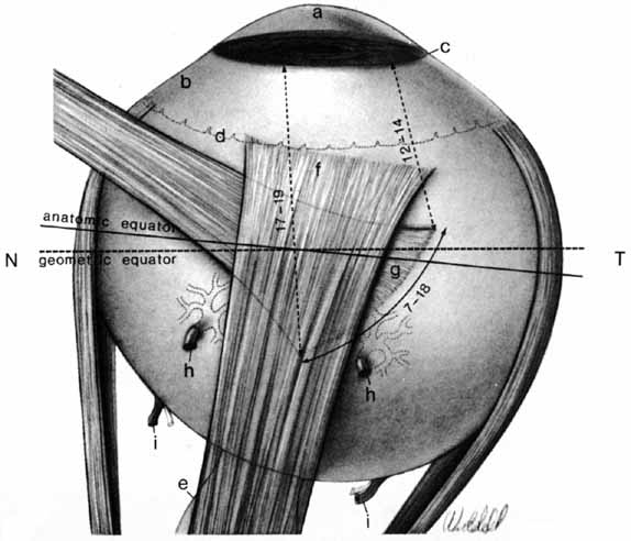
| Fig. 3 Drawing of the upper half of the eye. T, temporal; N, nasal. The contrasting degrees of curvature of the cornea (a) and sclera (b) are evident. At the limbus (c), where they join, is the external scleral sulcus. The relation of the ora serrata (d) to the surface is shown. The nasal displacement of the optic nerve (e) with respect to the posterior pole of the eye makes the three layers of the temporal eye longer than those on the nasal side. The slightly curved oblique insertion of the superior rectus muscle is at f, and the tendinous oblique insertion of the superior oblique muscle is at g. Two vortex veins are seen (h), and the long posterior ciliary arteries and nerves are at i. (Hogan M, Alvarado J, Weddell J: Histology of the Human Eye—An Atlas and Textbook. Philadelphia: WB Saunders, 1971:53) |