|
|
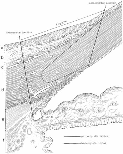
| Fig. 11 Drawing of a meridional section of the limbal area. The histologic limit of the corneolimbal junction is outlined by the dotted plane commencing at the termination of Bowman's layer and curving posteriorly toward Schlemm's canal, then extending forward to end at Descemet's membrane. Another definition of the limbus is that used by the pathologist; the anterior limit (corneolimbal junction) is formed by a plane joining the ends of Bowman's layer and Descemet's membrane; the posterior limit by a plane constructed 1.5 mm posterior to the corneolimbal junction at a right angle to the scleral surface in the superior and inferior limbus, and 2 mm in the horizontal meridian. The limbus has the following gross anatomic parts: conjunctival epithelium (a); conjunctival stroma (b); Tenon's capsule and episclera (c); and the limbal or corneoscleral stroma at (d). The longitudinal portion of the ciliary muscle is indicated at (e) and circular and radial bundles of the muscle at (f). (Reprinted with permission from Hogan MJ, Alvarado JA, Weddell JE. Histology of the Human Eye—An Atlas and Textbook. Philadelphia: WB Saunders, 1971) |