|
|
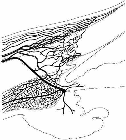
| Fig. 13 Meridional section of the eye showing the blood supply of the limbal area. An anterior ciliary artery (ACA) divides to form an episcleral (E) and a major perforating (MP) branch. The episclearl branches produce episcleral, conjunctival (C), and intrascleral (IS) nutrient vessels. The conjunctival vessels form the superficial marginal plexus of the cornea (SMP). Two sets of vessels arise from the superficial marginal plexus: one (1) extends forward to form the peripheral corneal arcades; the other forms recurrent vessels (2) that run posteriorly to supply 3 to 6 mm of the perilimbal conjunctiva. The latter eventually anastomose with the recurrent conjunctival vessels from the fornices. The major perforating artery passes through the sclera to join the major arterial circle (MAC) of the iris (note long ciliary arfery, LCA). At 3, a branch from the major perforating artery passes forward to form the intrascleral arterial channels of the limbus. This region often is supplied by a vessel that arises directly from the anterior ciliary artery as an episcleral vessel, such as the one indicated at 4. The major venous drainage from the limbus is into the episcleral veins, which then unite with the ophthalmic veins. The deep scleral venous plexus (5) is close to Schlemm's canal (SC). An aqueous vein (arrows) arises from the deep scleral plexus and joins the episcleral veins. The intrascleral venous plexus (6) forms an extensive network in the limbal stroma. An important part of the drainage from the ciliary plexus (CP) is into the deep and intrascleral venous plexuses. One of these channels is seen at 7. (Hogan M, Alvarado J, Weddell J: Histology of the Human Eye—An Atlas and Textbook. Philadelphia: WB Saunders, 1971:120) |