|
|
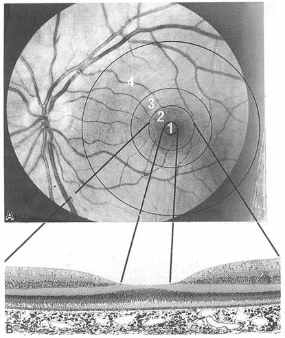
| Fig. 19 A. Fundus photograph of left human eye showing topographic demarcation of the area centralis that measures 5.5 to 6 mm in diameter (outermost circle) and its subdivisions (inner circles). From an anatomic standpoint, the zones demarcated are in fact horizontally elliptical rather than circular as depicted here. The central area of the macular region is represented by the fovea centralis (2), approximately 1.85 mm in diameter, which has a central pit, the foveola (1), 0.35 mm in diameter. The anatomically distinguishable retinal belts that surround the fovea centralis are the parafovea (3), 0.5 mm wide, and perifovea (4), 1.5 mm wide. B. Transverse section of the fovea retina matched to the fundus photograph show in (A). Photomicrograph, original magnification ×70. (From Tripathi RC, Tripathi BJ. In: Davson, H, ed. The Eye. Academic Press, 1984) |