|
|
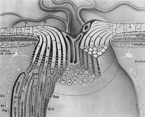
| Fig. 23 Three-dimensional drawing of the intraocular portion of the optic nerve and part of the orbital optic nerve. Where the retina terminates at the optic disc edge, the Müller cells (1a) are in continuity with the astrocytes, forming the internal limiting membrane of Elschnig (1b). In some specimens, Elschnig's membrane is thickened in the central portion of the disc to form the central meniscus of Kuhnt (2). At the posterior termination of the choroid on the temporal side, the border tissue of Elschnig (3) lies between the astrocytes surrounding the optic nerve canal (4) and the stroma of the choroid. On the nasal side, the choroidal stroma is directly adjacent to the astrocytes surrounding the nerve. This collection of astrocytes (4) surrounding the canal is known as the border tissue of Jacoby. This is continuous with a similar glial lining called the intermediary tissue of Kuhnt (5) at the termination of the retina. The nerve fibers of the retina are segregated into approximately 1000 bundles or fascicles by astrocytes (6). On reaching the lamina cribrosa (upper dotted line), the nerve fascicles (7) and their surrounding astrocytes are separated from each other by connective tissue. This connective tissue is the cribriform plate, which is an extension of scleral collagen and elastic fibers through the nerve. The external choroid also sends some connective tissue to the anterior part of the lamina. At the external part of the lamina cribrosa (lower dotted line), the nerve fibers become myelinated, and columns of oligodendrocytes and a few astrocytes are present within the nerve fascicles. The astrocytes surrounding the fascicles form a thinner layer here than in the laminar and prelaminar portion. The bundles continue to be separated by connective tissue all the way to the chiasm (Sep). This connective tissue is derived from the pia mater and is known as the septal tissue. A mantle of astrocytes (GI.M), continuous anteriorly with the border tissue of Jacoby, surrounds the nerve along its orbital course. The dura (Du), arachnoid (Ar), and pia mater (Pia) are shown. The central retinal vessels are surrounded by a perivascular connective tissue throughout its course in the nerve; this connective tissue blends with the connective tissue of the cribriform plate in the lamina cribrosa; it is called the central supporting connective tissue strand here. (Anderson D, Hoyt W: Ultrastructure of the intraorbital portion of human and monkey optic nerve. Arch Ophthalmol 82:506, 1969) |