Chapter 26
Degenerative Diseases of the Peripheral Retina
ROBERT E. ENGSTROM JR, BEN J. GLASGOW, ROBERT Y. FOOS and BRADLEY R. STRAATSMA
Main Menu   Table Of Contents

Search

TOPOGRAPHY AND RELATIONSHIPS
DEVELOPMENTAL VARIATIONS
TROPHIC AND TRACTIONAL PERIPHERAL RETINAL DEGENERATIONS
TRACTIONAL PERIPHERAL RETINAL DEGENERATIONS
REFERENCES

The peripheral retina may be defined somewhat arbitrarily as the anterior portion of the retina that begins about 3 mm posterior to the equator of the eye, where the conspicuous vortex veins pass from the choroid into the sclera and extend forward past the equator to the anterior termination of the retina at the ora serrata. This important segment of the retina and the structures lying anteriorly and posteriorly as well as internally and externally may be readily visualized with the techniques of ophthalmoscopy, biomicroscopy, and scleral depression. The physician's observations are recorded on an ocular fundus chart (Fig. 1) using a standardized color code (Table 1).

Fig. 1. Ocular fundus chart. Inner circle represents equator of eye; middle circle represents ora serrata; outer circle represents anterior border of pars plana ciliaris. Radiating lines represent meridians numbered according to clock hours. (Straatsma BR, Foos RY, Spencer LM: The retina: Topography and clinical correlations. In: Transactions of the New Orleans Academy of Ophthalmology: Symposium on Retina and Retinal Surgery. St Louis: CV Mosby, 1969.)


Examining the peripheral retina and recording the findings require accurate and detailed knowledge of normal topography, anatomic relationships, common developmental variations, significant degenerations, and the manifestations of specific disease entities.

Back to Top
TOPOGRAPHY AND RELATIONSHIPS
For topographic evaluation, the general size and shape of the peripheral retina and related structures are important. The average dimensions of the retina of the adult from the equator to the ora serrata are 5.07 ± 1.11 mm in the superior meridian, 4.79 ± 1.22 mm inferiorly, 5.81 ± 1.12 mm nasally, and 6.00 ± 1.22 mm temporally. From the ora serrata to Schwalbe's line, which constitutes the posteriorborder of the limbus, the distance is 6.14 ± 0.85 mmsuperiorly, 6.20 ± 0.76 mm inferiorly, 5.73 ± 0.81 mmnasally, and 6.53 ± 0.75 mm temporally (Fig. 2).1,2

Fig. 2. Peripheral retinal topography. Relationship of ora serrata to equator and to Schwalbe's line in the four principal meridians. Average measurements and standard deviations given in millimeters.

The retina expands from the optic disc to line the posterior pole and reach the equator, where it has an average diameter of 24.08 ± 0.94 mm in the vertical meridian and 24.03 ± 1.04 mm in the horizontal meridian. From the equator to the ora serrata, the retina diminishes in size so that at theora serrata the average retinal diameter is 20.41 ±1.09 mm vertically and 20.03 ± 1.04 mm horizontally (Fig. 3). Thus, the retina has the shape of a cup that is expanded to its greatest diameter at the equator and is considerably reduced in diameter at its serrated anterior margin.

Fig. 3. Retinal shape resembles a cup expanded to its greatest diameter at the equator and reduced in diameter at the ora serrata. Measurements and standard deviations given in millimeters.

The retina is considerably smaller in diameter and in circumference at the ora serrata than at the equator (see Fig. 3). Consequently, conventional charts of the ocular fundus, which depict the ora serrata as having a diameter and circumference greater than that of the equator, are misleading (see Fig. 1). These charts should be considered only as diagrammatic projections in which the retina anterior to the equator is disproportionately expanded, that is, features of the peripheral retina are actually closer together than they appear in the conventional fundus diagram.

Precise measurements of the optic disc diameter and the disc-to-fovea distance are also essential for correlation of topographic and clinical information. In the eye of the adult, the optic disc has an average vertical diameter of 1.86 ± 0.21 mm and an average horizontal diameter of 1.75 ± 0.19 mm. The center of the fovea is located an average distance of3.42 ± 0.34 mm temporal to and slightly below the temporal margin of the optic disc (Fig. 4). Inasmuch as the disc diameter (approximately 1.75 mm) is the standard for comparison in clinical examination of the retina, the distance from the equator to the ora serrata is approximately 3 disc diameters.

Fig. 4. Dimensions of optic disc and relationship between disc and center of the fovea. Average dimensions and standard deviations given in millimeters. (Straatsma BR, Foos RY, Spencer LM: The retina: Topography and clinical correlations. In: Transactions of the New Orleans Academy of Ophthalmology: Symposium on Retina and Retinal Surgery. St Louis: CV Mosby, 1969.)

The posterior margin of the peripheral retina is a line joining the points at which the vortex veins pass from the choroid to the sclera.3 This rather constant landmark is 3 mm (nearly 2 disc diameters) posterior to the equator of the eye.

The anterior edge of the peripheral retina is theora serrata, an irregularly scalloped border; the irregularities are more pronounced nasally than temporally and have extreme individual variations in contour (Fig. 5). Dentate or toothlike processes extend anteriorly from the main contour of this bor-der, and bays or indentations extend posteriorly from the main contour of the ora serrata. At theora serrata, those projections of the retina toward the vitreous body are termed meridional folds. There is a concentration of dentate processes, ora bays, and meridional folds in the superior nasal quadrant and a progressive decrease in these morphologic features in the inferior nasal, superior temporal, and inferior temporal quadrants (Figs. 6 and 7). Although individual variations in ora serrata contour may be extreme, studies of adult human eyes demonstrate that both of a patient's eyes are remarkably similar and that statistically, the “average” ora serrata has 16 dentate processes, 1 large or giant dentate process, 10 ora bays, and 1 double ora bay.1,2

Fig. 5. Composite scale drawing depicts peripheral retina, ora serrata, ciliary body, and lens as viewed from posterior aspect. Ora serrata have more dentate processes and ora bays in the nasal quadrants than in the temporal quadrants.

Fig. 6. Peripheral fundus showing preequatorial retina, ora serrata, and inner surface of ciliary body (smooth portion, pars plana; portion with ciliary processes, pars plicata). Ora serrata in this nasal sector shows largely short dentate processes that are typical; that is, they align with valleys between ciliary processes. On right, a giant dentate process is atypical; that is, it aligns with a ciliary process. Aligned with and posterior to the atypical dentate process is a focus of retinal thinning (peripheral retinal excavation). (× 14.)

Fig. 7. Dentate process, coronal section. Dentate process shows nonspecific degeneration (including microcystic change) and a cap of dense-staining glial cells on its central surface. Adjacent ciliary epithelium and photoreceptor cells underlying dentate process are largely unremarkable. (Hematoxylin-eosin; × 250.)

In clinical practice, morphologic variations in the contour of the ora serrata are most common in the superior nasal quadrant, least common in the inferior temporal quadrant, and subject to a wide range of individual irregularities. Thus, the ora serrata is depicted on the conventional ocular fundus diagram as a scalloped line straddling the middle circle on the chart and reflecting variations in the structure that are pertinent to the localization of clinically significant abnormalities.

Coursing through the peripheral retina are arterioles and venules that travel separately and are evenly distributed. Arterioles are smaller in caliber and lighter in color than venules; however, to distinguish the arterioles and venules it is usually necessary to trace the vessels posteriorly to an area where accurate differentiation is possible. As vessels extend anteriorly in the peripheral retina their size decreases; they occasionally may travel nearly parallel to the ora serrata and usually become invisible approximately 2 mm posterior to the ora serrata.

Externally, the retina is in contact with the retinal pigment epithelium and the choroid. Separation of the weak bond between the sensory retina and the retinal pigment epithelium is termed retinal detachment.

Internally, the retina contacts the vitreous body. This transparent, gel-like tissue fills two thirds of the globe, has a volume of 4 to 5 ml in adults, and is in contact with the optic disc, ciliary body, zonule, and lens. Although united to some extent with each of these structures in the normal eye of the young adult, the vitreous is joined most firmly with surrounding structures at the vitreous base, a circular band extending anteriorly and posteriorly from the ora serrata.

The anterior portion of the vitreous base is the area between the ora serrata and the origin of the anterior hyaloid membrane. In adult eyes stud-ied at autopsy, the anterior vitreous base usually conformed to the contour of the ora serrata and measured 0.26 ± 0.16 mm in the nasal horizontal meridian and 1.32 ± 0.29 mm in the temporal horizontal meridian.1,2

The posterior portion of the vitreous base is the zone of strong vitreoretinal attachment that extends posterior to the ora serrata; that is, it is the area between the ora serrata and the most anterior extent to which the vitreous may be detached without severely disrupting the inner retinal layers. In postmortem studies of adult eyes, the posterior vitreous base presented a relatively smooth posterior border; it measured 3.03 ± 0.84 mm in the nasal horizontal meridian and 1.81 ± 0.64 mm in the temporal horizontal meridian.4

The vitreous base is a major factor in the localization and clinical course of developmental variations, degenerations, and diseases. It involves the full circumference of the peripheral fundus, measures approximately 3.2 mm (nearly two disc diameters) anteroposteriorly, generally reflects the anterior contour of the ora serrata, presents a relatively smooth posterior border, extends onto the peripheral retina for a greater distance nasally than temporally, and usually is associated with visibly increased pigmentation in the corresponding portion of the retinal and ciliary body pigment epithelium.

These findings are characteristic of the peripheral retina and adjacent structures in the normal eye of the young adult; the peripheral retina actually undergoes changes throughout life. The peripheral retina in an infant has brilliant surface light reflexes and becomes increasingly white and translucent near the ora serrata. This translucency and some actual retinal redundancy in the infant accentuate the abrupt anterior termination of the retina at the ora serrata. However, Lange's fold, a detachment of the retina immediately posterior to the ora serrata, is a laboratory artifact seen during postmortem examination and in surgically enucleated eyes of infants as a result of the extremely strong vitreoretinal attachments in the posterior portion of the vitreous base in infants. Anterior to the ora serrata, the pars plana is underdeveloped in infants; therefore, the ciliary processes are relatively close to the ora serrata.

The peripheral retina becomes fully developed early in life. As the eye reaches full size by age 6 or 7 years, the peripheral retina, ora serrata, and pars plana assume essentially adult characteristics.

During the aging process following young adulthood, there is progressive loss of peripheral retinalsurface light reflexes; the peripheral retina invariably develops specific forms of degeneration; the retinal pigment epithelium becomes progressively more granular; and degenerative changes in the adjacent vitreous body are increasingly prevalent.

Back to Top
DEVELOPMENTAL VARIATIONS
Several developmental variations of the peripheral retina, defined as localized topographic and structural deviations related to ocular development, are superimposed on the normal topography, structure, and relationships.

MERIDIONAL FOLD

A meridional fold is a radially oriented, ridgelike elevation on the peripheral retina that projects into the vitreous, is aligned with a dentate process or with the middle of an ora bay, originates at theora serrata, and extends posteriorly for 0.6 to 6 mm.The surface of the fold is slightly irregular; the thickened retina contains irregular cystoid 
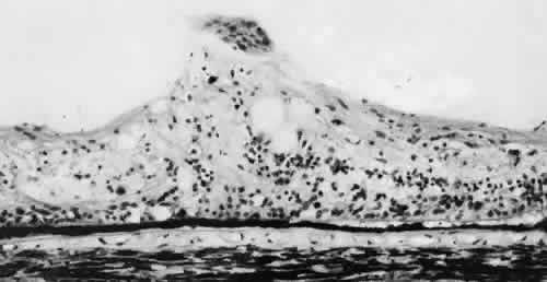
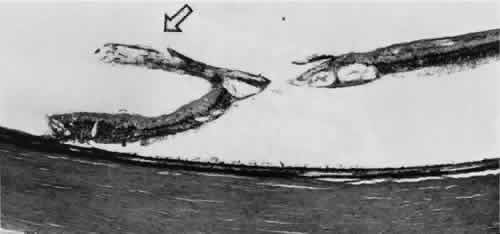
degeneration (Fig. 8).

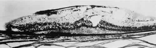
Fig. 8. Meridional fold in a young patient. Retina is thickened along course of fold, which shows microcystoid change near the surface and a cap of dense-staining glial cells along its surface. Middle and outer layers of the retina are largely unremarkable. Pigment epithelium shows focal redundancy anteriorly. (Hematoxylin-eosin; × 150.)

Meridional folds are present in 26% of the population and are bilateral in 55% of the affected patients; thus, they are present in 20% of all eyes (Table 2). Meridional folds are multiple in 27% of affected eyes; they are most common in the superior nasal quadrant.5,6


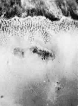
MERIDIONAL COMPLEX

A meridional complex is the occurrence of a dentate process and a ciliary process within the same meridian. When this abnormal alignment occurs, the den-tate process is exceptionally large, usually combined with a meridional fold, usually continuous with an enlarged ciliary process, and often associated with peripheral retinal excavation in the corresponding 
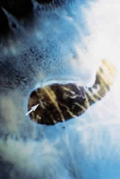
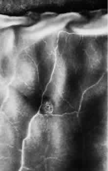
meridian (Fig. 9). The large dentate process and meridional fold are composed of excessive, disorganized, somewhat degenerated retinal tissue (Fig. 10).

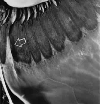
Fig. 9. Meridional complex (arrow). Note its basic constituent, an atypical dentate process, which aligns with and extends to an enlarged ciliary process. Complex also has a meridional fold which extends along the dentate process and posteriorly into the peripheral retina.

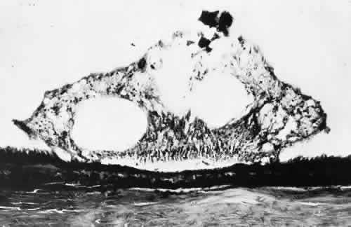
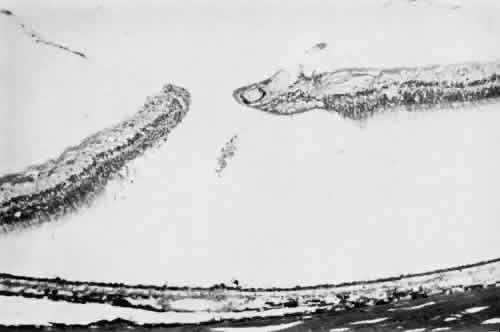
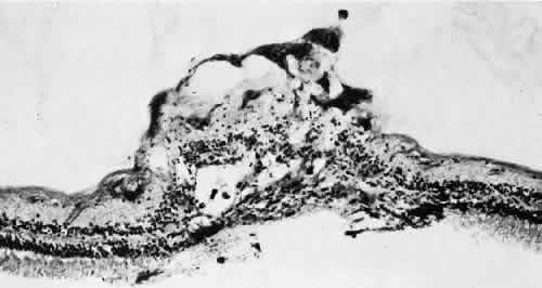
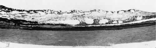
Fig. 10. Microsection of meridional complex through atypical dentate process and its meridional fold. Anteriorly (on the left) the complex shows marked redundancy of pigmented epithelium in its outer aspect and a dense glial plaque on its inner aspect. Posteriorly (on the right) there is microcystoid change, nonspecific degeneration, and dense-staining glial cells along its surface. (Hematoxylin-eosin; × 63.)

Meridional complexes are present in 16% of the population, are bilateral in 58% of affected patients, and thus are present in 12% of all eyes (see Table 2). The complexes are multiple in 45% of the affected eyes; they are most common in the superior nasal quadrant.

ENCLOSED ORA BAY

Enclosed and partially enclosed ora bays are relatively uncommon developmental variations. These are oval islands of pars plana epithelium located immediately posterior to the ora serrata and com-pletely or almost completely circumscribed by the peripheral retina (Figs. 11 and 12). The enclosed ora bay is composed of a thin layer of nonpigmented pars plana epithelium surrounded by neurosensory retina. Enclosed and partially enclosed ora bays are evident in 6% of patients, are bilateral in 8% of affected individuals, and are present in 3% of all eyes (see Table 2). These lesions are equally prevalent nasally and temporally near the horizontal meridian.6,7

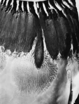
Fig. 11. Partially enclosed ora bay in a 20-year-old woman. Posteriorly, the ora bay extends 1.8 mm behind general line of ora serrata, and the retina shows a large area of typical cystoid degeneration. Anteriorly, the ora bay is embraced by two long dentate processes that converge toward, but do not meet, a prominent ciliary process of the pars plicata. (× 12.)

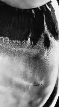
Fig. 12. Enclosed ora bay in a 35-year-old man. Anteriorly, two broad dentate processes converge and join to enclose a bay (island of pars plana). Posteriorly there is a focus of retinal thinning (peripheral retinal excavation; arrow). (× 12.)

PERIPHERAL RETINAL EXCAVATION

Peripheral retinal excavation appears as a rather small oval depression in the retina. Usually this lesion is aligned meridionally with a meridional fold or complex and located 1 to 7.2 mm posterior to the ora serrata (Fig. 13; see Figs. 6 and 12). The focal depression may be surrounded by margins that appear to be elevated; however, microscopic examination reveals that the depression corresponds to afocal loss of the inner retinal layers and that the surrounding 
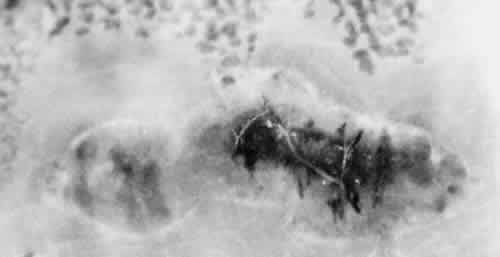
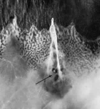
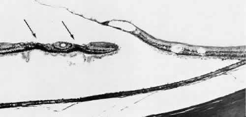
tissue is normal (Fig. 14).

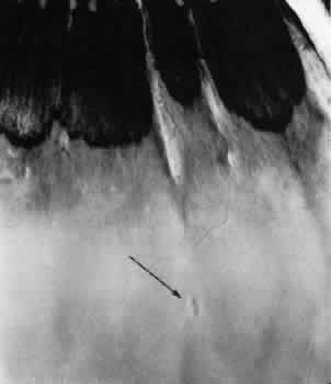
Fig. 13. Meridional complexes with peripheral retinal excavation. Two complexes can be seen anteriorly; both contain meridional folds (the fold of complex on the right is discontinuous). Peripheral retinal excavation (arrow) is aligned with the complex on the left (× 12.)

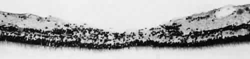
Fig. 14. Peripheral retinal excavation. The notable loss of tissue from the retinal surface extends to the inner nuclear layer. Centrally, the middle retinal layers show degeneration, but outer layers of retina are intact. (Hematoxylin-eosin; × 250.)

Peripheral retinal excavation is present in 10% of patients, is bilateral in 43%, and therefore is evident in 8% of all eyes (see Table 2). Half of the affected eyes contain two or more areas of focal excavation, and most of the excavations are located in the superior nasal quadrant.

Developmental variations of the peripheral retina are present in about 20% of eyes. These conditions have certain common features: they are present at birth, persist throughout life, tend to occur symmetrically in anatomically corresponding positions in both eyes, and are commonly associated with an abnormal alignment of a dentate and a ciliary process in the same meridian.5

Clinically, these developmental abnormalities are readily identified in the peripheral retina. The ridgelike elevation of a meridional fold is best seen by scleral depression combined with indirect ophthalmoscopy. A meridional complex should be suspected (even though the ciliary processes are not visible) when a large dentate process, a meridional fold, and peripheral retinal excavation occur in the same meridian. An enclosed ora bay appears as a depressed, bright red area that simulates a retinal hole. Although identification of an enclosed ora bay is aided by a thin epithelial layer extending across the base and by the absence of any elevation of the surrounding retina, in some instances, distinction between an enclosed bay and a retinal hole is impossible. Peripheral retinal excavation presents as a small retinal pit that may be adjacent to or up to 4 disc diameters posterior to the ora serrata. This form of focal depression probably has been often mistaken for a full-thickness retinal break; therefore, knowledge of developmental variations is essential to the appropriate diagnosis of a peripheral retinal lesion.

In clinical studies, retinal breaks posterior to meridional folds have been noted in a number of eyes with rhegmatogenous retinal detachment.8 Retinal tears may also develop at or near the posterior margins of enclosed ora bays.7 Therefore, when retinal detachment is present, the ophthalmologist must carefully search for meridional folds, meridionalcomplexes, enclosed ora bays, peripheral retinal excavations, and any associated retinal breaks.

TROPHIC PERIPHERAL RETINAL DEGENERATIONS

Various specific peripheral retinal degenerations augment these developmental variations. Degeneration (i.e., irreversible retrograde change) is evident in the peripheral retina of every adult. For classification, this degeneration may be regarded as trophic when the primary process is a loss of retinal tissue, tractional when the process is related primarily to tugging or pulling of vitreous or zonule on the retina, and trophic and tractional when both retinal tissue loss and vitreous-zonule traction are involved. The principal peripheral retinal degenerations are noted in Table 3.


TYPICAL CYSTOID DEGENERATION

The most common form of degeneration of the peripheral retina is typical cystoid degeneration. Spaces develop in the outer plexiform and inner nuclear layers and coalesce to form interlacing tunnels; they are separated by pillars that extend from the inner to the outer retinal layers, giving the inner surface a uniformly stippled appearance (Fig. 15). The stippled depressions correspond to retinal pillars; the intervening 
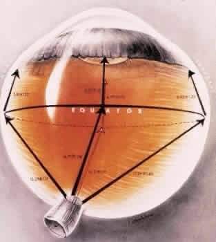
rounded domes result from the intraretinal cystoid spaces.9 Degeneration begins at the ora serrata, particularly at the base of dentate processes, and extends posteriorly and circumferentially to form a band that may encircle the eye and reach from the ora serrata to the equator (Fig. 16).

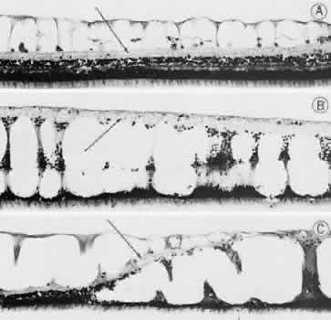
Fig. 15. Typical and retinal cystoid degenerations. A. Degeneration of nerve fiber layer with persistence of delicate vertical columns of Müller's cells. Superficial capillary plexus courses through the cystoid cavities, and surviving ganglion cells are subtended on the inner aspect of the inner plexiform layer (arrow). Outer retinal layers are well preserved. B. Extensive degeneration of middle retinal layers with broad cellular columns (between cystoid cavities) composed of Müller's cells and remnants of outer plexiform layer and inner nuclear layer (vertically stretched). Outer nuclear layer also shows degeneration. Inner plexiform layer (arrow) is intact. C. Overlapping reticular (on the left) and typical (on the right) cystoid degeneration. Note combined degenerative effect on the inner plexiform layer (arrow) of both types of cystoid degeneration. Superficial small arteriole retards progression of reticular cystoid degeneration. (Hematoxylin-eosin, × 250.) (Foos RY: Senile retinoschisis: Relationship to cystoid degeneration. Trans Am Acad Ophthalmol Otolaryngol 1970;74:33.)

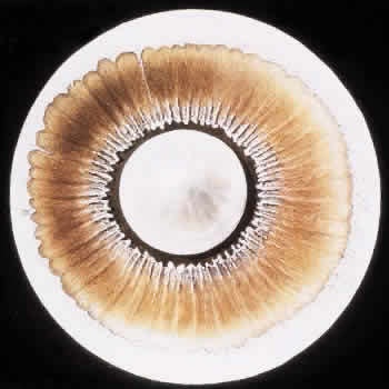
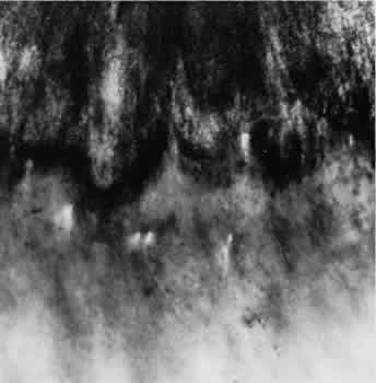
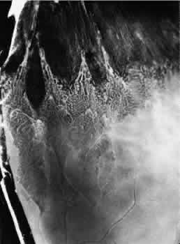
Fig. 16. Typical and reticular cystoid degeneration found immediately behind the ora serrata and about enclosed ora bay near cut edge of calotte. Posteriorly, note the conspicuous vascular pattern of degeneration (seen as gray background), finely stippled surface pattern, and angular free margins (related to limitation by surface vessels).

This degenerative process may be noted in infants at 1 year of age; it is always present in both eyes of patients over 8 years of age, usually increases in area with advancing age, and is most extensive in the superior and temporal quadrants (see Table 3).10,11

RETICULAR CYSTOID DEGENERATION

Reticular cystoid degeneration of the peripheral retina is almost invariably located posterior to and continuous with typical cystoid degeneration. It is characterized by a prominent linear or reticular pattern that corresponds to the retinal vessels and by a finely stippled internal surface. Areas of involvement are single or multiple, form the shape of an irregular angle, and are often demarcated posteriorly by retinal blood vessels (see Fig. 16).12 Spaces develop in the nerve fiber layer and are divided by delicate retinal pillars (see Fig. 15).

Reticular cystoid degeneration is present in 18% of adult patients, is bilateral in 41% of affected patients, and is thus evident in 13% of all adult eyes (see Table 3). The process is most prevalent in the inferior temporal quadrant.13

Clinically reticular cystoid degeneration is detected on contact lens biomicroscopy by noting the location, shape, prominent reticular pattern, and finely stippled appearance of the affected area.

DEGENERATIVE RETINOSCHISIS

Degenerative retinoschisis, a more extensive trophic process, presents as a round or ovoid area of retinal splitting with a smooth fusiform elevation of the inner layer (Fig. 17). The schisis is surrounded on all sides by typical cystoid degeneration; the retinal pillars of the cystoid degeneration as well as the broken pillars near the margin of the schists are prominent. Vessels are located in the inner retinal layer, the intraretinal cavity is optically empty, and the outer retinal layer is moderately irregular in contour.13,14

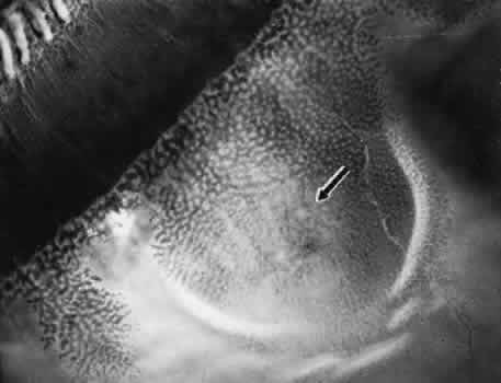
Fig. 17. Typical degenerative retinoschisis.Note extensive region of typical cystoid degeneration with a rounded and elevated posterior margin. In the center (arrow), radial columns are randomly disrupted, causing a disturbance in coarse surface pattern. (× 18.)

In one type of degenerative retinoschisis, the thin inner wall is composed of the internal limiting membrane, the nerve fiber layer, and retinal vessels (Fig. 18). The irregular outer wall contains portions of the inner nuclear, outer plexiform, outer nuclear, external limiting, and rod and cone layers. At the margin of the cavity, the retinoschisis blends with typical cystoid degeneration and may be relatively flat. Lesions with this appearance have been termed typical degenerative retinoschisis.

Fig. 18. Typical degenerative retinoschisis. There is extensive tissue loss in middle layers of the retina (typical cystoid degeneration); on the left, degeneration has also destroyed radial supporting columns. (Hematoxylin-eosin; × 250.)

Typical degenerative retinoschisis is present in 1% of adult patients and is bilateral in 33% of these patients; therefore, it is evident in 0.7% of adult eyes (see Table 3), with a predilection for location in the inferior temporal quadrant. A narrow band of typical cystoid degeneration is always present between the ora serrata and the anterior border of the schisis cavity; the involved area may extend to or somewhat posterior to the equator.

On clinical examination, typical degenerative retinoschisis appears as round or ovoid areas of retinal splitting with fusiform elevation of the inner layer (Fig. 19). The stippled pattern of surrounding typical cystoid degeneration extends on the inner layer for a variable distance; centrally the inner layer, which contains the blood vessels, is thin and smooth. On contact lens ophthalmoscopy, the inner layer is finely textured, some of the retinal vessels are attenuated, and there is a variable number of tiny, glistening, white dot opacities on the vitreous side. The outer layer, found external to the optically empty cavity, is best seen with indirect ophthalmoscopy when it becomes white on scleral depression. It is somewhat uneven, giving an appearance of finely hammered or beaten metal. Typical degenerative retinoschisis does not extend posteriorly to threaten the macula, and it is not often associated with breaks in either 
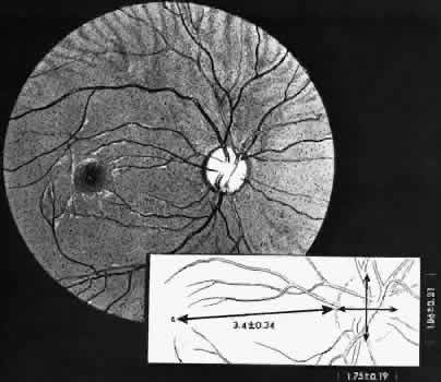
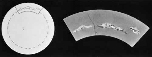
retinal layer; it rarely requires treatment.

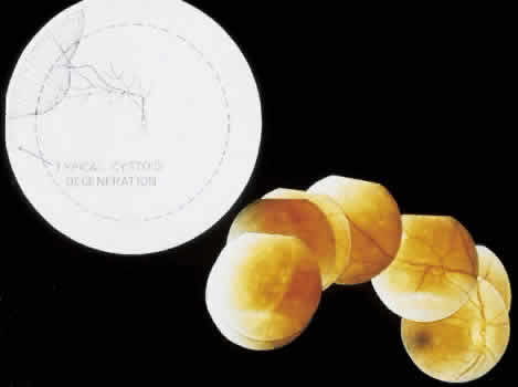
Fig. 19. Clinical appearance of typical degenerative retinoschisis: diagram of involved area and ocular fundus photographs showing optic disc, macula, and posterior portion of schisis. Within the schisis and adjacent to the margin is coarse stippling related to broken retinal pillars.

Retinoschisis associated with a bullous architecture and prominent reticular cystoid degeneration has been termed reticular degenerative retinoschisis. Reticular degenerative retinoschisis can be distinguished from typical degenerative retinoschisis by the large extent of retinal involvement, a round or ovoid configuration with bullous elevation of the extremely thin inner layer, and an irregular, pitted outer layer (Figs. 20 and 21). Typical cystoid degeneration is always present anterior to the schisis; reticular cystoid degeneration is usually prominent at some site in the involved eye. Blood vessels coursing through the inner layer give it an arborizing reticular pattern on contact lens biomicroscopy. The intraretinal cavity is optically empty; the outer wall is irregularly excavated to produce a pocked or honeycomb appearance. Round or ovoid holes are often present in the outer retinal layer; they are single or multiple, frequently large, and usually associated with a rolled posterior edge.13

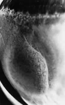
Fig. 20. Reticular degenerative retinoschisis. Note reticulated, highly elevated, inner wall with a conspicuous delicate vascular pattern. Radial columns of the retina are completely disrupted within the region of bullous elevation, and the retinoschisis extends posterior to the equator. (× 18.)

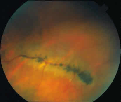
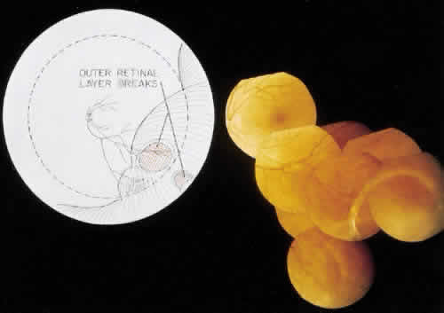
Fig. 21. Clinical appearance of reticulardegenerative retinoschisis: diagram of involved area and photographs of ocular fundus showing optic disc, macula, and posterior portion of the schisis. These illustrate outer layer retinal breaks, adjacent retinal pigment epithelium abnormality, and a lo-calized nonrhegmatogenous retinal detach-ment.

Microscopic sections demonstrate the extremely attenuated, blood vessel-containing inner layer composed of the internal limiting membrane and remnants of the nerve fiber layer (Fig. 22). The honeycomb appearance of the outer layer corresponds to irregular excavations. In some areas, the outer layer is made up of outer plexiform, outer nuclear, external limiting, and rod and cone layers; in other areas it is reduced to only the external limiting and the rod and cone layers; round or ovoid holes may be present (Fig. 23).

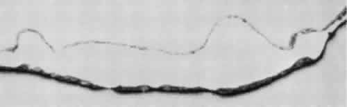
Fig. 22. Reticular degenerative retinoschisis. Note complete loss of radial supporting columns of retina and marked elevation of delicate inner wall, which contains fine blood vessels. Outer wall shows periodic exaggerated thinning. (Hematoxylin-eosin; × 60.)

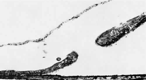
Fig. 23. Reticular degenerative retinoschisis with hole in outer wall and localized retinal detachment. Margins of hole are rolled and covered by a garland of degenerating photoreceptor outer segments. (Hematoxylin-eosin; × 250.)

Reticular degenerative retinoschisis is evident in 1.6% of adult patients, is bilateral in only 16% of these, and thus is noted in 0.95% of adult eyes (see Table 3). The lesion is found most commonly in the inferior temporal quadrant. A band of typical cystoid degeneration always separates the schisis from the ora serrata; the schists usually reaches the equator and often extends appreciably into the posterior retina.

On contact lens biomicroscopy, many retinal blood vessels present irregular contours, telangiectases, occluded segments, and microaneurysms. Between these vessels, the inner wall has a finely textured appearance and variable white, glistening particles on the vitreous side. The outer retinal wall is best seen when scleral depression produces a “white with pressure” phenomenon and reveals the honeycomb appearance. The retinal pigment epithelium often has a granular, salt-and-pepper appearance, and outer-layer retinal breaks are common. These breaks are particularly likely near the anterior and posterior margins of the schisis.

Treatment for reticular degenerative retinoschisis is rarely necessary.15 Treatment is indicated when the schisis cavity is symptomatic or progressive and associated with a rhegmatogenous component from both inner and outer wall retinal holes.15 Surgical management has included: external drainage ofsubretinal fluid with simultaneous intraocular gas injection,16 pars plana vitrectomy with either limited inner wall retinectomy and internal drainage through preexistent outer wall holes17 or with perfluorocarbon liquid assisted anterior displacement of subretinal fluid.18 No treatment is indicated for nonprogressive reticular degenerative retinoschisis that does not extend toward the macula. Photographic documentation of the posterior borders of the retinoschisis cavity may be useful in clinical monitoring for progression.

PAVING-STONE DEGENERATION

Paving-stone degeneration of the retina is characterized by one or more discrete, rounded foci of depigmentation and retinal thinning located between the ora serrata and the equator (Fig. 24). The lesions appear yellow-white, frequently reveal prominent underlying choroidal vessels, and often have a pigmented margin (Fig. 25). The basic lesion is rounded and 0.1 to 1.5 mm in diameter. Clusters of these rounded foci may merge to form larger lesions with convexly scalloped margins and incomplete pigmented septa;19 these features aid in distinguishing paving-stone degeneration from postinflammatory 
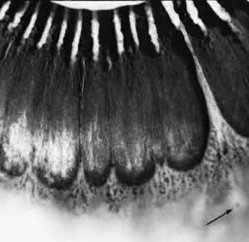
chorioretinal lesions.

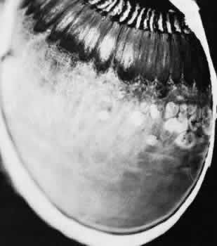
Fig. 24. Paving-stone degeneration. Note multiple round foci of depigmentation in temporal sector. Larger lesions show scalloped margins and linear pigmentation resulting from confluence of several smaller lesions. (× 65.)

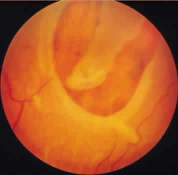
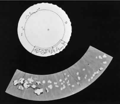
Fig. 25. Multiple discrete yellow-white lesions in the peripheral fundus in paving-stone degeneration.

Paving-stone degeneration is associated with sharply circumscribed retinal thinning due to loss of the rods and cones and external limiting membrane, absence of the pigment epithelium, adherence of the retina to Bruch's membrane, and alteration of the choriocapillaris (Fig. 26). The hyperpigmented cuffs and septa consist of proliferated pigment epithelial cells.

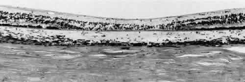
Fig. 26. Paving-stone degeneration. Within the lesion, there is selective loss of outer retinal layers,including photoreceptor cells, outer plexiform layer, and to a lesser extent, inner nuclear layer. The choroidand Bruch's membrane are intact. Retinal pigment epithelium cannot be discerned. (Hematoxylin-eosin; × 250.)

Paving-stone degeneration is present in 22% of adult patients, is bilateral in 38% of these, and thus is evident in 17% of adult eyes (see Table 3). Prevalence increases markedly with advancing age. In distribution, there is a preference for the inferior temporal and inferior nasal quadrants, with more than half of the lesions located in the inferior retina between the 5- and 7-o'clock positions.

Lesions are most numerous immediately posterior to the ora serrata; they rarely extend posterior to the equator.

Paving-stone degeneration does not predispose to retinal breaks or to retinal detachment and thus does not warrant any form of treatment. However, if a retinal detachment from some other cause involves an area of paving-stone degeneration, the retina may be torn at the site of the chorioretinal adherence. The resultant retinal breaks are often small, irregular in shape, inconspicuous, and best detected with contact lens biomicroscopy.

PERIPHERAL TAPETOCHOROIDAL DEGENERATION

With advancing age, the peripheral fundus usually develops a granular appearance, due to irregularity of the retinal pigment epithelium. Peripheral tapetochoroidal degeneration, a pronounced degree of this alteration, presents as a diffusely depigmented circumferential band that extends from the ora serrata to the equator. The anterior border is irregular because of splotchy pigmentation that remains within the area of the vitreous base, but the posterior border is usually smooth and well defined.11

Peripheral tapetochoroidal degeneration is associated with degeneration and loss of pigment granules in the retinal pigment epithelium, some loss of photoreceptor cells, diffuse thickening of Bruch's membrane, and a diminution of capillaries in the choriocapillaris.

Although minor degrees of degeneration of the retinal pigment epithelium are common in older patients, extensive alterations that meet the criteria of peripheral tapetochoroidal degeneration are present in 20% of patients over 40 years of age, are bilateral in all cases, and thus are present in 20% of eyes in this group (see Table 3). This age-related process does not predispose to retinal tear formation and requires no treatment.

RETINAL HOLE

Most retinal holes (76%) are secondary to lattice degeneration.20 Primary retinal holes, unrelated to lattice degeneration or other identifiable disorders, are rounded, full-thickness retinal breaks without a flap or a free operculum (Fig. 27). Usually located anteriorly within the area of the vitreous base, the holes have smooth margins; the adjacent retina usually appears normal; proliferative reactions are absent; and vitreous attachments are not
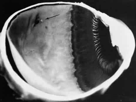
 unusual.11

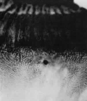
Fig. 27. Retinal hole with round, full-thickness break immediately behind the ora serrata in an area of relatively normal retina. Typical cystoid degeneration extensively involves retina on both sides of retinal hole. (× 16.)

Microsections of retinal holes confirm the complete retinal discontinuity and the smooth, rounded margins (Fig. 28). There is minimal reactive gliosis; no significant alteration is found in the adjacent vitreous body or in the pigment epithelium.

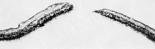
Fig. 28. Full-thickness retinal break in otherwise normal-appearing peripheral retina. Outer retinal layers are intact. (Hematoxylin-eosin; × 250.)

Primary retinal holes are present in 7.5% of adults, they are bilateral in 21% of patients, and thus are evident in 12% of adult eyes (see Table 3). Virtually all retinal holes occur within the vitreous base and give 
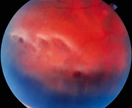
no evidence of quadrant predilection.

Retinal holes are detected clinically by indirect ophthalmoscopy and by contact lens biomicroscopy. Scleral depression is helpful in differentiating a retinal hole from a round retinal hemorrhage: the retinal hole develops a changing reddish color as it moves over the crest of the scleral depression, and the round retinal hemorrhage maintains a stable red color as it moves over the crest of the scleral depression. Asymptomatic round retinal holes with or without free opercula do not generally require treatment. A rhegmatogenous retinal detachment rarely originates from an untreated retinal hole.21 However, retinal holes located within a rhegmatogenous retinal detachment should be treated during the surgical repair as these can be a source of persistent subretinal fluid postoperatively.

Back to Top
TROPHIC AND TRACTIONAL PERIPHERAL RETINAL DEGENERATIONS

LATTICE DEGENERATION

Combining elements of trophic retinal degeneration and tractional degeneration related to vitreous pull on the retina, lattice degeneration of the retina is characterized by a sharply demarcated, circumferentially oriented, degenerative process that involves retinal thinning and abnormalities in the adjacent vitreous (Figs. 29 and 30). Other important distinguishing features include an arborizing network of fine white lines, which are often continuous with retinal blood vessels; alterations of retinal pigmentation, frequently with accumulations along the interlacing white lines; round, punched-out areas of retinal thinning or hole formation; small yellow-white particles at the margin of the surface of the lesion and in the adjacent vitreous; absence of overlying vitreous; exaggerated vitreoretinal attachments at the margin of the process; and a predilection for retinal tears to form along the posterior and lateral margins of the lesion. Although these general features of lattice degeneration are well recognized, there are many variations in their clinical appearance related to the stage or degree of the lesion, the extent or area of the process, and in the anteroposterior position of the degeneration.22

Fig. 29. Lattice degeneration presenting as a sharply demarcated, circumferentially oriented lesion anterior to the equator with retinal thinning and associated vitreous abnormalities.

Fig. 30. Lattice degeneration. Highermagnification of Figure 29 shows arborizing pattern of white lines (corresponding to retinal blood vessels within the lesion), white dots on the retinal surface of the lesion, and viteous condensation at the margin of the lesion.

Early stages of lattice degeneration have the characteristic spindle shape and circumferential orientation, but in general they are relatively narrow with a troughlike central retinal thinning; variable, round, punched-out areas of extreme retinal thinning; typical vitreous alterations; retinal blood vessels that appear normal; and mild pigment alterations (Fig. 31). Advanced stages of lattice degeneration may retain the characteristic shape and orientation but tend to be somewhat wider with a greater degree of overall retinal thinning, of arborizing white lines that are often continuous with blood vessels, and of conspicuous retinal pigment alterations. In addition, round or oval areas of extreme retinal thinning or holes are more common, liquefaction in the overlying vitreous is greater, and vitreoretinal attachments at the margin of the process are more pronounced.

Fig. 31. Latice degeneration demonstrating variable pigmentary changes to the underlying retinal pigment epithelium and circular atrophic hole (arrow)

No matter what the stage, the extent or area of involvement varies according to the size and number of the lesions. A single lesion may range from a small island encompassing less than 30 degrees on the circumference of the eye to a long band extending 120 degrees. There may be 20 or more discrete lesions, and these may be arranged in two, three, or even four rows parallel to the equator and affecting 270 degrees or more of the globe circumference.

In addition to variations related to stage and area, lattice degeneration has important features related to anteroposterior location. The usual location is slightly anterior to the equator. When located more anteriorly, within the area corresponding to the vitreous base, the lesion may be classified separately as vitreous base excavation, but it is more appropriately regarded as a variant of lattice degeneration that is likely to be oval or linear, precisely circumferential in orientation, and associated with somewhat exaggerated vitreous attachments at the margin. A retinal hole may be present, but a retinal tear never occurs within the vitreous locus.

Posterior to the equator, lattice degeneration tends to be wider than usual, oriented radially rather than circumferentially, and associated with much more extensive abnormal vitreoretinal attachments. Full-thickness retinal holes may be present, and retinal tears are particularly likely to occur (Fig. 32).

Fig. 32. Paravascular lattice degeneration with underlying retinal pigment epithelial attenuation and clumping.

Related to vitreous traction as well as to the overall stage, area, and anteroposterior location of the lattice degeneration is the tendency for tears to form along the posterior margin or end of the lesion where vitreous separation from the retina occurs. The tear, almost always associated with posterior vitreous detachment, assumes a linear form if it extends along the posterior margin, an L shape if there is anterior extension at one end, and a U shape if there is anterior extension at both ends (Fig. 33). Tears may be single or multiple and small or very large and may occur with or without associated rhegmatogenous retinal detachment.

Fig. 33. U-shaped retinal tear along the posterior and lateral borders of lattice degeneration. Bridging retinal vessel spans the full-thickness retinal defect (arrow).

Lattice degeneration is characterized by a circumscribed area of retinal thinning resulting from tissue loss that is greater in the inner retinal layers than in the outer (Fig. 34). Blood vessels in the lesion have thickened walls, the lumen may be decreased or completely obliterated, and pigment may be located in the paravascular space. Areas of extreme retinal thinning or retinal hole formation are not uncommon. An area in which all vitreous structure is lost overlies the retinal lesion; vitreous condensation and exaggerated vitreoretinal attachments may be seen at the margin of the liquefied area. Glial cells may extend from the retina into the vitreous along the interface between liquid vitreous and formed vitreous. Irregular degeneration and hyperplasia of the retinal pigment epithelium are usually evident in the pigment that is external to the retinal degeneration.

Fig. 34. Advanced stage of lattice degeneration with vitreous liquefaction overlying the lesion, gliosis at the margin of liquefied vitreous, full-thickness retinal hole formation, sclerosis of blood vessels with pigment in the paravascular space, and irregularity of retinal pigment epithelium.

Lattice degeneration is present in 6% of adult patients, it is bilateral in about 50% of affected patients, and is evident, therefore, in 4% of eyes. The prevalence is actually greater because the variant of vitreous base excavation, based on a separate tabulation, is present in 5% of adult patients, is bilateral only rarely, and thus is present in 3% of adult eyes (see Table 3). Allowing for the overlap of some patients who present with both lattice degeneration and the vitreous base excavation variant of lattice degeneration, the overall general process of lattice degeneration is evident in approximately 10% of adults, is bilateral in approximately 25% and thus is present in nearly 7% of adult eyes.23 Lattice degeneration is most prevalent in the superior temporal quadrant. As a more meaningful index of distribution, however, lattice degeneration is most common adjacent to the superior and inferior vertical meridians, less common adjacent to the temporal horizontal meridian, and least common in the vicinity of the nasal horizontal meridian.

Clinically, lattice degeneration has an appearance and general characteristics that correlate precisely with anatomic features (Fig. 35). Contact lens biomicroscopy and indirect ophthalmoscopy with scleral depression reveal the major features of retinal thinning, blood vessel abnormalities, focal thinning and retinal hole formation, vitreous abnormalities, pigment changes, and retinal tears. Small refractile yellow-white particles on the vitreous surface of the lesion are more conspicuous clinically than in laboratory studies, but the details of vitreous liquefaction, vitreous condensation, and increased vitreoretinal attachments at the margin of the lesion are seen with meticulous contact lens biomicroscopy.

Fig. 35. Clinical appearance of lattice degeneration. Diagram shows location; drawing shows sharply demarcated, circumferential process with prominent pigment irregularities.

Lattice degeneration may be responsible for rhegmatogenous retinal detachment when round holes within the degeneration permit the subretinal passage of fluid or when tears along the margin of the degeneration cause separation of the retina. Retinal detachment due to lattice degeneration with atrophic holes is more common in younger patients, with 50% being less than 30 years old.24 These detachments are not related to posterior vitreous separation, nor are they associated with communication between central vitreous degeneration (synchisis senilis) and the local vitreous liquifaction that is seen overlying these retinal lesions.25 Tractional retinal tears occur most often along the posterior or circumferential edges of lattice lesions where firm vitreoretinal attachments exist. Separation of the cortical vitreous during posterior vitreous detachment is the seminal event leading to the development of these retinal tears. A comparison of the prevalence of lattice degeneration, nearly 10% of adults, with the incidence of retinal detachment, 0.005% to 0.01% per year,26,27 emphasizes that most patients with lattice degeneration do not develop retinal detachment. Even so, lattice degeneration is detected in 20% to 35% of patients undergoing surgery for rhegmatogenous retinal detachment.8,22,26–28

The role of prophylactic retinal laser therapy in eyes with lattice degeneration remains controversial. Prophylactic treatment of lattice degeneration with or without atrophic holes in asymptomatic phakic eyes is not recommended because of the low risk of developing retinal detachment in these eyes.29 In a higher risk population of phakic eyes with lattice degeneration and retinal detachment in the fellow eye, laser prophylaxis to areas of lattice degeneration reduced, but did not eliminate, the risk of retinal detachment.30 This risk reduction was roughly equal to preventing a single retinal detachment every 2 years for every 100 patients treated. In aphakic fellow eyes of patients with retinal detachment, retinal tears developed in areas of normal appearing retina in 89% of eyes that developed a retinal tear indicating that targeted laser treatment alone would be inadequate at preventing most retinal detachments in this at-risk group.31

These studies suggest that laser application to prevent retinal detachment in eyes with lattice degeneration would need to be performed circumferentially throughout the vitreous base region, including areas of normal appearing retina, to be effective as a prophylactic measure. The adverse consequences of this confluent approach include large and giant retinal tear formation, premacular fibrosis, macular hole formation, and perhaps even an increased risk of retinal detachment.32 Evidenced-based analysis of prophylactic treatment rated only a “sometimes treat” recommendation for lattice degeneration in the fellow eye of a patient with retinal detachment. Furthermore, treatment was “rarely recommended” for eyes with symptoms of retinal tear formation that were found to harbor lattice degeneration alone, and treatment was rated as “never recommended” for asymptomatic lattice degeneration in phakic or aphakic eyes alone.33

Back to Top
TRACTIONAL PERIPHERAL RETINAL DEGENERATIONS
Peripheral retinal degenerations are considered tractional when primarily related to tugging or pulling of the vitreous or zonule on the retina. The general category of tractional tufts is subclassified by anatomic, pathogenetic, and clinical distinctions into noncystic retinal tufts, cystic retinal tufts, and zonular-traction retinal tufts.

NONCYSTIC RETINAL TUFT

A noncystic retinal tuft is a short, thin internal projection of retinal tissue that usually occurs in clustersand is almost invariably located within the vitreous base. From the vitreous attachment at the apex, the tuft extends to a base that is less than 0.1 mm in diameter and unassociated with cystic retinal degeneration (Fig. 36). Histologically, a noncystic tuft is composed of altered retinal cells and proliferated glial tissue (Fig. 37).27,34 Noncystic retinal tufts are not present at birth. They are present in 72% of adult patients, are bilateral in 50% of affected individuals, and thus are present in 59% of adult eyes (see Table3). Tufts occur in all sectors, but they are most common in the inferior nasal quadrant and are almost always located in the anterior portion of the peripheral retina corresponding to the vitreous base.

Fig. 36. Noncystic retinal tufts: cluster of small surface nodules of retina within vitreous base. Surrounding retina shows nonspecific degeneration and pigment dispersion. (× 20.)

Fig. 37. Microscopic features of the lesion seen in Figure 36. Entire retina within vitreous base shows nonspecific degeneration with gliosis. Centrally, the surface is elevated and shows a plaque of dense-staining glial cells. Retinal pigment epithelium also shows irregular thinning and pigment clumping, characteristically found within vitreous base. (Hematoxylin-eosin stain; × 500.)

On clinical examination, these tufts are commonly observed as small, pointed retinal projections within the area of the vitreous base. In later life, degenerative changes within the tufts may cause the tips of the tufts to break off, producing small spherical fragments that float in the vitreous adjacent to the base. These tufts are not associated with full-thickness retinal breaks and can be considered to be innocuous.

CYSTIC RETINAL TUFT

Larger than a noncystic tuft, the cystic retinal tuft is a nodular projection of retinal tissue that extends from a vitreous attachment at the apex to a base that is more than 0.1 mm in diameter and surrounded by cystic retinal degeneration (Fig. 38). Single ormultiple cystic retinal tufts may be located within or posterior to the vitreous base. On microscopic appraisal, the tuft, attached internally to a vitreous strand, is composed of degenerated and proliferated retinal cells that may contain a few pigment granules (Fig. 39). There may also be degeneration of the adjacent retinal pigment epithelium.

Fig. 38. Cystic retinal tuft in peripheral retina of a 14-year-old boy. Tuft measures 0.47 mm at its circular base, is 3.7 mm from the ora serrata, and contains many microcysts with dense walls. (× 19.)

Fig. 39. Microscopic features of the lesion seen in Figure 38. Surface of the tuft is irregular, with layer of dense-staining glial cells that partially surround subsurface microcysts. Degeneration of neurons, formation of microcysts, and pigment dispersion occur in deeper layers. Outer retina beneath lesion shows marked degeneration of photoreceptor cells. Patterns of vitreous over lesion are distorted, and coarse bundles of vitreous fibrils blend with irregular surface of tufts. (Periodic acid-Schiff; × 180.)

Cystic retinal tufts are present at birth; they are evident in 5% of adults, are bilateral in 6% of affected patients, and thus are detected in 2.5% of adult eyes (see Table 3). Although cystic retinal tufts show no quadrant predilection, 78% occur in the equatorial (extrabasal) zone, and 80% occur singly in eyes of affected patients.

On clinical examination, cystic retinal tufts are readily visualized and are distinguishable from noncystic tufts by size and other characteristics. Cystic retinal tufts are of importance because they may be avulsed by vitreous traction, with or without posterior vitreous detachment leading to retinal tear or retinal detachment.

Of patients with retinal detachment, an avulsed cystic retinal tuft is implicated in up to 7% of eyes. However, of all patients with a cystic retinal tuft, only 0.18% would develop retinal detachment due to this lesion. As such, prophylactic laser photo-coagulation for cystic retinal tuft is not recommended.35

ZONULAR-TRACTION RETINAL TUFT

The zonular-traction retinal tuft always projects from the retinal surface internally and anteriorly toward the zonule (Fig. 40). Usually single and located within the vitreous base, zonular-traction retinal tufts are joined to zonular fibers at the apex; the tufts, which vary in length and thickness, are associated with a broad range of trophic and tractional alterations at the retinal end of the process. Histopathologic changes include zonular attachments at the apex, neuroglial cells within the tuft, and degeneration with retinal thinning at the base (Fig. 41). Retinal holes and tears result from the combination of trophic changes at the base and traction resulting from zonular fibers. Within the vitreous base and posterior to it, zonular-traction tufts may be associated with partial-thickness or full-thickness retinal tears that can occur in the absence of posterior vitreous detachment.

Fig. 40. Zonular-traction tuft of the peripheral retina. Tuft is drawn at an acute angle from the retinal surface toward the ciliary body and shows microcystic degeneration anteriorly. Posteriorly, the tuft splays; the retina at its base shows marked trophic change, including three full-thickness holes (arrow). (× 20.)

Fig. 41. Zonular-traction tuft (arrow) drawn at an acute angle from the retinal surface toward ciliary body with dense-staining glial cells along surface. Base of tuft is microcystic and has full-thickness trophic hole. (Hematoxylin-eosin; × 150.)

Zonular-traction tufts are present at birth and detected with equal frequency in patients of all ages; they are noted in 15% of patients, are bilateral in 15% of affected people, and thus are evident in 9% of all eyes (see Table 3). These tufts are most common in the nasal quadrants, usually attached to the retina less than 0.5 mm posterior to the ora serrata and only infrequently attached to the retina posterior to the vitreous base.27,36

Clinical examination of the peripheral fundus reveals zonular-traction tufts in patients of all ages. These lesions are distinguished from noncystic and cystic retinal tufts by their greater size, sharp anterior angulation, and close proximity to the ora serrata. Zonular-traction tufts are a significant cause of small, round retinal holes in the extreme periphery of the retina. Although retinal holes within the vitreous base are considerably less likely to produce retinal detachment than holes posterior to the vitreous base, surgical aphakia or pseudophakia, which necessarily involves some form of zonular traction, is associated with an increase in the frequency of retinal detachment, which is likely to be related to small retinal breaks in the nasal periphery.36,37

PARTIAL-THICKNESS RETINAL TEAR

A partial-thickness retinal tear involves the inner layers of the retina and results in a thin flap or free operculum.38 Partial-thickness retinal tears are usually multiple and are classified into two types by distribution: paravascular tears occurring adjacent to peripheral retinal vessels and vitreous base tears aligned circumferentially along a segment of the posterior border of the vitreous base. Both forms, which sometimes exist concurrently in the same eye, reflect anatomic features and mechanisms that are of practical importance in ophthalmology.

Paravascular partial-thickness retinal tears stem from exaggerated vitreoretinal attachments at and immediately adjacent to peripheral arterioles and venules. These relatively firm attachments often produce multiple, discrete foci of cystic degeneration adjacent to peripheral retinal vessels. When posterior vitreous detachment occurs, the inner retinal layers at these foci of paravascular rarefaction remain attached to the vitreous, and clusters of partial-thickness retinal tears are formed. After this avulsion, microscopic studies of the retina show focal absence of the internal limiting membrane and absence of varying amounts of the inner retinal layers.39

Paravascular partial-thickness retinal tears occur in 17% of adults, are bilateral in 27% of affected patients, and thus are present in 11% of adult eyes (see Table 3). Posterior vitreous detachment is present in 100% of affected eyes. The superior quadrant is the area most commonly involved, and the tears are always located posterior to the vitreous base.

On clinical examination by contact lens biomicroscopy, paravasealar retinal rarefaction is seen as a subtle irregularity of the retina adjacent to the peripheral retinal vessels. Paravascular partial-thickness retinal tears are visible as small, usually multiple retinal craters adjacent to peripheral retinal vessels. These tears assume substantial clinical significance because of the occasional association with retinal vessel avulsion and the frequent association with partial-thickness vitreous base tears and full-thickness peripheral retinal tears.

Partial-thickness retinal tears also develop in association with posterior vitreous detachment as multiple flap tears aligned circumferentially along a segment of the posterior border of the vitreous base. Inner retinal layers are partially avulsed and attached to the detached vitreous body.27

Partial-thickness retinal tears at the posterior border of the vitreous base are present in 12% of adults, are bilateral in 5% of affected patients, and thus are noted in 7% of adult eyes (see Table 3). The process is equally prevalent in all quadrants, is always located at the posterior margin of the vitreous base, and is invariably associated with posterior vitreous detachment.

On clinical examination, vitreous base partial thickness tears are visible as translucent shreds of tissue partially avulsed from the retina adjacentto the posterior border of the vitreous base. Likeparavascular tractional lesions, vitreous base partial-thickness retinal tears are clinically significant because of the occasional association with paravascular partial-thickness tears and vessel avulsion and the frequent association with full-thickness peripheral retinal tears. Treatment of paravascular partial-thickness retinal tears, peripheral retinal vessel avulsion, and vitreous base partial-thickness retinal tears should be considered to decrease the risk of recurrent vitreous hemorrhage and rhegmatogenous retinal detachment.

FULL-THICKNESS RETINAL TEAR

A full-thickness retinal tear is a complete traction-related break in the sensory retina. The tear is usually U or V shaped with the broad base directed anteriorly, a tapered flap extending into the vitreous, and condensed vitreous strands attached to the apex and inner surface of this flap (Figs. 42 to 44). In some instances, the flap is partially or completely avulsed and can be identified as a free operculum in the overlying vitreous. This free operculum retains its attachment to the vitreous body, and it is almost always located anterior to the retinal tear (Fig. 45). As time passes, the margins of the retinal tear become smooth or rounded and the retinal flap becomes shrunken and degenerated. Seen through the retinal break, the retinal pigment epithelium often appears stippled or granular.

Fig. 42. Tears in nasoinferior quadrant of peripheral retina of a 76-year-old man. Large full-thickness tear has shriveled flap (arrow) to which vitreous fibrils are attached. There are many small partial tears of the surface of adjacent retina, most notable along vessels. (× 8.)

Fig. 43. Flap of large full-thickness retinal tear shows microcystic degeneration; it is attached to condensed hyaloid of the posteriorly detached vitreous body. Posterior hyaloid also contains fragments from surface of the underlying retina, corresponding to partial retinal tears (arrows) behind the full-thickness retinal break. (× 150.)

Fig. 44. Tractional full-thickness retinal tear with avulsed retinal blood vessel (white arrow). Edges of tear are beneath the elevated retinal flap (black arrows)

Fig. 45. Retinal tear at equator of eye of a 48-year-old woman. Avulsed spheric fragment of retina (operculum; arrow) is subtended over round full-thickness retinal break. (× 5.5.)

Full-thickness retinal tears unassociated with lattice degeneration, cystic retinal tuft, zonular-traction tuft, or other identifiable retinal abnor-mality are likely to occur at sites of exaggeratedparavascular vitreoretinal attachment and along the posterior border of the vitreous base. When posterior vitreous detachment occurs, full-thickness as well as partial-thickness tears may occur at one or more of these locations.

On histologic examination, full-thickness retinal tears demonstrate vitreous attachment to the degenerated retinal flap or free operculum, gliosis and degeneration adjacent to the smooth, rounded margins of the tear, a variable degree of associated retinal detachment, and a combination of cell breakdown and hyperplasia in the underlying retinal pigment epithelium.27

Retinal breaks occur in about 13% to 14% of autopsy cases.40 Excluding retinal tears at the ora serrata and tears related to lattice degeneration or other identifiable disease processes, full-thickness retinal tears are found in 12% of adults at autopsy, are bilateral in 5% of affected adults, and are present in 7% of adult eyes (see Table 3). These lesions are most prevalent in the inferior temporal quadrant, are always located posterior to the vitreous base, and are universally associated with posterior vitreous detachment. Approximately half such full-thickness retinal tears are associated with at least a localized retinal detachment; this is especially likely to be present where the tear is located in the superior temporal or superior nasal quadrant.

Clinical identification of full-thickness retinal tears is particularly significant. In the presence of posterior vitreous detachment, attention may be focused on abnormal retinal areas by a free operculum, projecting retinal flap, hemorrhage, or localized retinal pigment epithelium alteration.41

With or without these diagnostic clues, full-thickness retinal tears are visible on contact-lens biomicroscopy or on indirect ophthalmoscopy and scleral depression. As the scleral depressor creates translucency in the adjacent sensory retina, the tear is seen as a contrasting red area. A retinal tear and a retinal hemorrhage may be differentiated by the fact that the intensity of the contrasting red color of a tear varies with the position of the scleral depressor, but the red color of a retinal hemorrhage is unaffected by movement of the depressor. Full-thickness retinal tears should be carefully evaluated because these tears often warrant prophylactic therapy in an effort to prevent retinal detachment, and they require treatment when associated with rhegmatogenous retinal detachment.

GENERALIZED RETINAL DISEASES

Superimposed on normal morphology, developmental variations, and common degenerative processes of the peripheral retina is a wide array of diseases capable of affecting all parts of the retina. Among these diseases are developmental malformations (e.g., coloboma of the retina and choroid and familial hyaloidal vitreoretinopathies); vascular disorders (e.g., vasculitis and sickle cell diseases); metabolic diseases (e.g., diabetic retinopathy); infectious diseases (e.g., toxoplasma retinochoroiditis); neoplasms (e.g., retinoblastoma); degenerative disorders (e.g., retinitis pigmentosa); and traumatic disorders (e.g., traumatic retinitis and traumatic retinal dialysis). Pathologic features and clinical manifestations of these diseases are influenced by the specific anatomy of and relationships in the peripheral retina, the developmental variations, and the spectrum of degenerative changes that occur in the peripheral retina.

Back to Top
REFERENCES

1. Straatsma BR, Foos RY, Spencer LM: The retina: Topography and clinical correlations. In: Transactions of the New Orleans Academy of Ophthalmology: Symposium on Retina and Retinal Surgery. St Louis: CV Mosby, 1969:1–26

2. Straatsma BR, Landers MB, Kreiger AE: The ora serrata in the adult human eye. Arch Ophthalmol 1968;80:3

3. Rutnin U, Schepens CL: Fundus appearance in normal eyes. II: The standard peripheral fundus and developmental variations. Am J Ophthalmol 1967;64:840

4. Heller MD, Straatsma BR, Foos RY: Detachment of the posterior vitreous in phakic and aphakic eyes. Mod Probl Ophthalmol 1972;10:23

5. Spencer LM, Foos RY, Straatsma BR: Meridonal folds and meridional complexes of the peripheral retina. Trans Am Acad Ophthalmol Otolaryngol 1969;73:204

6. Spencer LM, Foos RY, Straatsma BR: Meridional folds, meridional complexes, and associated abnormalities of the peripheral retina. Am J Ophthalmol 1970;70:697

7. Spencer LM, Foos RY, Straatsma BR: Enclosed bays of the ora serrata: Relationship to retina tears. Arch Ophthalmol 1970;83:421

8. Dumas J, Schepens CL: Chorioretinal lesions predisposing to retinal breaks. Am J Ophthalmol 1966;61:620

9. Rutnin U, Schepens CL: Fundus appearance in normal eyes. III. Peripheral degenerations. Am J Ophthalmol 1967;64:1040

10. O'Malley PF, Allen RA: Peripheral cystoid degeneration of the retina. Incidence and distribution in 1,000 autopsy eyes. Arch Ophthaumol 1967;77:769

11. Foos RY, Spencer LM, Straatsma BR: Trophic degenerations of the peripheral retina. In: Transactions of the New Orleans Academy of Ophthalmology: Symposium on Retina and Retinal Surgery. St Louis: CV Mosby, 1969:90–102

12. Foos RY, Feman SS: Reticular cystoid degeneration of the peripheral retina. Am J Ophthalmol 1970;69:392

13. Straatsma BR, Foos RY: Typical and reticular degenerative retinoschisis. Am J Ophthalmol 1973;75:551

14. Foos RY: Senile retinoschisis: Relationship to cystoid degeneration. Trans Am Acad Ophthalmol Otolaryngol 1970;74:33

15. Byer NE: Long-term natural history study of senile retinoschisis with implications for management. Ophthalmology 1986;93:1127

16. Ambler JS, Meyers SM, Zegarra H et al: The management of retinal detachment complicating degenerative retinoschisis. Am J Ophthalmol 1989;107:171

17. Sneed SR, Blodi DF, Folk JC et al: Pars plana vitrectomy in the management of retinal detachments associated with degenerative retinoschisis. Ophthalmology 1990;97:470

18. Lomeo MD, Diaz-Rohena R, Lambert HM: Use of perfluorocarbon liquid in the repair of retinoschisis retinal detachments. Ophthalmic Surg Lasers 1996;27:778

19. O'Malley PF, Allen RA, Straatsma BR et al: Paving-stone degeneration of the retina. Arch Ophthalmol 1965;73:169

20. Foos RY: Retinal holes. Am J Ophthalmol 1978;86:354

21. Byer NE: What happens to untreated asymptomatic retinal breaks, and are they affected by posterior vitreous detachment? Ophthalmology 1998;105:1045

22. Straatsma BR, Allen RA: Lattice degeneration of the retina. Trans Am Acad Ophthalmol Otolaryngol 1962;66:600

23. Straatsma BR, Zeegen P, Foos RY et al: Lattice degeneration of the retina. Trans Am Acad Ophthalmol Otolaryngol 1974;78:87

24. Tillery WV, Lucier AC: Round atrophic holes in lattice degeneration: An important cause of phakic retinal detachment. Trans Am Acad Ophthalmol Otolaryngol 1976;81:509

25. Foos RY, Simons KB: Vitreous in lattice degeneration of the retina. Ophthalmology 1984;91:452

26. Michaelson IC, Stein R, Barkai S et al: A study in the prevention of retinal detachment. Ann Ophthalmol 1969;1:49

27. Spencer LM, Straatsma BR, Foos RY: Tractional degenerations of the peripheral retina. In: Transactions of the New Orleans Academy of Ophthalmology: Symposium on Retina and Retinal Surgery. St Louis: CV Mosby, 1969:103–127

28. Lorentzen SE: Frequency of lattice degeneration and retinal breaks in the fellow eye in retinal detachment. Acta Ophthalmologica 1988;66:157

29. Byer NE: Long-term natural history of lattice degeneration of the retina. Ophthalmology 1989;96:1396

30. Folk JC, Arrindell EL, Klugman MR: The fellow eye of patients with phakic lattice retinal detachment. Ophthalmology 1989;96:72

31. Benson WE, Grand MG, Okun E: Aphakic retinal detachment: Management of the fellow eye. Arch Ophthalmol 1975;93:245

32. Byer NE: Rethinking prophylactic treatment of retainal detachment. In Stirpe M (ed): Advances in Vitreoretinal Surgery. New York: Ophthalmic Communications Society, 1991:399–411

33. Wilkonson CP: Evidence-based analysis of prophylactic treatment of asymptomatic retinal breaks and lattice degeneration. Ophthalmology 2000;107:12

34. Foos RY, Allen RA: Retinal tears and lesser lesions of the peripheral retina in autopsy eyes. Am J Ophthalmol 1967;64:643

35. Byer NE: Cystic retinal tufts and their relationship to retinal detachment. Arch Ophthalmol 1981;99:1788

36. Foos RY: Zonular traction tufts of the peripheral retina in cadaver eyes. Arch Ophthalmol 1969;82:620

37. Ashrafzadeh MT, Schepens CL, Elzeneiny II et al: Aphakic and phakic retinal detachment. I: Preoperative findings. Arch Ophthalmol 1973;89:476

38. Foos RY: Postoral retinal tears. Ann Ophthalmol 1974;6:679

39. Spencer LM, Foos RY: Paravascular vitreoretinal attachments: Role in retinal tears. Arch Ophthalmol 1970;84:557

40. Foos RY, Simons KB, Wheeler NC: Comparison of lesions predisposing to rhegmatogenous retinal detachment by race of subjects. Am J Ophthalmol 1983;96:644

41. Rutnin U, Schepens CL: Fundus appearance in normal eyes. IV. Retinal breaks and other findings. Am J Ophthalmol 1967;64:1063

Back to Top