|
|
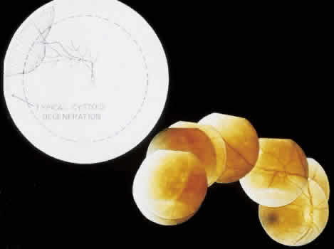
| Fig. 19. Clinical appearance of typical degenerative retinoschisis: diagram of involved area and ocular fundus photographs showing optic disc, macula, and posterior portion of schisis. Within the schisis and adjacent to the margin is coarse stippling related to broken retinal pillars. |