|
|
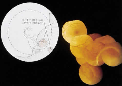
| Fig. 21. Clinical appearance of reticulardegenerative retinoschisis: diagram of involved area and photographs of ocular fundus showing optic disc, macula, and posterior portion of the schisis. These illustrate outer layer retinal breaks, adjacent retinal pigment epithelium abnormality, and a lo-calized nonrhegmatogenous retinal detach-ment. |