|
|
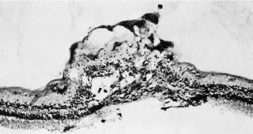
| Fig. 39. Microscopic features of the lesion seen in Figure 38. Surface of the tuft is irregular, with layer of dense-staining glial cells that partially surround subsurface microcysts. Degeneration of neurons, formation of microcysts, and pigment dispersion occur in deeper layers. Outer retina beneath lesion shows marked degeneration of photoreceptor cells. Patterns of vitreous over lesion are distorted, and coarse bundles of vitreous fibrils blend with irregular surface of tufts. (Periodic acid-Schiff; × 180.) |