|
|
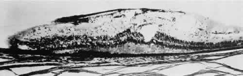
| Fig. 8. Meridional fold in a young patient. Retina is thickened along course of fold, which shows microcystoid change near the surface and a cap of dense-staining glial cells along its surface. Middle and outer layers of the retina are largely unremarkable. Pigment epithelium shows focal redundancy anteriorly. (Hematoxylin-eosin; × 150.) |