|
|
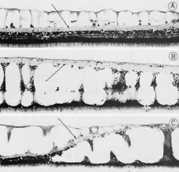
| Fig. 15. Typical and retinal cystoid degenerations. A. Degeneration of nerve fiber layer with persistence of delicate vertical columns of Müller's cells. Superficial capillary plexus courses through the cystoid cavities, and surviving ganglion cells are subtended on the inner aspect of the inner plexiform layer (arrow). Outer retinal layers are well preserved. B. Extensive degeneration of middle retinal layers with broad cellular columns (between cystoid cavities) composed of Müller's cells and remnants of outer plexiform layer and inner nuclear layer (vertically stretched). Outer nuclear layer also shows degeneration. Inner plexiform layer (arrow) is intact. C. Overlapping reticular (on the left) and typical (on the right) cystoid degeneration. Note combined degenerative effect on the inner plexiform layer (arrow) of both types of cystoid degeneration. Superficial small arteriole retards progression of reticular cystoid degeneration. (Hematoxylin-eosin, × 250.) (Foos RY: Senile retinoschisis: Relationship to cystoid degeneration. Trans Am Acad Ophthalmol Otolaryngol 1970;74:33.) |