|
|
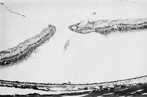
| Fig. 34. Advanced stage of lattice degeneration with vitreous liquefaction overlying the lesion, gliosis at the margin of liquefied vitreous, full-thickness retinal hole formation, sclerosis of blood vessels with pigment in the paravascular space, and irregularity of retinal pigment epithelium. |