|
|
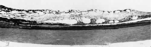
| Fig. 10. Microsection of meridional complex through atypical dentate process and its meridional fold. Anteriorly (on the left) the complex shows marked redundancy of pigmented epithelium in its outer aspect and a dense glial plaque on its inner aspect. Posteriorly (on the right) there is microcystoid change, nonspecific degeneration, and dense-staining glial cells along its surface. (Hematoxylin-eosin; × 63.) |