|
|
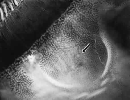
| Fig. 17. Typical degenerative retinoschisis.Note extensive region of typical cystoid degeneration with a rounded and elevated posterior margin. In the center (arrow), radial columns are randomly disrupted, causing a disturbance in coarse surface pattern. (× 18.) |