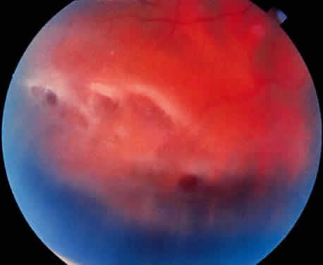
Fig. 31.
Latice degeneration demonstrating variable pigmentary changes to the underlying retinal pigment epithelium and circular atrophic hole (arrow)