|
|
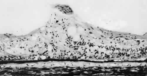
| Fig. 37. Microscopic features of the lesion seen in Figure 36. Entire retina within vitreous base shows nonspecific degeneration with gliosis. Centrally, the surface is elevated and shows a plaque of dense-staining glial cells. Retinal pigment epithelium also shows irregular thinning and pigment clumping, characteristically found within vitreous base. (Hematoxylin-eosin stain; × 500.) |