|
|
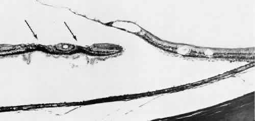
| Fig. 43. Flap of large full-thickness retinal tear shows microcystic degeneration; it is attached to condensed hyaloid of the posteriorly detached vitreous body. Posterior hyaloid also contains fragments from surface of the underlying retina, corresponding to partial retinal tears (arrows) behind the full-thickness retinal break. (× 150.) |