Chapter 13
Pediatric Neuro-Ophthalmology: General Considerations and Congenital Motor and Sensory Anomalies
R. MICHAEL SIATKOWSKI and JOEL S. GLASER
Main Menu   Table Of Contents

Search

THE PATIENT ENCOUNTER
VISUAL DEVELOPMENT
THE BLIND INFANT
ACQUIRED VISUAL LOSS IN CHILDHOOD
CONGENITAL MOTOR ANOMALIES
NYSTAGMUS AND RELATED DISORDERS
DYSLEXIA
HEADACHE IN CHILDREN
OCULAR MOTOR CRANIAL NEUROPATHIES
MYASTHENIA GRAVIS IN CHILDHOOD
SYMPTOMATIC SENSORY AND NONPARETIC STRABISMIC ABNORMALITIES
OCULAR TORTICOLLIS
FUNCTIONAL VISUAL LOSS
REFERENCES

This faculty of searching for the object is slowly acquired in the child: and, in truth, the motions of the eye are made perfect, like those of the hand, in slow degrees. In both organs there is a compound operation: the impression on the nerve of sense is accompanied with an effort of the will, to accommodate the muscular action to it.

The Hand, Its Mechanism and Vital Endowments

Sir Charles Bell, 1840

In Bell's early treatise on the function of the hand, he suggests that both the hand and the eye perfect their motor function through practice, meaning the brain teaches itself to coordinate visual and tactile perceptions. Thus, the brain actively orients the retinal receptors (or the hand) toward a target of interest, and then moves them with increasing precision. Perhaps it is not too far-fetched to compare the fine sense of touch at the fingertips, especially the thumb and index finger, to the fine sense of seeing at the macula. For development of effective eye–hand coordination, the head must become a stable platform for the visual system; however, in infants the neck muscles are weak. In an interesting experiment, it was found that, when the head is supported in an upright position, babies of 5 to 8 weeks of age reach with the accuracy of infants 20 weeks old.1 So, the visual system waits for a stronger neck.

The elegant complexity of the developing visual system is a recurring theme throughout the practice of pediatric neuro-ophthalmology, a unique subspecialty because it straddles three medical fields. Ophthalmologists are at the forefront of diagnosing and managing patients with combined visual, neurologic, and systemic abnormalities. Neuro-ophthalmologic problems of congenital origin or childhood onset may come to diagnostic attention in infancy (the blind baby), later in childhood (the grade school child with migraine), or in some cases, not until adulthood, when a congenital motor or sensory defect may be attributed to an acquired abnormality. For example, Duane retraction syndrome may be confused with a sixth nerve palsy, or anisometropic amblyopia may be interpreted as an acquired optic neuropathy. This chapter reviews and summarizes many of the important entities that fall into these potentially confounding categories.

Back to Top
THE PATIENT ENCOUNTER
Children are not little adults and infants are not even little children. Accordingly, the neuro-ophthalmologic history in infancy and childhood must be focused selectively; inquiries often must be directed toward the details of the pregnancy and postpartum period, including the gestational age, maternal illnesses, medications used during pregnancy, infant birth weight, Apgar scores, and use of perinatal oxygen. A detailed family history of ocular, neurologic, or musculoskeletal problems, as well as parental consanguinity must be explored. Neurodevelopmental milestones (Table 1) should be documented when indicated; height, weight, and head circumference should be charted on standard nomograms.

TABLE 1. Neurodevelopmental Milestones

Behavioral Event Age
Has sucking, rooting, and swallowing reflexes Neonatal period
Lifts head in sitting position4 mo
Rolls over6 mo
Sits up9–10 mo
Crawls10–11 mo
Walks unassisted12–15 mo
Walks up and down stairs holding on 18 mo
Can stand on one foot3 yr


Because the attention span of infants and children is quite limited, the physical examination must be conducted in a relatively brief time period. Simple inspection may allow the examiner to assess skull, facial, and other physical anomalies; note the presence of lid malposition or globe displacement; evaluate spontaneous ductal eye movements or the presence of nystagmus; and assess cognitive and to some extent, visual function.

Vision in infants is evaluated by noting response to light or hand–motion threat, the accuracy of the fixation reflex toward a hand light or other attractive object, and the capacity of each eye to follow during contralateral occlusion. In many cases, an infant's gross objection to occlusion of one eye, by crying or avoiding or pushing away the occluder, provides the first clue that vision in the uncovered eye is significantly impaired. Elicitation of optokinetic nystagmus assesses a visual sensory, as well as a motor, response.

The measurement of grating acuity by forced preferential looking tests (Teller acuity cards) is arguably the most useful method of obtaining reliable, reproducible, quantifiable information on visual acuity in infants and nonverbal children (Fig. 1).2–4 However, some have questioned the definition of “normal limits.” Kushner et al5 noted the often poor correlation between Teller (grating) acuity and Snellen (optotype recognition) acuity, the former yielding significantly better results, particularly when Snellen acuity is less than 20/70. It has been suggested that Teller acuity card testing results are imprecise and should not be used by social service agencies to determine legal blindness.6 Vernier acuity also may be measured in infants and children using preferential looking techniques.7 This may be more sensitive than grating acuity in detecting visual changes resulting from amblyopia. Nevertheless, grating acuity measurement is a practical method to assess visual function in the outpatient office setting; it is a valuable asset for determining whether vision is in a normal range for age, and it is useful for following progression of disease or visual development. Various clinical strategies for measuring visual acuity in children are summarized in Table 2.

Fig. 1. A. The examiner, masked to orientation of the stripes in the cards, evaluates visually directed eye movements during Teller acuity card testing. B. Cards may be presented at closer distances for children with low vision; vertical presentation is particularly helpful for patients with horizontal nystagmus.

TABLE 2. Clinical Applications of Vision Testing in Children


Neonatal
Phototropism (turning toward light)
Avoidance of bright light
 
Infancy
Fixation and following behavior
Optokinetic responses
Forced preferential looking test (Teller acuity cards)
Pattern VEP
 
Childhood
Allen pictures
Lea symbols
Snellen “E”
Landolt “C”
H_O_T_V letter game
Snellen acuity

VEP, visual evoked potential


Infants and children present particular challenges for successful visual evoked potential (VEP) recording, including their sleep–wake status, often poor postural positioning of the patient, and the inability to control and monitor ocular fixation and accommodation. Overestimation of acuity by VEP, particularly in amblyopia, may occur8; thus, for each laboratory, normative data for age group must be established. In many centers, VEP testing for the purpose of measuring acuity is unrewarding and inaccurate. However, in standardized laboratories with experienced personnel who are comfortable and familiar with administering the test, the visual evoked response, particularly pattern VEP, may be useful for monitoring visual development in both preterm and full-term infants.9

The visual field in infants may be assessed by gross confrontation mechanisms, such as visually evoked eye movements or withdrawal or fright to threatening maneuvers (see Chapter 2, Figs. 13, 14, and 15). Hemifield defects are easily detected when a child regularly fails to look at an object of interest (e.g., a toy) held in the nonseeing field. The patient's response to simultaneous presentation of objects in both hemifields can be assessed. Visually elicited eye movements are noted quite easily by the examiner. For more quantitative visual field measurements in infants, a variant of arc perimetry can be used. The work of Mohn et alsupplies useful normative data regarding normal development of binocular and monocular visual fields during the first year of life.10 By 3 years of age, children are able to comply with finger-mimicking tests. An experienced examiner frequently is able to perform simple kinetic perimetry in children older than 5 years, and many children aged 7 years and older are able to perform suprathreshold automated static perimetric testing, which may be presented as a “video game” with rewards for “good scores.”

The American Academy of Pediatrics has established guidelines for visual assessment.11 Children from birth through 3 years of age should have an eye evaluation in the physician's office to include: ocular history, vision assessment, external inspection of eyes and lids, ocular motility assessment, pupil examination, and red reflex examination. After age 3 years, evaluations should include all of the former, as well as more age-appropriate visual acuity assessments and an attempt at ophthalmoscopy. Appropriate testing strategies and referral criteria for distance acuity, ocular alignment, and assessment of ocular media clarity are detailed in the preceding reference.

Back to Top
VISUAL DEVELOPMENT
Visual and ocular developmental milestones are summarized in Table 3. The “critical period” of visual development refers to the period early in infancy when the visual system is sensitive to deprivation. It is believed that two different types of visual processing occur. Magnocellular (M) pathways deal with perception of motion, whereas parvocellular (P) neurons are more attuned to image shape and color (see Chapter 4). These two pathways are separated at the retinal ganglion cell level through to the lateral geniculate nucleus, and are represented in different areas in layer V-1 of the striate cortex. The parieto-occipital extrastriate cortex deals more with motion perception, whereas the temporo-occipital region is concerned with image shape and color. There is direct neural communication between these two areas. Maldevelopment of the magnocellular system occurs with infantile esotropia, latent and congenital nystagmus, impairment of gross stereopsis, and motion VEP deficits. Maldevelopment of the P system is associated with anisometropic and strabismic amblyopia, deficits in fine stereopsis, and spatial sweep VEP deficits.12

TABLE 3. Visual and Ocular Developmental Milestones


Event Age
Pupillary light reaction30 wks' gestation
Lid closure and response to bright light30 wks' gestation
Visual fixationBirth
Bliunk response to visual threat2–5 mo
Well-developed fixation6–9 wk
Visual following well developed3 mo
Accommodation well developed4 mo
Visual acuity potential at adult level by VEP6–12 mo
Grating preferential (Teller) acuity at adult level3 yr
Snellen acuity at adult level2–5 yr
End of critical period for monocular visual deprivation8–12 yr
Conjugate horizontal gaze well developedBirth
Conjugate vertical gaze well developed2 mo
Ocular alignment stable1–3 mo
Fusional convergence well developed6 mo
Stereopsis well developed6 mo
Stereoacuity at adult level7 yr
Foveal maturation completed4 mo
Myelination of optic nerve completed7–24 mo
Iris stromal pigmentation well developed6–12 mo

VEP, visual evoked potential
(Modified from Greenwald MJ: Visual development in infancy and childhood. Pediatr Clin North Am 30:977, 1983.)


Although normal binocular vision requires aligned eyes with intact sensory and motor fusion mechanisms, in the first few months of life some children have intermittent or constant ocular deviations, most commonly exotropia. Nevertheless, as the strength of the neck muscles permits (see earlier) in normal infants at about 3 months of age, there is a relatively rapid maturation of binocularity, including fusional convergence and sensory fusion (stereopsis). It has been hypothesized that refinement of cortical circuits, particularly those in the ocular dominance columns, occurs during this period of instability.13 It is well documented that children with esotropia, amblyopia, or early visual deprivation have asymmetric monocular pursuit noted on optokinetic nystagmus testing, favoring targets moving in a temporal-to-nasal direction; however, the presence of normal stereoacuity and binocular function is not always sufficient to prevent this bias.14 The relationship among binocularity, fusion, visual deprivation, and monocular optokinetic nystagmus asymmetry is complex and not precisely understood.

Visual development in infants with severe ocular disorders (e.g., retinal dystrophies, congenital optic nerve anomalies) may show delayed improvement of assessable visual function later in childhood, even up to the end of the first decade of life.15 Continuing posterior visual pathway maturation may be the mechanism responsible for late visual improvement in some cases. Of course, significant improvement in visual acuity as a result of amblyopia therapy also may occur in patients with either unilateral or bilateral, asymmetric structural abnormalities.16 It is imperative that the clinician exercise restraint when attempting to predict final levels of visual function in such children. In clinical practice frequently it is not possible to estimate visual function from either fundus appearance or neuroradiographic status of the anterior and posterior visual pathways.

Back to Top
THE BLIND INFANT
Assessment of the infant with subnormal or absent visual responses usually distinguishes between two major groups: those with and those without congenital (idiopathic infantile) nystagmus. Congenital nystagmus (see the following) may appear as a jerk or pendular, horizontal, uniplanar eye movement disorder, which frequently diminishes on convergence and is absent during sleep. In the past, congenital nystagmus has been divided into “sensory” and “motor” types. Although these concepts may be helpful clinically, they do not accurately reflect the underlying pathophysiology or the complex foveation strategies that the ocular motor system employs. It should be noted that a variety of other clinical features may be encountered in congenital nystagmus, including inversion of the optokinetic response and vertical or torsional movements (see Chapter 11).

Congenital nystagmus occurs in 1 in 1,000 to 6,550 births, and it is frequently associated with bilateral visual loss of prechiasmal or chiasmal origin.17 When occurring in the presence of bilateral corneal or lens opacities, optic nerve disorders, or macular disease, the diagnosis is straightforward. In the infant population, Gelbart and Hoyt18 demonstrated anterior visual pathway disease in 119 of 152 patients with congenital nystagmus, including diagnoses of bilateral optic nerve hypoplasia, Leber's congenital amaurosis, and ocular or oculocutaneous albinism. Likewise, Cibis and Fitzgerald19 noted bilateral retinal disease in 56% of patients with congenital nystagmus. Unilateral anomalies, however, allow development of normal foveation in the sound eye and, with rare exception, are not associated with congenital nystagmus. Often, congenital nystagmus does not present until the second to fourth month of life, when central, steady fixation and foveation should be established in normal infants.

In an infant with poor vision and nystagmus, but with an apparently otherwise normal eye examination, electroretinography (ERG) should be performed. Lambert et al20 reviewed this clinical scenario. Leber's congenital amaurosis is an autosomal-recessive retinal rod/cone disorder that causes poor vision from birth. At least seven different genetic defects have been shown to cause a similar clinical picture, and there is good evidence that photoreceptor degeneration actually starts prenatally.21 Although initial fundus examination may be unremarkable, pigmentary retinopathy, vessel attenuation, and optic atrophy occur over time. ERG shows nonrecordable or grossly attenuated electrical potentials of both rods and cones (“flat ERG”) (Fig. 2). Although initial acuity is quite poor, it is unlikely to deteriorate substantially over the course of the patient's life.22 No treatment is available. In exceptionally rare cases, spontaneous visual improvement may occur23; pharmacologic treatment in a mouse model has also been reported to be effective in improving vision.24

Fig. 2. Electroretinography from patient with Leber's congenital amaurosis demonstrates absent rod and cone responses.

Achromatopsia (rod monochromatism) is an autosomal-recessive or X-linked disease characterized by a partial or complete absence of retinal cone function. Acuity generally is better than in Leber's amaurosis and frequently exceeds 20/200. In this condition, the rod ERG is normal, whereas the cone ERG is significantly impaired (Fig. 3). Photophobia, which may be marked, is quite common, and these children show marked preference for dim lighting. Other hereditary retinal disorders may be seen in Joubert's syndrome (cerebellar vermis hypoplasia), congenital stationary night-blindness, Refsum's disease, neonatal adrenoleukodystrophy, neuronal ceroid lipofuscinosis, Jeune's syndrome, and osteopetrosis.

Fig. 3. Electroretinography from a patient with achromatopsia. Rod (top) and mixed (second) tracings are normal, but the cone flash and flicker responses (bottom two tracings) are severely reduced or absent.

Patients with albinism (Fig. 4) and aniridia, both of which may be associated with foveal (and optic nerve) hypoplasia, may present with congenital nystagmus. Anomalous (excessive) chiasmal crossing defects and cortical miswiring25 have been reported in ocular albinism. VEP in albinism demonstrates that the majority of visual fibers decussate at the chiasm, rather than about half as in normal individuals.

Fig. 4. Fundus of patient with albinism. Note marked absent of melanin in the RPE, with prominent choroidal vessels and foveal hypoplasia with lack of foveal reflex.

Note that the nystagmus in association with disorders of bilateral anterior visual pathway disease should not be confused with the bilateral “manifest latent nystagmus” seen in patients with monocular decreased vision, which acts as an occluder, thus manifesting what otherwise would have been truly latent nystagmus (see Chapter 11).26

Optic nerve hypoplasia, unless subtle, usually is diagnosed on fundus examination. Although most cases are idiopathic, maternal gestational diabetes and use of phenytoin are well-known risk factors; more recent epidemiologic studies have also suggested that young maternal age, first parity, smoking, and use of fertility and antidepressant drugs may also play a role.27 If bilateral, this condition may be associated with congenital nystagmus. De Morsier's syndrome (septo-optic dysplasia) refers to the constellation of bilateral optic nerve hypoplasia, absence of the septum pellucidum (Fig. 5), thinning or absence of the corpus callosum, dysplasia of the anterior third ventricle, and pituitary dysfunction (see Chapter 5, Fig. 9). Brodsky and Glasier28 broadened the spectrum of this condition. In a study of 40 children, some optic nerve anomalies were isolated, but in other children, midline craniofacial defects, hemispheric gray matter dystrophic anomalies, and posterior pituitary ectopia were noted. Of 21 cases of optic nerve hypoplasia described by Zeki et al,29 there were midline central nervous system defects in six and endocrine deficiencies in nine. In the series of 35 patients with bilateral optic nerve hypoplasia described by Siatkowski et alet al30 neuroradiographic abnormalities were seen in 46% and endocrinopathies in 27%. Growth hormone deficiency was the most common endocrine abnormality. The visual spectrum ranged from 20/20 in one case to no light perception in 34% of patients; 80% were legally blind (20/200 or less in both eyes). Absence of the septum pellucidum and corpus callosum, with panhypopituitarism, occurred in only 11.5% of all patients with bilateral optic nerve hypoplasia. Recently, a sporadic mutation in the HESX1 gene has been reported to cause optic nerve hypoplasia with pituitary insufficiency.31

Fig. 5. Magnetic resonance image of patient with bilateral optic nerve hypoplasia demonstrating absence of septum pellucidum with single midline ventricle.

A variant of this condition has been termed “superior segmental optic nerve hypoplasia,” wherein central acuity may be spared, but affected patients have inferior altitudinal field defects and superior optic pallor with nerve fiber layer dropout; maternal diabetes may be an associated factor.32 Additionally, it has been proposed that mild cases of optic nerve hypoplasia may indeed be misdiagnosed as amblyopia. Lempert demonstrated a significant difference in mean disc area and axial length in presumed amblyopic eyes compared to non-amblyopic controls.33

Optic disc colobomata (Fig. 6), if bilateral, may be seen in association with congenital nystagmus. Of greater import, however, is the association of colobomas or other dysplastic optic discs with basal encephaloceles, particularly in conjunction with hypertelorism, midfacial anomalies, or tongue-shaped retinochoroidal pigmentary disturbances.34 The morning glory disc syndrome, although typically unilateral, does not present in association with nystagmus, but rarely may be seen with midline central nervous system defects and endocrinopathies as well.35

Fig. 6. Spectrum of congenital disc anomalies. A. Large coloboma involving optic nerve and inferior retina. B. Dysplastic disc, central cup filled with fibroglial tissue. C. Morning glory disc anomaly. D. Markedly hypoplastic optic nerve. E. Coloboma of optic nerve without retinal involvement.

The infant with poor vision, no nystagmus, and a normal eye exam is likely to have cortical visual impairment. Good et al36 extensively reviewed this entity. These patients have normal pupillary light reaction, unremarkable fundi (at least initially), and absence of nystagmus in all but the rarest cases; however, they do have various neurobehavioral signs of cortical visual impairment:

  1. They have a preference for brightly colored objects and stare at bright lights (although up to one-third may have photophobia).
  2. They adopt peculiar head turns when attempting to look at or reach for an object of interest, because peripheral visual field, rather than central vision, is used.
  3. They may use extrageniculate vision, or “blindsight,” in some cases enabling them to localize targets or to see colors. This concept remains much in debate however, and many instances of “blindsight” may, in fact, result from preservation of some portions of visual cortex.
  4. They exhibit varying degrees of alertness and employ idiosyncratic visual “strategies” to use patches of visual field remnants.

Prognosis for recovery depends on etiology, age of onset, and severity of brain damage (Fig. 7). Although such children often remain visually handicapped, dramatic recovery may ensue occasionally.36 Cortical visual loss from perinatal hypoxia/ischemia has a particularly poor prognosis if congenital, but up to a 70% recovery rate if acquired.37 Although hydrocephalus, brain dysgenesis, and infections may produce cortical visual loss, by far the most common cause of CVI in the developed world is perinatal hypoxia. In term infants, damage primarily affects the watershed areas of the cerebral cortex (frontal and parieto-occipital regions). In premature babies, however, the damage is predominantly periventricular. Age-related differences in the developing intracranial vascular systems may account for the disparate sites of injury. The watershed zone in term infants (and adults) is at the parieto-occipital junction, whereas in preterm infants it is in the subcortical area.38

Fig. 7. Computed tomography of child with cortical visual impairment resulting from congenital hydrocephalus. Note significant ex vacuo dilatation of the ventricles posteriorly, with shunt catheter in place.

Although initially unremarkable in appearance, the optic disc subsequently may become abnormal in some cases of cortical visual impairment. Periventricular leukomalacia (PVL), which selectively involves the optic radiations near the trigone of the lateral ventricle, as well as the anterior corticospinal fibers, may produce abnormalities of visual acuity and visual field, disorders of higher cortical visual function, as well as spastic diplegia. Likely resulting from trans-synaptic degeneration across the lateral geniculate nucleus, PVL may produce secondary optic disc cupping, which may be confused with normal-tension glaucoma.39,40 As PVL primarily involves the retrogeniculate white matter, Brodsky has suggested that it be reclassified as “subcortical visual loss,” noting that such patients may have optic disc pallor and apparent hypoplasia in addition to cupping. In contrast, preferential involvement of the gray matter in the visual cortex carries with it a much lower incidence of optic disc abnormalities. Children with subcortical visual loss (PVL) also are more likely to exhibit tonic downgaze (sunsetting) and esotropia, whereas those with gray matter (cortical) disease have a higher incidence of exotropia and dysconjugate horizontal gaze.39,40 As PVL primarily often are present early in infancy, optic atrophy and cupping may not become apparent until later in the first, or seven second, year of life, as transsynaptic degeneration ensues.

Delayed maturation of vision is a diagnosis of exclusion and retrospection.41 This pertains to visually impaired infants with normal ocular examinations, normal neuroimaging and ERG, and no nystagmus. The exact etiology is unknown. Delayed myelination of the posterior visual pathways has been implicated, but in most cases, magnetic resonance imaging (MRI) shows age-appropriate myelination. Another hypothesis is that eye movement abnormalities (apraxia) may be misinterpreted as poor vision, as assessment of visual behavior in infants is to large degree interpreted based on visually directed ocular movements. Thus, care should be taken in making a diagnosis of delayed visual maturation in patients with abnormal saccades. More recent psychophysical studies suggest that delayed development of the subcortical (extra-geniculostriate) pathways may be the primary site of the defect.42–44 Alternatively, Hoyt has suggested that there is no evidence for failure of any primary visual system, and that a temporary visual inattention is the cause of this problem.44A

In this entity, visual function spontaneously improves, often quite rapidly over several days, suggesting a discrete rather than a widespread structural abnormality. This retrospective diagnosis should be made only when visual function indeed spontaneously improves to normal or near-normal levels, as best ascertainable by the end of the first year of life. However, careful visual testing later in childhood often discloses less than 20/20 acuity and various levels of binocular dysfunction; neurologic milestones may be delayed and cognitive function often is below average.

Children with unilateral hemianopic defects may adopt a head turn toward the side of the field defect so that the eyes are rotated toward the objects of visual interest. In addition, many of these children develop an exotropia,45,46 possibly to increase the panoramic visual field in compensation for the underlying hemianopic defect. It has been suggested that strabismus surgery in these children, therefore, should be avoided.

Back to Top
ACQUIRED VISUAL LOSS IN CHILDHOOD
Hereditary optic atrophies, neuroretinitis, optic nerve tumors, and trauma are discussed in Chapter 5 of this text. However, a few statements are in order regarding several entities.

Optic neuritis has notably different characteristics in the pediatric population than in adults. Various authors have reviewed this entity.47–52 Childhood optic neuritis, particularly in prepubescents, commonly presents as simultaneous bilateral papillitis (see Fig. 8, also see Chapter 5, Fig. 22); unlike adults, there is no clear sex predilection in this age group. Antecedent vaccinations or viral illnesses, ranging from upper respiratory infections to varicella, mumps, and mononucleosis, may precede visual loss by 2 to 6 weeks; some clinicians regard pediatric optic neuritis as a forme fruste of acute disseminated encephalomyelitis (ADEM). Initial visual acuity is typically poor, 20/200 or less, in many instances. Cerebrospinal fluid pleocytosis and elevated protein are common, but the eventual association with multiple sclerosis is less frequent in children than in adults.53 Prompt responses (within days) to systemic corticosteroids in doses ranging from 1 mg/ kg prednisone to high-dose intravenous methylprednisolone are the rule. Steroid taper over several weeks is recommended to reduce rebound inflammation. Although good visual outcomes are achieved in the majority (50% to 75%) of cases, there appears to be a higher rate of permanent visual deficits than in the adult population. Although MRI may demonstrate optic nerve enhancement or cortical white matter lesions in some cases, the results of the Optic Neuritis Treatment Trial (see Chapter 5) are not applicable to children. Later in childhood and in adolescence, optic neuritis begins to take on more of the characteristics seen in adults (i.e., female predilection, unilateral disease, retrobulbar neuritis, final visual acuity greater than 20/40 in over 90% of cases, and increasing likelihood of developing multiple sclerosis).52

Fig. 8. Fundus photo of 10-year-old girl with bilateral optic neuritis demonstrates mild optic nerve edema without hemorrhages or exudates. The fellow eye appeared similar. Vision was 20/400 in each eye.

Leber hereditary optic neuropathy (Fig. 9) commonly presents in the second decade of life,35 but several cases occurring within the first decade also have been reported.54–57 There is some evidence that those patients with the mtDNA 14484 mutation presenting in the second decade may show a higher incidence of spontaneous visual recovery, generally within 12 to 18 months of the initial visual loss. Because the initial fundus appearance in such patients often is unremarkable (especially if the typical telangiectatic peripapillary vessels are not present and there is no relative afferent pupillary defect in bilateral disease), children with Leber hereditary optic neuropathy may erroneously be misdiagnosed as functional visual loss at first presentation. Serial examinations are appropriate in such cases. In addition to Leber type, both dominant and recessive hereditary optic neuropathies may present in the first or second decades of life (see Chapter 5).

Fig. 9. Sixteen-year-old boy with Leber hereditary optic neuropathy. Vision was 20/400 with large central scotoma. Note minimal disc swelling and peripapillary telangiectatic vessels. The fellow eye became involved 1 year later.

Patients with both dominant and recessive hereditary optic neuropathies may complain of a slow deterioration of vision throughout the first and second decades of life. Dominant disease rarely causes acuities less than 20/200, but in recessive disease it may be 20/400 or worse. Another hereditary condition with optic neuropathy is Wolfram syndrome, also called “DIDMOAD,” for diabetes insipidus, diabetes mellitus, optic atrophy, and deafness (Fig. 10). Further information on hereditary optic neuropathies is found in Chapter 5.

Fig. 10. Six-year-old boy with Wolfram (DIDMOAD) syndrome. Note symmetric temporal pallor of both optic discs. Vision was 20/60 in each eye.

Anterior visual pathway gliomas account for the majority of intrinsic optic nerve tumors in childhood (Figs. 11, 12). Although they are true neoplasms, malignant features are extraordinarily rare in the pediatric population (see Chapter 5). Dutton58 provided a thorough review of this subject. When the glioma is initially confined to the optic nerve alone, the mortality rate is 5%. However, when the hypothalamus is involved, survival is less than 50% in some series. With a typically indolent course, these tumors generally can be managed conservatively, especially when confined to the optic nerve. Hoffman et al59 reviewed 62 cases of optic pathway/hypothalamic gliomas over a 14-year period, with 48 of these exhibiting relative stability with only visual defects: six patients had significant neurologic abnormalities, and eight died. Gayre et al60 reported a series of 42 patients with optic gliomas seen over 28 years at a single institution. Two-thirds were female, and slightly over half had neurofibromatosis type I (NF-1). Presenting signs and chiasmal involvement were similar in both NF (+) and NF (–) groups, the latter usually occurring within the first year after diagnosis. Regardless of treatment, the eye with better vision tended to remain stable over the long-term (and vision in the poorer eye often declined). Spontaneous regression of optic gliomas with visual improvement also has been reported.61,62

Fig. 11. Magnetic resonance imaging scans of optic gliomata. A. Marked enlargement of optic chiasm on coronal image. B. T1-weighted, gadolinium-enhanced image showing enlargement and marked enhancement of the right optic nerve and chiasm.

Fig. 12. “Bow-tie” atrophy of the optic nerve in a patient with NF-1 and diffuse optic glioma involving the optic tracts (see Chapter 5).

Tumors abutting the visual system that may cause progressive visual loss by extrinsic compression, including craniopharyngiomas, are discussed in Chapter 5. Pituitary tumors in childhood, although reported, are extremely rare.

Other causes of progressive visual loss in childhood are those syndromes associated with retinal pigmentary abiotrophies (tapetoretinal degenerations). These have a later onset than the congenital photoreceptor dystrophies, which profoundly affect vision at birth, and generally are stable. Included among these progressive retinal degenerations are the Bardet-Biedl syndrome (an autosomal-recessive disorder of polydactyly, obesity, hypogenitalism, renal disease, and tapetoretinal degeneration); Refsum's disease; Bassen-Kornzweig's syndrome (abetalipoproteinemia with intestinal malabsorption); and Kearns-Sayre's syndrome (progressive external ophthalmoplegia [see Chapter 12]).

Aicardi's syndrome consists of agenesis of the corpus callosum, chorioretinal lacunae (Fig. 13), and infantile spasms with seizures. The condition is limited to females and, with one exception, is lethal in males. Additional ocular features may include microphthalmia, optic nerve colobomas, peripapillary glial tissue, and variants of persistent fetal vasculature.63,64 Affected children rarely achieve developmental milestones beyond 12 months, and vision is decreased because of combined anterior and posterior visual pathway disease.65

Fig. 13. Fundus of a 3-year-old girl with Aicardi's syndrome. Note dysplastic optic discs surrounded by hypopigmented retinal lacunae. (Courtesy of John T. Flynn, MD)

The neurometabolic syndromes may present with progressive visual loss in infancy and childhood. These include, but are not limited to, Tay-Sachs', Niemann-Pick's, and neuronal ceroid lipofuscinosis (Batten's disease). The majority of these conditions also produce various other neurologic symptoms, with developmental regression in virtually all. Visual loss may be on the basis of optic atrophy, retinal degeneration, cortical factors, or a combination of both (see Chapter 5).

The most commonly encountered neurometabolic disease in pediatric neuro-ophthalmology is the group of neuronal ceroid lipofuscinoses (NCLs). These are autosomal recessive conditions characterized by brain and retinal atrophy. Infantile NCL produces generalized neurologic deterioration before visual problems occur, but in juvenile NCL (Batten's disease), visual symptoms are frequently the first sign of the disorder. Acuity declines to hand motion or worse in half of cases before other problems manifest. Fundus examination reveals optic atrophy and maculopathy, which may range from a subtle grainy appearance to the typical bull's-eye maculopathy. ERG shows depressed a and b wave responses under both photopic and scotopic conditions. Although many of the gene products responsible for this disorder have been identified in the laboratory, the exact mechanism of disease remains elusive, and treatment is unavailable.66,67

For further information on NCL and other metabolic disorders, the reader is referred to Brodsky et al

Back to Top
CONGENITAL MOTOR ANOMALIES

ANOMALIES OF INNERVATION AND ORBITAL MECHANICS

For many decades, clinical descriptions of eye movements, coupled with gross observations made during surgery, have generated a preoccupation with malformations of extraocular muscles, fascial connections, tendon sheaths, or check ligaments. In addition, the phenomena of “overacting” and “underacting” single ocular muscles are invoked by way of “explanations” of defective motor patterns. Electromyography (EMG) provides a technique whereby the electrical activity of extraocular muscles may be sampled. More recently, special orbital MR imaging techniques under both stationary and dynamic conditions have allowed greater understanding of orbital anatomy and extraocular muscle pulley systems. Thus, an interest in the neural and mechanical bases of paradoxic ocular motor defects has been renewed.

Hoyt and Nachtigaller68 reviewed the evidence for peripheral anomalies in the number and distribution of human ocular motor nerves, and have catalogued examples of omission, substitution, and duplication. In the dissection room, absence of the abducens nerve(s) has been documented, as well as agenesis of the sixth nerve nucleus, despite the presence of normal lateral rectus muscles. The lateral rectus indeed may be aggrandized by the oculomotor nerve in the absence of an abducens nerve, or it may receive a dual innervation from both the abducens and oculomotor nerve. In none of the foregoing anatomic studies was clinical information available, and the incidence of such anomalies of innervation cannot be assessed. Phillips et al69 reported pathologic correlation in a 5-year-old girl with bilateral abducens palsy, who died during surgical correction of convergent strabismus; neither abducens nerve was present, and the corresponding nuclei were diminutive. Rarely is such clinicoanatomic correlation possible, but sufficient material has accrued to validate the concept of “congenital miswiring,” including both failure of innervation and anomalous annexation of extraocular muscles by inappropriate nerve branches.

Simultaneous electrical activity in agonist–antagonist pairs of extraocular muscles (cofiring, cocontraction) has been documented on EMG, further substantiating the role of anomalous innervation in the production of paradoxic eye movements. For example, since the early work of Blodi,70 such paradoxic innervation of one of the horizontal rectus muscles is the most outstanding and consistent feature of Duane's retraction syndrome.

The outstanding work of Demer et al71–77 has used dynamic orbital MRI and elegant orbital histoanatomy studies to reformulate many concepts of ocular motor physiology. Each of the six extraocular muscles consists of global and orbital layers, the former contiguous with the tendon and inserting on the globe, the latter inserting on an intraorbital connective tissue ring, forming the muscle pulley. These pulleys guide the path of the muscles and serve as their functional origin. It is these pulleys that shift position during vergence eye movements to stimulate stereopsis, and account for the viability of Listing's law. The global and orbital layers have different innervations, fiber types, and cellular metabolism. Ectopic or unstable muscle pulleys may result in various types of incomitant strabismus, sometimes simulating cranial neuropathies, overacting muscles, or even supranuclear motor defects. Conventional strabismus surgery often is successful in part by its secondary effects on the muscle pulley systems. Pulley position also may be influenced by the placement of retroequatorial sutures to attach the eye muscles to the globe (retroequatorial myopexy).

Duane's Retraction Syndrome

Clinicians are quite familiar with the most common form of Duane's retraction syndrome (DRS): limited abduction of one eye, with globe retraction posteriorly into the orbit and narrowing of the lid fissure on adduction. Both autopsy and electrophysiologic studies are in concordance on the etiology of this condition.78 Matteucci79 reported a case of unilateral Duane's syndrome with a normal medial rectus, but a hypoplastic lateral rectus and hypoplastic abducens nucleus and nerve. In a patient with bilateral Duane's syndrome, Hotchkiss et al80 found bilateral absence of abducens nuclei and nerves; both lateral recti were partially innervated by branches from the inferior division of the oculomotor nerve and appeared histologically normal in innervated areas, but fibrotic in areas not innervated. Miller et al81 in a Duane patient who had had eye-movement recordings before death, demonstrated that the left abducens nerve was absent and, as in the previous case, the lateral rectus was innervated by branches from the inferior division of the third nerve, with fibrosis in areas not innervated, but normal in innervated areas. These studies provide elegant support for the hypothesis derived from previous EMG studies showing cofiring of the horizontal rectus muscles on attempted abduction (Fig 14).70,78

Fig. 14. Duane's retraction syndrome. A. Type I. Forward gaze, eyes straight with slightly narrowed left fissure. Right gaze, retraction of left globe with downward oblique deviation. Left gaze, failure of abduction. B. Type II. Forward gaze, slight exotropia. Right gaze, retraction of left globe with upward oblique deviation. Left gaze, normal abduction.

This variant of DRS was labeled type I, with type II described as an isolated adduction deficit, and type III with deficits of both abduction and adduction. However, newer concepts of embryology, coupled with the realization that there are many more twists and turns of aberrant innervation possible, has weakened the traditional classification of DRS.

The review by Mims82 documents that, in the fourth week of embryogenesis, the posterior portion of the abducens nerve in the pons innervates structures of the branchial clefts, which later atrophy as development continues. In most cases of DRS, all or most of the sixth cranial nerve (instead of just the posterior portion) is attracted caudally to these structures. Later in embryogenesis, most or all of the nerve and its nucleus atrophy, leaving the developing fetus either with no sixth nerve or very few residual fibers. The uninnervated (or hypoinnervated) lateral rectus provokes a neurotropic competition among the extraocular muscles in the orbital apex, attracting nerve fibers from the medial rectus subnucleus of the oculomotor nuclear complex. Although this phenomenon is what most likely occurs in the majority of DRS cases, the proximity of these structures during embryogenesis may lead to various other innervational and developmental anomalies, resulting in other motility deficits (e.g., adduction problems, synergistic divergence, abnormal vertical movements), associated synkinetic conditions (e.g., jaw-winking, crocodile tears), cranial nerve dysfunction (e.g., sensorineural deafness), and branchial arch disorders (e.g., Goldenhar's syndrome). An interesting variant of motility defect has been termed “synergistic divergence,” in which both eyes abduct on attempted lateral gaze.82A

In all types of Duane's syndrome, vertical deviations of the adducting eye may take the form of upshoots and downshoots (Fig. 15). Scott and Wong83 recorded increased activity in both the inferior oblique and superior rectus muscles during upshoot in adduction, most consistently in the superior rectus, suggesting another variant of aberrant innervation. However, a tightly fibrosed lateral rectus insertion also may act as a mechanical leash, causing dynamic vertical overaction and a so-called flipping mechanism. Meticulous clinical examination and intraoperative assessment of anatomy and forced ductions help to estimate the relative contributions of mechanical and innervational anomalies in DRS-related upshoots and downshoots; then the most effective surgical strategy is possible.84

Fig. 15. Electromyography in Duane retraction syndrome. Simultaneous recordings of left lateral rectus (LR) and medial rectus (MR). A. Type I. Peak innervation of lateral rectus on right gaze (paradoxic innervation) and minimal innervation on left gaze. Normal electrical behavior of medial rectus throughout. B. Type II. Lateral rectus innervation on both right and left gaze. Normal electrical behavior of medial rectus throughout. C. Type III. Synchronous and strikingly similar electrical activity in both recti, with intense firing in right gaze (black arrow) and marked inhibition in left gaze (open arrow). (From Huber A: Electrophysiology of the retraction syndromes. Br J Ophthalmol 58:293, 1974)

When describing individual patients with DRS, five clinical characteristics are important: (a) the type of deviation in primary gaze (esotropic versus exotropic DRS); (b) any compensatory face turn to promote fusion; (c) degree of co-contraction (especially noting globe retraction, adduction deficits, and upshoots and downshoots); (d) type of strabismus present in the reading position (downgaze at near); and (e) degree of abduction deficit. Such details more precisely characterize the condition than the previous nomenclature, and also allow more appropriate planning of corrective surgical strategies.82

According to Raab85 and others, Duane's syndrome occurs more commonly in women (about 2:1 over men), and the left eye is involved perhaps three times more commonly than the right eye. Approximately 10% to 18% of cases are bilateral. Primary gaze strabismus (usually esotropia) and secondary ocular torticollis are present in approximately 50% of patients. Familial cases are not uncommon, and bilateral retraction has been reported in monozygotic twins.86 Duane syndrome also has been reported in association with abnormalities on chromosomes 4q.22,87,88

Patients with DRS may demonstrate a peculiar sensory adaptation, with excellent binocular function in directions of gaze where visual axes are aligned, and suppression without diplopia in the field of the affected lateral rectus (i.e., a facultative suppression scotoma, as is employed in intermittent exotropia). Occasionally, mild amblyopia may be present in the involved eye, but the reported incidence of amblyopia varies greatly, as does the incidence of anisometropia.89 A few patients who as children became accustomed to avoiding the diplopic field may present as adults complaining of an “acute” onset of diplopia. However, microtropia or larger angles of strabismus may be present, convergence often is poor, and sensory adaptations are complex and sometimes tenuous. Age-related deterioration in fusion ability and in convergence, as well as progressive fibrosis of the rectus muscles, can produce symptoms of diplopia and asthenopia. Surgical approaches to the various presentations of DRS are beyond the scope of this chapter, but standard muscle recessions and transposition procedures for primary gaze strabismus and ocular torticollis, and Y-splitting of the lateral rectus for upshoots and downshoots, all may play a role. Surgery on the fellow eye often is required to make ductions and versions more symmetric.

Retraction of the globe somewhat mimicking Duane's syndrome occasionally may be seen with acquired orbital lesions that cause mechanical restriction. The chronic fibrotic changes of extraocular muscles in thyroid-related orbitopathy may cause a leash effect, most typically observed on abduction attempts, but possible on gaze opposite the field(s) of action of any involved muscle(s). The tethering also prevents full ductional range, and forced-duction testing (see Chapter 12) confirms the restrictive process. Proptosis and other orbital congestive signs generally are present. Similarly, infiltration of muscles by granulomatous inflammation (orbital pseudotumor) or by carcinoma may cause restrictive contraction, but rarely to the degree observed in Duane's syndrome.90 Medial orbital wall fractures can entrap the medial rectus muscle, with variable abduction limitation and globe retraction,91 and retraction also has been reported with muscle fixations resulting from orbital metastases.92

Möbius' Syndrome (Congenital Bulbar Palsies)

The discovery in 1888 of the condition of facial diplegia of variable degree associated with paralysis of lateral gaze, and usually esotropia, is attributed to Möbius. Although traditionally considered simply as palsies of the facial and abducens nerves, EMG and neuropathologic evidence indicates a more complex situation, as does involvement of other body systems. Variable associations include tongue hemiatrophy; head and extremity deformities; abnormalities of lower cranial nerves, producing hearing, speech, and swallowing difficulties; chest malformations; congenital heart defects; limb disorders, including club feet; micrognathia and tongue malformations; Poland anomaly; and Kallmann's syndrome (hypogonadism and anosmia).93–95 Clinical studies also have noted a relatively high incidence of mental retardation and cognitive dysfunction96 as well as autism.97 Although generally considered a static neonatal disorder, there have been reports of progressive, increasing motor deficits during later life.

Clinically, children present with feeding problems because of their inability to suck, and their lack of facial expression is noted, especially when crying (Fig. 16). Because vertical gaze and convergence typically are preserved, the ocular motor anomaly mimics bilateral abducens palsy, usually with convergent squint. Convergence is used as a substitution phenomenon to “cross-fixate” (i.e., the left eye adducts to look rightward, and the right eye adducts to look leftward). Vertical gaze typically is preserved, but total ophthalmoplegia may be present. Möbius' sequence is infrequently familial, with evidence of autosomal transmission, but many involved members show only facial palsies with intact eye movement, possibly a forme fruste of Möbius' sequence. In rare instances, thalidomide use during pregnancy has been implicated.98 A report found maternal exposure to misoprostol in almost 20% of cases.96 Both of these reports are consistent with a vascular etiology, but the exact cause of Mobius' sequence is not precisely known. Neuroimaging may be completely normal or may demonstrate profound abnormalities, including diffuse brainstem hypoplasia and straightening of the floor of the fourth ventricle.99

Fig. 16. Möbius syndrome. Esotropic deviation with bilateral deficit of horizontal gaze. Vertical gaze is intact. Note facial diplegia.

The clinician also should be aware of central respiratory dysfunction and sleep disorder in infants with Möbius' sequence.100,101 Additionally, these children may have a higher incidence of psychological and emotional difficulties as a result of their inability to smile and communicate their affect noticeably.102 Strabismus surgery can have a profound visual and psychological effect on these patients. Additionally, newer plastic surgery procedures may restore some facial animation and the ability to smile in Mobius patients.103,104 Use of gracilis muscle transplantation to the face using facial vessels for revascularization and the motor nerve to the masseter for reinnervation has had notable success in the medical and lay press.105 In affected children with speech difficulty, gracilis transplantation also may improve function in this regard.106 It is important to consider all of these surgical procedures in Möbius sequence, as affected children may be regarded erroneously as unintelligent or autistic because of difficulties in facial and oral expression.

Elevator Deficiencies

Congenital vertical eye movement anomalies may be the result of supranuclear, nuclear, or cranial nerve defects, mechanical restrictions of the globe itself, or variable combinations of these factors. Ziffer et al107 delineated three such groups of patients: (a) those with primary inferior rectus restriction; (b) those with primary superior rectus paresis; and (c) those with supranuclear elevation deficiency. The latter two types also may develop secondary contracture of the ipsilateral inferior rectus, producing combined supranuclear/nuclear and infranuclear deficits. Monocular elevation deficiency usually takes the form of double elevator palsy, with inability to elevate the globe in both adduction and abduction, simulating apparent weakness of superior rectus and inferior oblique muscles. The involved eye may be without deviation in the primary position or may be hypotropic, but diplopia generally is absent and the pupil categorically is uninvolved. Mild to moderate homolateral ptosis frequently is observed (Fig. 17).

Fig. 17. Double elevator palsy. A. Defective elevation of the right eye is demonstrated in abduction and adduction. B. In similar situation right eye does not elevate in volitional upward gaze. C. Upward deviation of right eye on forced lid closure (Bell's phenomenon) indicates supranuclear nature of defect.

Jampel and Fells108 reported that unilateral, discrete vascular lesions involving the contralateral pretectal, supranuclear fibers destined for the superior rectus subnucleus in the oculomotor nuclear complex may produce acquired monocular elevation paresis. Ford et al,109 however, reported on a case of acquired monocular elevation paresis caused by a tumor metastatic to the ipsilateral dorsal mesodiencephalic junction. Munoz and Pager110 reported progressive monocular double elevator palsy in an 8-year-old girl complaining of diplopia. MRI disclosed a pineocytoma, and her strabismus and ocular motility improved postoperatively.

Surgery for double elevator palsy is indicated when there is a large hypotropia in the primary position or significant chin-up posture in order to maintain fusion. If forced ductions are positive, recession of the inferior rectus should be performed. Otherwise, vertical transposition of the horizontal rectus muscles (Knapp procedure) offers good, long-lasting effects.111

That the anatomic pathways for the control of vertical eye movements are not completely understood should be evident (see Chapter 10). For acquired monocular elevation paresis, consensus dictates that the lesion must lie immediately adjacent to the oculomotor nucleus on the opposite side, because the motor fibers to the superior rectus muscle arise in the contralateral superior rectus subnucleus. Such cases have been documented with unequal, large pupils showing light-near dissociation, and with miotic, Argyll Robertson–type pupils also with light-near dissociation.

Monocular depressor deficiency is quite rare, and it has been recorded as a congenital defect. Acquired forms occur with restrictive orbital lesions and “inferior rectus”–type oculomotor palsies (see Chapter 12) or skew deviation (see Chapter 10).

Acquired bilateral downgaze palsies are the result of bilateral lesions located dorsomedial to the red nuclei, with variable rostral and caudal extension, and involving the rostral interstitial nucleus of the medial longitudinal fasciculus. Jacobs et al112 reported an unusual cause of bilateral downgaze palsy (i.e., a bilateral lesion of the dorsolateral mesencephalic periaqueductal gray matter), remote from the rostral interstitial nucleus of the medial longitudinal fasciculus.

Vertical Retraction Syndrome

Vertical retraction syndrome, comparable to horizontal retraction seen with Duane's syndrome, is rarely encountered. One sporadic unilateral case,113 as well as familial bilateral cases,114 have been reported. Typically, there is limited elevation or depression associated with retraction of the globe and narrowing of the lid fissure on either upward or downward gaze. There may be an associated esotropia or exotropia more marked in the direction of the restricted field of vertical movement. Forced duction test was positive, but EMG data are not available.

In an atypical case, there was poor abduction and elevation of the right eye associated with retraction of the right globe on leftward, upward, and downward gaze.115 EMG showed cofiring of the right vertical recti in three gaze directions; eye movement recordings also suggested anomalous innervation involving fibers to the right vertical recti and right lateral rectus. A miswiring anomaly during embryogenesis is likely.

Marcus Gunn Jaw-Winking Synkineses

Synkinesis is defined as a simultaneous movement or a coordinated sequence of movements of muscles supplied by different nerves or by separate peripheral branches of the same nerve. Normally occurring cranial nerve synkineses are exemplified by sucking, chewing, conjugate eye movements, or Bell's phenomenon, all of which are mediated by supranuclear pathways.

Abnormal cranial nerve synkineses may be either acquired or congenital. Intrafacial dyskinesia may follow peripheral facial nerve palsies (the “inverted” Marcus Gunn phenomenon, or Marin Amat syndrome116 [see Chapter 8]), and aberrant oculomotor misdirection is commonly a sequel to trauma (see Chapter 12). Marcus Gunn jaw-winking (trigemino-oculomotor synkinesis) and, indeed, the Duane retraction syndrome (see earlier) are examples of pathologic congenital synkineses. There are also a number of peculiar yet physiologically associated movements, including the coupling of volitional horizontal gaze shift with involuntary ear wiggle (oculoaural) or brow elevation (oculofrontal).117

Using EMG in normal subjects, Sano118 demonstrated distinct cofiring of the extraocular muscles innervated by the oculomotor nerve, and of the muscles of mastication innervated by the trigeminal nerve. Sano believes that such findings support Wartenberg's hypothesis of “release phenomena”; that is, synkineses result from damage to cranial nerve nuclei incurred secondary to peripheral nerve injury (réaction á distance), and that the secondary nuclear lesion “releases phylogenetically older [neural] mechanisms with their tendency toward associated movements.”119 Therefore, release phenomena represent failure of inhibition of primitive reflexes, as exemplified by palmomental, suck, snout, and primitive grasp-feeding reflexes, and by the dysraphic “mirror movements” seen occasionally in Duane's syndrome or in the lid-facial-oral dyskinesia of Meige's syndrome.

Although not necessarily associated with disturbances of ocular motility, the congenital trigemino-oculomotor synkineses involving the jaw muscles and the lid levator are included here as examples of anomalous innervational patterns. Originally described by Gunn in 1883, this phenomenon presents as unilateral ptosis of variable degree, usually noted shortly after birth. As the infant nurses, the ptotic lid rhythmically jerks upward. In a series of nearly 1,500 cases of congenital ptosis,120 80 patients (5%) displayed the Marcus Gunn phenomenon. There was no distinct laterality preference, and three patients had bilateral Marcus Gunn phenomenon. There was no gender preponderance, and only two cases were familial. Amblyopia was present in 54%, anisometropia in 26%, and some degree of strabismus in 56%, including 19 cases each of superior rectus palsy and “double elevator palsy” and two cases of Duane syndrome. The question of spontaneous amelioration with aging has not been clarified. Other authors have commented that the synkinesia does become less conspicuous, but it is possible that adults learn to minimize lid excursions consciously.121

Sano118 extensively reviewed the subject of trigemino-oculomotor synkinesis. Two major groups are classified: (a) external pterygoid-levator synkinesis (i.e., elevation when jaw is thrust to opposite side [homolateral external pterygoid], when jaw is projected forward [bilateral external pterygoid], or when mouth is opened widely); and (b) internal pterygoid-levator zsynkinesis (i.e., lid elevation on teeth clenching). The first group is by far the more common. Regarding pathogenesis, Sano presented EMG evidence that the jaw-winking phenomenon is an exaggeration of a normally existing, but barely detectable, physiologic co-contraction (associated movement), which a congenital brainstem lesion has “released” from higher central control. Sano also demonstrated that direct stimulation of the pterygoid muscle results in lid retraction; however, damage to the trigeminal motor fibers relieves the lid activity. Mrabet et al122 described two families with Marcus Gunn jaw-winking, demonstrating an autosomal-dominant type of inherited pattern with incomplete penetrance.

The surgical management of Marcus Gunn jaw-winking phenomenon is complex, and the reader is referred elsewhere for discussions of the various proposed surgical approaches.123,124

A rare condition in which the lid falls as the mouth opens has been dubbed the inverse Marcus Gunn phenomenon. Lubkin125 used EMG to demonstrate inhibition of the affected levator palpebrae superioris concurrent with external pterygoid contraction, but no associated activity of the orbicularis oculi. Therefore, in this condition, trigeminal innervation to the pterygoids is associated with inhibition of the oculomotor branch to the levator, whereas in the true Marcus Gunn phenomenon it is associated with excitation of the oculomotor branch to the levator.

A peculiar case of torsional diplopia associated with the initiation of swallowing (i.e., deglutition trochlear synkinesis) has been reported,126 incriminating a dyskinesia that couples the fourth cranial nerve with bulbar musculature, which is innervated by the trigeminal, facial, or hypoglossal nerves. The cases of two sisters, each with monocular lid retraction on adduction or downward gaze, also have been reported.127 Another variant is jaw-winking with pseudo overaction of the inferior oblique muscle.128

RESTRICTIVE SYNDROMES

The term “restrictive” refers to mechanical tethering of the globe because of congenital or acquired anomalies of the extraocular muscles or fascial attachments, rather than to supranuclear or infranuclear synkinesias or peripheral neural miswiring patterns. The mechanical nature of the motor defect is proved by resistance to passive (forced) duction of the eye when the conjunctiva or muscle insertions are used in attempts to rotate the globe (see Chapter 12 and Chapter 3, Fig. 6).

Brown Tendon Sheath Syndrome

In 1950, Brown 129 described patients with apparent congenital paresis of the inferior oblique muscle, with restricted elevation on forced duction testing when the globe was in the adducted position (see Chapter 12). At surgery, thickening of the superior oblique tendon sheath was observed, and passive movement was restored after the sheath was stripped. The positive forced duction test clearly distinguishes this disorder from inferior oblique palsy; however, other common features can be observed, including increasing exotropia in upgaze (V-pattern), downward displacement of the eye on attempted adduction (superior oblique overaction), and occasional widening of the palpebral fissure in adduction. Most patients enjoy good binocular function with or without compensatory head position, and amblyopia is uncommon. The syndrome may be bilateral, rarely is familial, and has been documented with mirror symmetry in monozygotic twins.130

Catford and Dean-Hart131 demonstrated normal reciprocal firing patterns in the oblique muscles of three patients with tendon sheath syndrome, and concluded that the syndrome was caused strictly by a local mechanical defect rather than paradoxic innervation with cofiring. Sevel132 provided a useful review of etiologic considerations and added the suggestion, derived from this investigation of the trochlear region in embryonic life, that there may be persistent fine trabeculae between the superior oblique tendon and the cartilaginous trochlea.

Although the majority of patients with congenital Brown syndrome do not require surgery, correction should be considered when the eye is significantly hypotropic in the primary position, or a disfiguring or otherwise unsatisfactory head position is present. A superior oblique weakening procedure (e.g., tenotomy, tenectomy, or silicone or nonabsorbable suture tendon expander) is preferred. Although these maneuvers effectively free the mechanical restriction so that the globe can elevate in adduction, additional surgery may be necessary to correct an induced superior oblique palsy (e.g., inferior oblique recession).

Restriction of elevation in adduction may result from acquired lesions localized to the superior oblique muscle or the trochlea itself. This acquired form of tendon sheath syndrome can be a rare complication of rheumatoid arthritis (adult or juvenile form),133–135 orbital trauma,136 sinusitis, sinus surgery,137 encircling scleral buckles, blepharoplasty,138 and superior oblique tuck procedures, or may occur as an idiopathic form of painful tenosynovitis.139 An isolated metastasis to the superior oblique muscle also has been reported.140 Several authors have advocated administering a trial of local steroid injection when acute tenderness and swelling in the region of the trochlea indicate an inflammatory cause.133,135,139 However, both oral nonsteroidal and steroidal anti-inflammatory drugs often afford relief as well.

Both congenital and acquired forms of tendon sheath syndrome may be momentarily intermittent, and numerous instances of audible “clicks” or “snaps” have been reported at the instant of restoration of full elevation.

Congenital Fibrosis Syndromes

Replacement of extraocular muscles by fibrous tissue is a relatively rare form of fixed congenital ophthalmoplegia. Variable motility restrictions are dependent on the number and location of involved muscles. Rarely, all of the extraocular muscles and lid levator may be fibrosed. These frequently familial conditions are clinically characterized by downward or inward fixation of both eyes, moderate to marked ptosis, and jerky convergent movements on attempted upgaze. The head is thrown backward to compensate for fixed downward gaze. Astigmatism, amblyopia, and less frequently, a degree of enophthalmos are present. Strictly unilateral cases are recognized, and may be seen in association with ptosis and enophthalmos. Eye muscle surgery is indicated for symptomatic stable deviations; as in other types of restrictive myopathies, muscle recessions rather than resections are the procedure of choice. Ptosis surgery often is required as well, but care must be taken to avoid exposure keratopathy in patients with postoperative lagophthalmos and poor to absent Bell's phenomenon.141

Notable work by Engle et alhas now defined a genetic and molecular basis for many of the congenital fibrosis syndromes.142–144 Several distinct phenotypes are described, with evidence that the genetic defects result in aberrant development of all or portions of the oculomotor and trochlear nuclei and their cranial nerves. Congenital fibrosis of the extraocular muscles (CFEOM) type 1 have bilateral ptosis and inability to elevate eyes beyond the midline. This phenotype is inherited in an autosomal dominant fashion and maps to the FEOM1 locus on chromosome 2. Neuropathologic correlation shows absence of the superior division of the oculomotor nerve and its mesencephalic neurons, suggesting a primary developmental abnormality of the oculomotor nucleus. CFEOM2 presents with ptosis and exotropia with severe limitation of horizontal and vertical gaze. This phenotype is inherited in an autosomal recessive fashion and maps to the REOM2 locus on chromosome 11. Affected subjects have mutations in the transcription factor gene ARIX, which is known to be essential to development of both the oculomotor and trochlear nuclei in mice and zebra fish. CFEOM3 phenotypes are more variable and map to chromosome 16. Additional reports demonstrate a variety of other neurologic abnormalities in these patients,145 further supporting a genetic mechanism affecting neural tissues.

Back to Top
NYSTAGMUS AND RELATED DISORDERS
Although the following entities are discussed in Chapter 11, they are reviewed here with particular attention to the pediatric population.

TRANSIENT IDIOPATHIC NYSTAGMUS OF INFANCY

Nystagmus in children always raises clinical concern for visual or neurologic disease; however, transient neonatal nystagmus is generally benign. Good described 11 infants in whom nystagmus developed at a mean age of 2/7 months.146 No cause was found despite extensive workup, and the nystagmus had disappeared by the end of the first year of life in all cases. The nystagmus pattern was horizontal in 50%, but vertical and monocular forms also were seen. Some patients had delayed visual maturation or retinopathy of prematurity. They hypothesized that an unstable, plastic ocular motor system in infancy could be affected by any transient disruption of afferent visual input and could potentially lead to nystagmus.

CONGENITAL NYSTAGMUS

Congenital (idiopathic infantile) nystagmus frequently is seen in association with bilateral anterior visual pathway disease, although it may be isolated as well and occur in patients with normal vision. The waveforms are typical and have been well described, consisting of a velocity-increasing slow phase (see Chapter 11). The clinical findings and eye movement recordings of congenital nystagmus are identical in cases with and without visual loss. The nystagmus usually is conjugate, horizontal, and uniplanar, although small vertical or torsional movements may occur. Congenital nystagmus presents typically at 2 to 4 months of life and may diminish somewhat in amplitude and frequency in adulthood. Some patients develop a null point and adopt compensatory anomalous head postures to reduce ocular oscillations and maximize visual acuity. Although many consider congenital nystagmus to be the result of a primary fixation defect, Dell'Osso et al147 have demonstrated accurate and maintained target-foveation in patients with congenital nystagmus. Furthermore, they demonstrated that eye velocity matched target velocity during foveation intervals and concluded that the smooth-pursuit mechanisms in patients with congenital nystagmus were relatively normal.148

Most investigators now believe that congenital nystagmus results from instability of the brainstem neural integrator responsible for gaze-holding, although apparently normal gaze-holding mechanisms have been demonstrated in at least some congenital nystagmus patients.149 Abnormal histoanatomy of the extraocular muscle tendon has been reported in humans with congenital nystagmus, but the clinical or causative implications of this finding remain unclear.150 Oscillopsia is not a typical feature in cases of congenital nystagmus, but has been described by Abel et al,151 who postulated that suppression of oscillopsia is operable only within certain limits of foveation stability: When nystagmus exceeds these, oscillopsia results. Additionally, Gresty et al152 described six patients who had eye-movement recordings characteristic of congenital nystagmus, but who presented in adulthood with symptoms of oscillopsia; no etiology was uncovered, and the process was believed to be benign. Hertle et alreported a set of similar patients and suggested that oscillopsia in patients with congenital nystagmus may be precipitated by concomitant visual sensory conditions (e.g., decompensating strabismus or retinal degeneration).153

Various therapeutic modalities have been attempted in congenital nystagmus. Contact lenses are known to dampen nystagmus, although worsening (rebound phenomena) may occur after their removal.154 Base-out prisms may be helpful for patients whose nystagmus dampens during fixation, provided there is no strabismus. In those patients with a null point and consequent anomalous head position, Anderson-Kestenbaum strabismus surgery yields improvement in head position as well as visual acuity.155 Alternatively, for patients without a null point, Helveston156 reported improvement in nystagmus and near visual acuity after large retroequatorial recessions of all four horizontal recti. Dell'Osso has reported that four horizontal rectus muscle tenotomy (without recession) dampens congenital nystagmus in a dog model and in some humans.157 Using the hypothesis that the nerve endings at the tendino-scleral interface innervate afferent fibers of the first division of the trigeminal nerve, and by so doing influence eye-movement control, Hertle has performed four-horizontal muscle tenotomy in 10 subjects with congenital nystagmus. He reports improvement of binocular visual acuity in 50%. Despite future potential, firm conclusions are currently hampered by the small numbers of patients, and the hour-to-hour and day-to-day variability of congenital nystagmus, which limits data analysis and interpretation. At this time, such surgery does not enjoy widespread support and remains controversial.

In the authors' experience, eye muscle surgery in cases of congenital nystagmus is most beneficial when a significant anomalous head posture is present or the nystagmus is of very high amplitude. Underlying structural ocular anomalies or amblyopia often precludes significant improvement in visual acuity. However, early surgery may reduce or resolve the nystagmus in infants with bilateral congenital cataracts.

LATENT NYSTAGMUS

Clinically, latent nystagmus is defined as nystagmus that appears when one eye is covered, the fast component (jerk phase) occurring away from the covered eye. This form is common in children with congenital strabismus, particularly with esotropia. It is not an acquired condition and does not require extensive neurologic investigation. Latent nystagmus may become manifest if visual acuity is decreased in one eye (e.g., because of injury or amblyopia), which effectively acts as an “occluder” over that eye. On eye movement recordings, latent nystagmus demonstrates linear or exponentially decreasing slow phase velocity. Although conventional wisdom holds that a nasotemporal optokinetic imbalance occurs with congenital strabismus and latent nystagmus, because of failure to develop binocular vision, Gresty et al159 believe that both latent and congenital nystagmus arise from a genetic or acquired embryologic disorder. Kommerell and Zee160 described two patients who were able to release or suppress latent nystagmus at will, presumably on the basis of voluntary control of visual input contributed by the amblyopic eye. Most patients with latent nystagmus are asymptomatic and do not require treatment, but in patients with cosmetically bothersome oscillations, strabismus surgery or injection with botulinum toxin type A (BOTOX, Allergan, Inc., Irvine, CA;Myobloc) may be helpful.161 The primary clinical importance of latent nystagmus is that its presence always signifies a congenital or infantile abnormality of the ocular motor and binocular fusion systems, requiring no further neurological workup.

PERIODIC ALTERNATING NYSTAGMUS

Periodic alternating nystagmus (PAN) is a conjugate horizontal jerk oscillation, during which there are regular reversals of the direction of the fast phase, separated by brief, quiet intervals. The time period of each cycle is variable, ranging from 1 to 5 minutes, and may be asymmetric. During the beating phases, the amplitude, frequency, and velocity progressively change. Therefore, a periodically shifting null point is seen commonly, with compensatory face turning to alternate sides. PAN may be an acquired condition after caudal brainstem or cerebellar disease, especially tonsillar herniation resulting from Chiari malformations. However, there have been a few reports of congenital periodic alternating nystagmus, particularly in patients with albinism.162 It is likely that PAN goes undiagnosed in a large number of instances. Shalloh-Hoffman noted PAN in over one-third of patients with congenital nystagmus.163 To definitively exclude this condition, the examiner must observe patients' nystagmus for a minimum of three minutes (164).

Both congenital and acquired cases reportedly respond well to oral Lioresal (Baclofen tablets). If surgery is indicated, large recessions of all four horizontal rectus muscles are likely the best approach. Kestenbaum-Anderson type procedures are contraindicated, as the null point and anomalous head position are constantly shifting in this condition.

HEIMANN-BIELSCHOWSKY PHENOMENON

The Heimann-Bielschowsky phenomenon is an unusual motility pattern that develops after monocular visual loss, potentially many years later, and therefore is frequently incorrectly interpreted as a sign of acquired neurologic disease. The phenomenon consists of strictly monocular, coarse, pendular, primarily vertical oscillations occurring only in the poorly seeing eye. It is more prominent at distance and is inhibited by convergence or fixation. The oscillations vary from 1 to 5 cycles/second, and vertical amplitude from a few to more than 25°.165 It has been suggested that there is a correlation among the amplitude, oscillations, and duration of visual loss.166 As with latent nystagmus, the presence of these movements does not require neuroimaging or further laboratory investigation unless the cause of the visual loss remains unclear. Strabismus surgery is beneficial if the movements are cosmetically or socially disturbing.

CONGENITAL OCULAR MOTOR APRAXIA

Originally described by Cogan in 1952, congenital ocular motor apraxia is characterized by a deficiency in the generation of voluntary horizontal saccadic eye movements despite the presence of spontaneous saccades (see Chapter 10). Vertical saccades are unaffected and are completely normal. Affected infants may present with delayed visual or psychomotor development or even may appear to be blind. During the later half of the first year, compensatory head thrust movements become apparent. The ocular motor abnormalities tend to improve with increasing age, and there is a clear tendency for natural resolution between the first and second decades of life,167 but often subclinical abnormalities of the horizontal saccades are noted on careful examination or eye movement recordings. The cause of congenital ocular motor apraxia is unknown. More recent studies have shown clinical evidence of cerebellar vermis abnormality on detailed neurologic examination,168 and MRI evidence of interior vermian hypoplasia may be seen in up to 25% of cases.169 Anecdotal cases have also been reported with hydrocephalus and various metabolic abnormalities.170,171 Acquired ocular motor apraxia may occur in ataxia-telangiectasia172 or Leigh's syndrome.173 In adults, infarcts or demyelinating disease may produce a similar clinical picture. Familial incidence is unusual.

SPASMUS NUTANS

Spasmus nutans is a triad of asymmetric nystagmus, head nodding, and torticollis, usually in infants, but without a known neural substrate. The syndrome is noted clinically in infancy or during the first several years of life and is often self-limited, resolving by the mid- to late first decade. Young et al174 emphasized that spasmus nutans is a condition occurring during critical stages of visual development (amblyogenic period), and is associated with strabismus and amblyopia in the eye with the greater amplitude of nystagmus. Gottlob et al175 showed that, in some cases, the head nodding in spasmus nutans is a compensatory mechanism oscillating at the same amplitude in phase and 180° out of phase to the head movements, as a result of a normal compensatory vestibulo-ocular reflex. Long-term follow-up reveals that good acuity is expected in these patients, and one-third have normal binocular function. However, subclinical nystagmus persists until at least the second decade.176 Interestingly, patients with spasmus nutans tend to have lower birth weights and come from a lower socioeconomic status than children with other forms of infantile nystagmus.177 Retinal diseases, particularly congenital stationary night blindness and optic nerve or chiasmal gliomas, may produce an identical eye movement abnormality. Therefore, clinically directed neuroimaging and electroretinography should be considered in all patients with spasmus nutans.178,179

OCULAR FLUTTER/OPSOCLONUS

These eye movement disorders consist of bursts of conjugate saccades with no intersaccadic intervals, ocular flutter being horizontal and opsoclonus being multidirectional. Smooth pursuit and optokinetic mechanisms generally are normal, but ocular dysmetria, primarily consisting of saccadic overshoots, is quite common; this suggests that these abnormal eye movements are of cerebellar origin. Shawkat et al,180 believe that the cerebellar flocculus is spared, as evidenced by the persistence of normal smooth pursuit and absence of gaze-paretic, downbeat, or rebound nystagmus; they proposed the origin to be in the cerebellar fastigial nuclei. In children, this entity may be seen after acute encephalitis,181 demyelinating disease, or as a paraneoplastic syndrome,182 particularly in association with neuroblastoma.183 These tumors may be quite difficult to diagnose, escaping initial body imaging studies, and urine and blood catecholamine assays. Anti-Hu, anti-Ri, and anti-Yo antibodies have been present in some adult cases, but appear to be less frequent in children, suggesting individually unique autoantibodies.184,185 Opsoclonus-myoclonus syndrome also has been described in association with Beckwith-Wiedemann's syndrome and hepatoblastoma.186 Treatment with intravenous adrenocorticotropic hormone (ACTH) often is helpful; indeed, antibodies to ACTH have been shown in patients with opsoclonus and myoclonus.187 When the opsoclonus is a result of paraneoplastic disease, long-term neurologic and behavioral deficits may accrue despite effective tumor treatment and normalization of eye movements, suggesting a more diffuse encephalopathy.188 Interestingly, neuroblastoma patients with opsoclonus tend to have more histologically benign tumors than patients with no eye movement abnormalities.

Volitional control of saccadic oscillations that resembles ocular flutter has been described in apparently otherwise normal individuals,190 and often is referred to as “voluntary nystagmus.” It is not truly nystagmus, but rather back-to-back saccades, often initiated following volitional eyelid flutter or convergence maneuvers. These voluntary movements rarely can be maintained for longer than 30 seconds and frequently are displayed as “party tricks.”

Back to Top
DYSLEXIA
Dyslexia is used to describe a variety of learning disorders presenting as difficulty with reading in persons with otherwise normal intelligence. The definitive cause is unknown, and the clinical spectrum is quite variable, resulting in the absence of a universally accepted definition, even by those who routinely diagnose and treat this disorder. A wide variety of intracranial abnormalities has been described on both structural and functional MRI, including increased or decreased frontal lobe activity and size differences in dyslexic frontal gyri, cerebella, temporal lobes, and thalamic nuclei. Increasing evidence is accumulating that a large percentage of dyslexics (perhaps a clear majority) have defects in phonologic processing.191 Successful reading requires two processes: decoding and comprehension. The former is the site of the phonologic deficit. The dyslexic reader has an inability to segment words into their phonemic constituents. For example, the word “cat” is not readily analyzed as three phonemes (“cah” “aah” “tuh”), nor is it reassembled as a recognizable concept. The dyslexic patient is a slow reader, but yet possesses intact higher-order cognitive abilities, with good comprehension, syntax, and reasoning capacities. The dyslexic patient is barely able to read, but written material is perfectly understood if it is read aloud to him or her. The variety of different activation patterns on functional MRI and single photon emission computed tomography (SPECT) imaging in dyslexics may indicate attempts to shift neurologic processing from primary dysfunctional areas to ancillary regions.

In the 1980s the use of Irlen lenses was suggested based on the assumption that many dyslexic children suffer from a condition called “scotopic sensitivity syndrome.”192 A large cohort study by Menacker et al,193 however, showed no correlation between the use of tinted lenses and the reading performance of dyslexic children. Some optometrists recommend “visual training” therapy to treat reading disability, but the literature contains no well-designed, case-control, randomized, prospective studies to demonstrate or evaluate any efficacy in this condition. Abnormal and repetitive saccades in poor readers often are used to justify “tracking exercises” and eye muscle therapy; however, the explanation for these eye movements is not poor motor control or muscular dysfunction, but rather the necessity to go over previously read material in repeated attempts to decode its phonologic constituents and decipher its meaning.

The hazards of endorsing any therapy for this condition without having preliminary information from well-controlled clinical trials are obvious. The duty of the ophthalmologist is to perform a careful clinical examination on these children, treating refractive errors, anisometropia, muscle imbalance, and fusional deficits to the greatest extent possible. A joint statement by the American Academies of Ophthalmology and Pediatrics and the American Association of Pediatric Ophthalmology and Strabismus in 1998 reiterated this approach and did not support the use of any optical or pharmaceutical therapy for this condition. Emphasis is placed on early detection and a multidisciplinary approach to the problem. Until the etiology of dyslexia is better understood, therapy for this condition should rest with professional teachers, educational psychologists, and neurodevelopmental experts. Currently, most reading problems are not diagnosed until age 8 or 9 years, but there is good evidence that early diagnosis (at age 4 or 5 years), followed by phonics instruction, tutoring, and reading comprehension strategies can yield significant beneficial results.191 The clinician should also recommend baseline auditory evaluation in these children, because hearing difficulty may contribute to or exacerbate a variety of types of learning disorders. Similarly, attention deficit/hyperactivity disorder may be a confounding comorbidity, and effective treatment of such may significantly improve function in these children. For older dyslexics, measures of accommodation such as increased time for standardized testing, orally administered examinations, and tape recorded notes are helpful and warranted.

Back to Top
HEADACHE IN CHILDREN

PEDIATRIC MIGRAINE

The International Headache Society has developed criteria for the diagnosis of pediatric migraine.194 In addition to the common and classic forms in the adult, patients with pediatric migraine may present with gastrointestinal distress alone (i.e., without cephalgia)195 or with somnambulism196; pediatric migraine also may be associated with transient oculosympathetic paresis or episodic pupillary dilation.197 Motion sickness may be a forme fruste of migraine as well, particularly in the pediatric population. Complicated migraines, such as those resulting in ophthalmoplegia (see Chapter 12) or hemiplegia (see Chapter 16), are discussed elsewhere.

Treatment strategies may be prophylactic or abortive. Lifestyle modification includes assuring proper rest and minimizing stress. Avoidance of migraine triggers such as dehydration and overconsumption of chocolate, cheese, nuts, and processed meats often is notably beneficial. In the authors' experience, when attacks are frequent or severe enough to warrant medical prophylaxis, the use of cyproheptadine (Periactin) 2 to 4 mg nightly has been quite effective, particularly in the prepubertal population. Propanolol and calcium channel blockers also may help in some cases, and low-dose tricyclic agents (e.g., amitriptyline 5–10 mg q HS) often yield improvement as well. Additionally, the comorbidity of migraine and epilepsy has been recognized in recent years198 and should not be overlooked because of the frequent clinical overlap of these disorders. Abortive therapies are similar to those in adults, with the triptans and their derivatives at the forefront. Injection of Botulinum A toxin is also being studied as both prophylactic and abortive migraine therapy in children (see Chapter 16).

PEDIATRIC PSEUDOTUMOR CEREBRI

The literature contains several noteworthy reviews, case series, and a meta-analysis of pseudotumor cerebri (idiopathic intracranial hypertension) in children.199–204 Additional details on diagnosis, etiology, associated conditions, and treatment are discussed in Chapter 5. Important points in assessing children with idiopathic intracranial hypertension include the often rather minimal headache because infants and youngsters are more likely to present with irritability, apathy, somnolence, dizziness, or ataxia. Some children present with visual loss resulting from long-standing papilledema, often with secondary maculopathy, discovered during routine examination (Fig 18). Unlike the adult form, pediatric pseudotumor cerebri in the prepubescent population has no strong sex predilection and often is not associated with obesity. With progressing sexual maturation and adolescence, the disease takes a form similar to adults, occurring much more commonly in obese women.

Fig. 18. Pediatric IIH. A, B. High-grade papilledema in 10-year-old boy weighing 300 lbs. C. Twelve-year old girl with papilledema and retinal folds through the macula.

So-called “secondary causes” or associated factors are more frequent in children than in adults. Especially in children, mastoiditis with lateral dural sinus thrombosis must be considered. Cases have been reported in association with both oral and topical tetracycline,205 minocycline,206 vitamin A and its derivatives, growth hormone supplementation,207 the chemotherapeutic agent cytabarine,208 Lyme disease, and malnutrition. Several cases in patients eventually diagnosed with aplastic anemia also are reported.209,210 Management strategies are similar to those in adults.

Back to Top
OCULAR MOTOR CRANIAL NEUROPATHIES
Kodsi and Younge211 reported on 160 pediatric patients with third, fourth, or sixth nerve palsies, as follows: there were 88 isolated sixth nerve palsies, 35 oculomotor palsies, 19 trochlear palsies, and 18 multiple cranial neuropathies. Trauma accounted for 40% of cases of oculomotor palsies; 17.1% of cases were idiopathic, 14.3% resulted from neoplasm. Ophthalmoplegic migraine was responsible for 8.6% of cases. No aneurysms were encountered in their series. Trauma and congenital causes comprise the largest group of fourth nerve palsies; trauma and intracranial neoplasms were responsible for the bulk of isolated abducens and of multiple cranial neuropathies. No microvascular lesions were encountered in this series.

In children with third cranial nerve palsies, neuroimaging is mandatory in virtually all cases, regardless of whether the paresis in full or incomplete, and whether the pupil is involved or spared. There is a relatively high likelihood of detecting underlying pathologic lesions. Norman et al212 have reported five children with presumably idiopathic congenital third nerve palsies, all with initially normal neuroimaging. However, when symptoms progressed, repeat studies showed enhancing lesions of the subarachnoid or intracavernous segment of the ipsilateral third nerve consistent with benign neuroma (Fig. 19). These lesions are often missed if high-quality 1- to 2-mm axial cuts along the course of the nerve are not performed. The authors speculated that such lesions may account for a large proportion of “idiopathic” congenital third nerve palsies. Hamed213 also noted the frequent phenomenon of pupillary miosis as a result of aberrant regeneration in congenital third nerve palsies (Figs. 20 and 21). Ing et al214 studied 54 children with oculomotor nerve palsy, the majority of whom had acquired lesions as a result of trauma or bacterial meningitis rather than congenital cases. Although aneurysms causing oculomotor palsy are rare in children, Wolin and Saunders215 and Branley et al,216 reported two such cases, in an 11-year-old boy and a 7-year-old girl, respectively. Review of the literature yields a few such cases occurring in the first decade of life. All reported lesions thus far have been at least 5 mm in size (presumably detectable with high-quality MR angiography), and some may represent congenital conditions.

Fig. 19. Axial and coronal T1-weighted images in a 4-month-old boy with complete, pupil-involving right third nerve palsy resulting from presumed neuroma. Note enhancement of oculomotor nerve with gadolinium.

Fig. 20. Aberrant regeneration of third nerve palsy OD in a 15-year-ld boy. Note misiwirng of inferior rectus to levator palpebrae, causing upper lid retraction in attempted downgaze (“pseudo-von Grafe sign”).

Fig. 21. Axial magnetic resonance image showing large meningeal tumor in right cavernous sinus in a 15-year-old child who presented with third and fourth nerve palsies.

Other causes of third cranial nerve palsy in childhood include ophthalmoplegic migraine and cyclic oculomotor palsy. Nazir also has reported a case of recurrent third nerve palsy resulting from parainfectious systemic viral disease in a child. These entities are discussed in more detail in Chapter 12. It is important to note, however, that acute phase MRI may appear virtually identical in third nerve palsy caused by neuromas, ophthalmoplegic migraine, cyclic oculomotor palsy, and parainfectious demyelinating disease. As always, careful history and physical examination as well as assessment of the clinical course are needed to confirm the diagnosis.

Fourth cranial nerve palsies in children result from either congenital causes or trauma in the vast majority of cases. Neuroimaging may be required only in cases of trauma, or with associated neurologic signs and symptoms, and in some situations where review of antecedent photographs does not provide evidence of a long-standing ocular torticollis characteristic of congenital paresis.

Pediatric sixth cranial nerve palsies also require imaging if associated neurologic signs or history of trauma is present. The most common intracranial mass producing abducens pareses in children is pontine glioma (see Fig. 21). Cases of presumed parainfectious disease should begin to show improvement within 3 weeks, and if such does not occur, MRI is indicated in these cases. Ear examination also is prudent in children with sixth nerve disease, because severe otitis may cause mastoiditis or contiguous inflammation in the petrous ridge. Care should be taken in children with presumed abducens palsies to rule out mimickers of myasthenia gravis, Duane's syndrome, Mobius' syndrome, infantile or accommodative esotropia with medial rectus fibrosis, and spasm of the near reflex (see Fig. 20).

In all cases of pediatric ocular motor nerve paresis, consideration must be given to subsequent amblyopia and binocular dysfunction. The use of patching, prisms, and BOTOX injections to antagonists of affected muscles should be contemplated, with the goal of preserving acuity and bifoveation in adulthood.

Although not a cranial neuropathy per se, Hamed et al218 noted the high frequency of A-pattern strabismus and superior oblique overaction in children with neurologic diseases such as hydrocephalus or cerebral palsy. They speculated that superior oblique overaction may be a clinical marker for associated neurologic dysfunction and may represent a form of skew deviation in some cases. In their companion paper,219 with seven children who had alternating skew on lateral gaze, all subjects had tumors involving the level of the cervicomedullary junction or cerebellum.

Back to Top
MYASTHENIA GRAVIS IN CHILDHOOD
Although this disorder is covered in greater detail in Chapters 12 and xx.pediatric myasthenia carries several considerations not pertinent to adults. Neonatal myasthenia gravis (MG) occurs in approximately 10% of infants to myasthenic mothers because of transplacental crossing of acetylcholine receptor antibodies. Symptoms develop hours to days after birth and last several weeks until the antibodies are cleared. Ocular involvement occurs in a small minority of cases, and symptoms in general often are subclinical. In severe cases with respiratory compromise, plasmapheresis is effective.

The congenital form of myasthenia has a prevalence of less than 1/500,000 and comprises approximately 10% of all cases of pediatric MG. A variety of defects are reported, including defective ACh synthesis or release, deficient receptor formation, and abnormal acetylcholinesterase. The clinical appearance is similar to the other forms of the disease.

Juvenile MG is the most common form of the disease in childhood, comprising 80% of all pediatric disease, and 15% of all MG. The mean age of onset is 14 years (75% after age 10) and there is a 4:1 female predominance (Fig. 22). Half or more of all patients develop generalized disease, usually within the first 2 years after onset. Although long-term damage to the extraocular muscles is rare, affected children may develop apnea, aspiration, and failure to thrive; rarely, death may ensue because of severe respiratory compromise.

Fig. 22. Ocular myasthenia gravis in a 5-year-old girl. Note bilateral asymmetric ptosis and exotropia. Ductions and versions were diffusely limited.

In contrast to adults, vertical strabismus and exotropia resulting from lateral rectus paresis may be more common in children. Amblyopia remains a concern as well, occurring in approximately one-fifth of patients.

Treatment strategies are similar to those in adults. Ptosis is more responsive to medical therapy than ophthalmoplegia, and many patients require immunosuppression in addition to pyridostigmine. When thymectomy is indicated, a new technique of thoracoscopic removal may be considered. This approach seems as effective as open surgery, but has the advantages of shorter hospitalization, less postoperative pain, and improved cosmesis.

Back to Top
SYMPTOMATIC SENSORY AND NONPARETIC STRABISMIC ABNORMALITIES
The business of the ocular sensory and motor systems is to provide a clear, stable image precisely at the fovea of the two eyes. Binocular sensory input from the retinas is aligned by higher cortical centers that determine a “fusional reflex,” providing a single panoramic view with the advantage of depth perception (stereoacuity, stereopsis). The neural pathways for binocular visual coordination are incompletely understood, but are partially under volitional control. Moreover, most persons harbor a constant tendency toward imprecise ocular muscle balance, and if fusion is insufficient, the eyes may momentarily diverge (i.e., an exophoria bias becomes a frank exotropic deviation); converge (i.e., an esophoria bias becomes a frank esotropic deviation); or engage in vertical slippage (i.e., a hyperphoria bias becomes a frank hypertropia deviation, because “hyper-” refers to the higher eye).

The fusion reflex holds these tendencies toward muscular imbalance in check to a greater or lesser degree: strongly in childhood and adolescence, and progressively less firmly with increasing age, systemic illness, or psychological stress. That is not to say that many persons are actually troubled by symptomatic diplopia caused by failure of fusional mechanisms. Double vision is itself the greatest stimulation for the fusional mechanism to correct the intolerable sensory situation of simultaneous perception of two dissimilar images. In infancy, when one eye receives a defective image by virtue of, for example, an uncorrected refractive error, the less clear image is suppressed and, if uncorrected, the eye becomes amblyopic, with variable degrees of reduction in acuity. Alternately, a muscular imbalance that would produce diplopia in adults leads to a failure of neuronal maturation in the deviating eye—a state of strabismic amblyopia. Thus, the cortical mechanism of “suppression” rids the infantile brain of diplopia (identical images falling on noncorresponding retinal areas are perceived as double vision) and confusion (dissimilar images falling on each fovea, simultaneously perceived as two different objects in the same place), but at the expense of permanent failure of visual maturation of one eye and, hence, absence of normal binocularity.

The adult is much less likely to employ suppression of a deviated image, and thus symptomatic diplopia heralds the onset of acquired ocular imbalance or, less frequently, of spontaneous “decompensation” of central fusional reflexes, or of fusional deficiencies evoked by fatigue, sedatives, even small amounts of alcohol, or seemingly trivial head trauma. However, in adults with acquired ocular deviations of large magnitude, the two images may be sufficiently separated such that it is possible for the subject to “suppress” or otherwise ignore the more peripheral image. Typically, even if paretic, the dominant eye is preferred for fixation, and the secondary deviation of the nonfixing eye carries the second image toward the periphery of visual space. In long-standing monocular visual deprivation (e.g., with monocular cataract or corneal opacity), when vision is restored, the adult fusional mechanism may be insufficient to re-establish binocularity (i.e., a single fused image) despite full ocular ductions.

The concept of normal retinal correspondence implies that corresponding retinal points (e.g., the two foveas) have a common visual direction in space, and that an object in space is appreciated as a single image so long as corresponding retinal points are simultaneously stimulated. Any disparity in image location on the retina results in diplopia, itself a strong sensory stimulus for fusion. Motor fusion is a combination of both saccadic and vergence eye movements (see Chapter 10), the first being conjugate and the latter being disjugate or disjunctive (i.e., the eyes move in opposite directions, either toward or away from one another). Although retinal image disparity at the fovea is the principal stimulus for fusion (particularly stereopsis), the retinal periphery also plays an important role (e.g., extrafoveal correspondence).

Fusion of retinal images is an essential characteristic of overlapping visual fields. The uncrossed (temporal) retinal fiber projection and segregation of afferent fibers in the chiasms provides the neural substrate to allow successful fusion of a binocular panorama (see Chapter 4). Very little is known of the more central cortical and interhemispheric neural mechanisms for vergence and fusion. Functional MRI and SPECT scanning hopefully will yield more information in this regard. The putative supranuclear defects of fusion, aside from surgically created stereotactic lesions of the midbrain that principally affect convergence,224 are not precisely localized. One may infer empirically that major or minor lesions of the midbrain disrupt neural substrates for convergence (and divergence) that will result in loss of fusional amplitudes. Nevertheless, the vast majority of adults with disruption of convergence and divergence show little evidence of brainstem dysfunction, and a benign course is the rule. Children with vergence disorders, however, have a higher incidence of concomitant neurologic abnormalities (see the following).

Convergence and divergence amplitudes may be measured by determining the amount of base-out prism (provoking convergence) or base-in prism (provoking divergence) that the patient's versional system can overcome by realigning and fusing the prismatically separated images. Vertical vergence may be evaluated similarly. However, when there is central loss of fusion, even prismatic correction of image malposition (either resulting from cranial nerve palsy or vergence deficiency) fails to gain image superimposition, except briefly and intermittently.

The variety of tests that assess fusion, stereopsis, and other features of binocularity are beyond the scope of this presentation; the reader is referred to works dealing principally with the theory and management of strabismus, especially the excellent text of von Noorden.

FAILURE OF FUSIONAL MECHANISMS

Acquired Central Defects

Acquired central disruption of fusion is most commonly associated with moderate to severe closed head trauma (although diplopia after head trauma in the face of nonparalytic heterotropia is most commonly caused by convergence or accommodative insufficiency).225 The patient with central fusional disruption displays loss of fusional convergence and divergence, and an inability to sustain superimposition of images. Ocular ductions generally are normal, but even when existing ocular motor nerve palsies are compensated by prismatic balance (e.g., left abducens palsy is corrected by appropriate strength base-out prism), fusion is absent or incomplete. At best there is a single distance at which fleeting, unsustainable fusion occurs, resulting in rather constant diplopia that requires occlusion of an eye. Patients often complain of vertical slippage of one image, moving through the other, but never with constant fusion. Pratt-Johnson and Tillson226 pointed out that bilateral trochlear palsies especially, which cause considerable excyclotorsional diplopia, may mimic central disruption of fusion, but such patients can indeed hold fusion when any vertical, horizontal, and torsional defects are neutralized on, for example, the synoptophore (amblyoscope).

Central disruption of fusional amplitude also has been reported in association with cerebrovascular accidents and brain tumors, and after neurosurgical procedures.227 Another well-known etiology is that of prolonged monocular occlusion, as from a dense unilateral cataract or uncorrected aphakia. It is unclear how long the disruption of binocularity by such occlusion must be present, but in most cases it is probably several years.226

Given that vergence movements and accommodation are linked and under significant volitional control, it may be a difficult task to distinguish the dissembler from the unfortunate patient with true central disruption of fusion. Orthoptics evaluation may aid in this regard, with careful attention to the consistency of patient responses on a variety of binocular function tests. This condition is infrequent enough that prevalence data and natural history are not well established. Unfortunately many patients have permanent intractable diplopia, but reports of spontaneous improvement do exist, often with recovery of high-grade stereopsis.228 On the other hand, spontaneous recovery is unusual enough229–231 that resolution following legal settlements suggests a functional, nonstructural cause.

Kushner232 noted the phenomenon of unexpected cyclotropia (e.g., in association with prior ocular surgery, corneal scarring, or long-standing strabismus) simulating disruption of fusion. In these patients, diplopia could not be eliminated by prisms, but fusion was restored after appropriate eye muscle surgery for the cyclodeviation. Miller and Guyton233 hypothesized that loss of fusion, particularly “sensory torsion,” predisposes patients to the development of A or V patterns in strabismus. Of 21 patients with consecutive esotropia after surgery for intermittent exotropia, 43% had an A or V pattern, versus only 5% of bifoveating patients after similar surgery. Indeed, continuing clinical observations have shed light on the complex mechanism of fusion, and many dogmatic concepts are being challenged. Of 24 patients with strabismus in the first 2 years of life (13 in the first 6 months of life), 50% (two-thirds in the congenital group) achieved stereopsis of 200 seconds of arc or better after undergoing strabismus surgery in adulthood.234 Kushner and Morton235 reported development of binocularity as measured with the Bagolini lens test in 86% of 359 adults who underwent surgery for long-standing strabismus. Obviously, a great deal of plasticity must exist in the visual cortex; the reader is directed to Daw's236 1994 Friedenwald Lecture for further details.

Congenital Anomalies

Rarely, congenital (or infantile) lack of fusional amplitudes may be the cause of long-standing, but intermittent, diplopia in children or adults. Failure of fusion, whether primary (central) or secondary to muscle imbalance, excessive accommodation, or visual malfunction of an eye, usually results in infantile esotropia. That is, as a rule, anomalies of fusion produce manifest eye deviations (heterotropias), regardless of a central versus peripheral, or a motor versus sensory cause. Such patients, however, are free of diplopia, possibly resulting from suppression (see earlier) or other sensory anomalies (e.g., anomalous retinal correspondence).

Microtropia (monofixation syndrome) represents one of these anomalous states, characterized by amblyopia (often minor) and a very small angle (8 or less prism diopters horizontally) of manifest deviation. Thus, “visual loss of unknown cause” may become the clinical dilemma. Although some authors comment that microtropia is asymptomatic, this is clearly not always the case for adults with microtropia.237 The use of the 4-diopter base-out prism test238 is quite helpful in identifying these patients. In a normal person, when a 4-diopter base-out prism is placed over one eye, both eyes will deviate toward the side of the eye without the prism (the prism-covered eye because of displacement of the visual image, and the fellow eye because of Hering's law), followed by a secondary refusional movement of the fellow eye only toward the prism-covered eye. In a patient with microtropia or monofixation syndrome, only one eye foveates at a time, and there is a central suppression scotoma in the fellow eye. If the prism is placed over the preferred (foveating) eye, both eyes will deviate toward the eye without the prism, but no refixation movement of the other eye will be noted; if, however, the prism is placed over the eye with the suppression scotoma, no movement will occur in either eye. Additionally, stereopsis will be diminished (no better than 60 arc seconds and often at much coarser levels), and the typical response on Worth 4-dot testing is fusion at near and suppression of the nondominant eye at distance. Although the monofixation syndrome is almost always congenital in origin (typically occurring after surgical correction for infantile esotropia or caused by anisometropia), it may be acquired; for example, in patients with a very small macular lesion. Competence of the fusion reflex may deteriorate with time in these patients, their underlying phorias become manifest, and symptoms may appear in the teenage years or later in life; diplopia often is initially intermittent. Treatment is directed toward restoring the original microtropic state, with either prisms or surgery; only very rarely, if ever, can bifoveal fixation be established.

Acquired Visual Loss

Because binocular sensory input is so vital, being a requisite for coordinated ocular muscle balance, it is surprising how infrequently loss of central visual acuity produces diplopia. In some patients with macular diseases that distort retinal topography (e.g., epiretinal membrane), foveal photoreceptors are shifted to a noncorresponding point, with good levels of acuity but with diplopia. This situation is not to be confused with monocular diplopia, which is detailed in Chapter 1. Burgess et al239 suggested that such patients (e.g., those with subretinal neovascular membranes) acquire a form of “rivalry” between central and peripheral fusional mechanisms. Small vertical deviations are regularly present, and prisms can correct this form of fusional disturbance.240 In other situations where visual acuity is reduced in one or both eyes, peripheral retinal fusional capacity may be insufficient to control ocular motor balance, with resultant diplopia of peripheral visual space. The authors have seen patients with dense unilateral (after optic neuritis) or bilateral (ethambutol optic neuropathy) central scotomas, with severe diplopia, who required monocular occlusion.

Other patterns of visual loss may be associated with nonparetic forms of diplopia. With chiasmal interference and bitemporal field loss, the remaining nasal field sectors may be insufficiently overlapped by depressed temporal hemifield function, leading to defective fusion and a hemifield slide phenomenon (see Chapter 6). Usually acuity is also depressed in one or both eyes. Latent deviations (phorias) then permit the “free-floating” nasal hemifields to slip horizontally or vertically. Similarly, profound visual field loss in one or both eyes because of advanced glaucoma may rid the fusion system of corresponding retinal points, resulting in variable ocular deviations and diplopia.

SPECIAL FORMS OF ACQUIRED ESOTROPIA

Acute Acquired Comitant Esotropia

The sudden onset of laterally comitant esotropia in patients with no prior history or evidence of strabismus excludes a neurologic cause. In children, more commonly than adults, otherwise benign strabismus may follow febrile illnesses, emotional or psychological trauma, or even incidental short-term patching; sometimes no obvious inciting cause is uncovered.241 Preverbal children will not complain of diplopia, nor will a head-turn suffice, but usually one eye is habitually closed. An accommodative anomaly may be present, and fusional reserves often are deficient.242

Two additional groups of adults may develop sudden, nonparetic diplopia: those with accommodative esotropia who stop wearing the corrective lenses as teenagers and patients with microtropia/monofixation syndrome (see earlier). Microtropia that decompensates by manifesting a large-angle esotropia (unlike the situation described earlier, where the original ability to fuse is lost) can be corrected by prisms or surgery. Although abnormal fusion generally ensues, these patients are free of diplopia.

Divergence Insufficiency Esotropia

Divergence insufficiency-type esotropia is a subtype of acute acquired esotropia; that is, a comitant or almost comitant esotropia greater at distance than at near. Often, there is no frank near deviation at all, or only a small well-controlled esophoria. Ocular ductions and versions are normal and symptoms are almost invariably present for distance viewing only. Divergence fusional amplitudes may or may not be decreased. In most cases of divergence insufficiency in adults, senescence plays a role. In children, however, posterior fossa abnormalities (most commonly Chiari I malformation) may be present.243–245 Neurosurgical decompression may or may not relieve the diplopia; eye muscle surgery often is required.246,247

Cyclic Esotropia

This rare circadian form of strabismus usually occurs in children 2 to 4 years old, but it has also been reported in adults.248 It should not be confused with sixth nerve palsy. Usually a temporal rhythm occurs, during which a 24-hour period of 30- to 50-prism diopter esotropia is followed by a 24-hour period of normal binocular vision. Other rhythms of 72- and 96-hour cycles have been reported, however. Generally, the condition occurs spontaneously, but has followed surgery for intermittent exotropia and retinal detachment.249 Treatment consists of surgery to correct the maximum deviation; the cycles stop after surgery. Although Friendly et al250 could find no associated cyclic phenomena after monitoring numerous psychologic and physiologic functions, Roper-Hall and Yapp251 reported cyclic changes in behavior, frequency of micturition, and electroencephalographic abnormalities. Troost et al249 recorded normal eye movements in both eyes when straight or esotropic. Therefore, recurrent cranial nerve dysfunction is extremely unlikely, and the mechanism of cyclic esotropia remains unknown. Most strabismologists recommend bimedial rectus recession for these cases; surprisingly, postoperative exotropia is rarely seen. It is intriguing that other cyclic heterotropias do occur, such as the cyclic oculomotor palsy of Bielschowsky and a rare case of cyclic superior oblique paresis following trochlear trauma,252 although these cases differ in that they represent paretic disease with incomitant deviations.

Progressive Esotropia with Myopia

The gradual onset of esotropia in adults with significant myopia of –7.00 to –35.00 diopters has been documented.253 The esotropia progresses until the eyes are markedly adducted, often with positive forced ductions found in the later stages. Surprisingly (as in chronic progressive external ophthalmoplegia), diplopia is not usually disturbing to these patients. Instead, the presenting complaint is of cosmetically unacceptable, large-angle esotropia; thus, the early stages of this syndrome are not well described. No underlying neurologic disease is found, but orbital MRI may demonstrate abnormal displacement of the horizontal rectus muscles and pulleys secondary to a large globe254; muscle surgery is required to restore fusion.

PROBLEMS AT NEAR

Convergence Insufficiencies

Symptomatic convergence insufficiency occurs when fusional convergence is poor, but accommodation is normal. Often the patient is a young adult in good general health, but with the complaint of “eyestrain” and other asthenopic symptoms, or even frank diplopia at near. Examination reveals the following: (a) normal near point of accommodation; (b) remote near point of convergence; (c) exophoria at near, made worse by +3.00 lenses; and (d) poor convergence (base-out prism) amplitudes. Such patients respond well to orthoptic exercises aimed at improving fusional vergence amplitudes. In adults, convergence insufficiency generally is related to decompensation (breakdown) of a long-standing intermittent exotropia or may accompany senescence. However, in children it has been suggested that development delay and attention deficit/hyperactivity disorders are much more frequent in these children (Granet DA, personal communication).

The availability of home computer orthoptic exercises has aided those patients not close to formal orthoptic training facilities. Symptomatic improvement may occur in up to 50% of young or highly motivated patients. Prisms or surgery rarely are necessary, and often yield less than dramatic results. However, patients with a manifest exotropia of smaller magnitude at distance often do well with horizontal recess–resect procedures, with proportionately more surgical displacement of the medial than lateral rectus. Unfortunately, for patients who do not have significant deviation at distance, bilateral medial rectus resections are required, but result in significant distance esotropia, which may be present for months or longer.

Convergence insufficiency may also be accompanied by a remote near point of accommodation, especially in the following patients: myopes, patients accustomed to presbyopic corrections, generally debilitated patients, patients taking any of a variety of psychoactive medications (see Chapter 15), or patients with an accompanying condition (e.g., neurologic disease, endocrine disease, head trauma, systemic infections, decompression sickness in divers).255

Accommodative Effort Syndrome

The so-called accommodative effort syndrome is a less common problem that also causes asthenopia and diplopia at near. Like convergence insufficiency, the patient often is young and in good health. Examination reveals: (a) normal near point of accommodation; (b) normal near point of convergence; (c) esophoria at near lessened by +3.00 lenses; and (d) poor divergence amplitudes. Treatment includes plus lenses (or phospholine iodide) as well as orthoptic exercises, as in other cases of increased accommodative convergence-accommodation (AC/A) ratio.

Back to Top
OCULAR TORTICOLLIS
Torticollis refers to habitual abnormal head position that may result from congenital musculoskeletal anomalies, such as fibrous shortening of the sternocleidomastoid muscle, occipitocervical stenosis, or Klippel-Feil's syndrome; or to acquired traumatic or inflammatory cervical myositis. Ocular torticollis indicates an unnatural head posture assumed to maintain binocularity in incomitant strabismus (as with face-turn and head-tilt that compensates for superior oblique palsies), or to optimize visual acuity in congenital or acquired nystagmus by adopting a head-turn that places the eyes in a conjugate gaze direction (i.e., the null point) that dampens the nystagmus (see Chapter 11). The nystagmus “blockage” syndrome is a type of congenital nystagmus that is reduced or absent with adduction of the fixating eye, so that a convergent position is assumed.256 Nystagmus increases on attempts at abduction of either eye and, when an eye is occluded, the face turns in the direction of the fixating eye. Also, patients with ocular motor apraxia (see the preceding) may use head thrusts in order to generate saccades

Children may also adopt an anomalous head position in the presence of a homonymous hemianopia, with the head turn toward the blind hemifield. Blepharoptosis and uncorrected cylindrical refractive errors are other possible causes of ocular torticollis. Finally, there are some patients with head tilt that disappears with monocular occlusion, but who have ano cyclovertical strabismus; small-amplitude congenital nystagmus is present in some. This condition has been termed idiopathic ocular torticollis, and the anomalous head position may resolve after horizontal transposition of the vertical rectus muscles.

For more information on ocular torticollis, the reader is referred to Rubin and Wagner's257 comprehensive review.

Back to Top
FUNCTIONAL VISUAL LOSS
By functional it is understood that structural or organic disease has been ruled out by meticulous examination techniques. The terms malingering or hysteria may apply, but less precisely so in children than in adults, where the rewards of dissembling may be more far-reaching. The emotional stresses of puberty and adolescence frequently enough affect sense of body image, social standing, and sibling and peer relationships. These nervous tensions considered, the common occurrence of symptoms of visual disturbances where no true ocular disease is present is not that remarkable. Of course, it is the parents who are often more disturbed than the child. That is not to say that true psychiatric disorders requiring specialized management may not be present, especially when complaints are very chronic. Mild or minimal organic disease may be enlarged, resulting in disproportionate disability (functional overlay).

It is estimated that functional visual loss constitutes up to 5% of ophthalmologic patients, pediatric patients presenting usually at 9 to 12 years of age, with a female predominance.258,259 Visual acuity is moderately reduced, frequently only in one eye, but frank blindness is rare. Headaches are common complaints, often debilitating at school, but not at home or play.

Likely circumstances that precipitate functional visual complaints include sibling rivalry. Often the younger child feels jealous or less successful in social situations. (For example, the older sister is prettier, a cheerleader, boys call her; the younger sister is the ugly duckling with glasses or braces. One brother has athletic skills; the other is clumsy and noncompetitive.) There may be family disturbances at home (e.g., marital tensions, divorce, parental discipline, new step-parents or step siblings, sexual abuse, illness); school pressures (e.g., advanced placement classes!), scholastic failures; loss of boyfriend or girlfriend, or pet; or a desire to wear glasses. Frequently, the stressful conditions are surprisingly obvious and, indeed, often suspected by a parent, revealed in confidence with the child out of the room.

The assessment of visual function begins with subtle observations of how the patient walks into the room, where and at what he looks, and whether an extended handshake is acknowledged. Does the child seem truly troubled or nonchalant? Are the eyes straight? Special tricks may be employed, such as monocular acuity testing starting with small optotypes announced as large ones: “Read these big letters”; children may be asked to draw rather than read optotypes; at the phoropter with both eyes viewing, progressively fogging the good eye with plus lenses while the child is encouraged to “keep reading down the chart”; tests of stereoacuity, high-grade stereopsis being consistent with excellent vision in each eye; the use of weak or plano lenses hyped as a strong lens; a red lens placed over the good eye while the child is asked to read small words written lightly with a red pencil on white paper (the writing is invisible to the good eye); accurate fixation of each eye during alternate cover testing; objective refixation of the bad eye when a 4-prism diopter lens base-out is placed before it; when the field is spuriously constricted, performing confrontational testing at 3 and then 6 feet, and leading the patient to believe the “tunnel” will shrink even smaller. Simultaneously varying the distance between the patient and the visual target while changing the size of the optotypes may yield mathematically impossible results (e.g., a child reads the 20/80 line at 20 feet but the 20/40 line at only 2 feet). A normal eye examination and lack of a relative afferent pupillary defect in purported severe monocular visual loss is consistent with nonorganic disease. When all else fails, of course, resorting to electrophysiologic testing (electroretinography, visual evoked potential) is reassuring. It goes without saying that many patients are subjected to inappropriate neuroimaging procedures.

Occasionally the child presents with normal acuity in each eye but complains of peripheral visual field loss. Demonstration of spiraling or crossing of isopters on Goldmann perimetry indicates functional disease. Binocular perimetry is also useful in this situation (Fig. 23). For example, a patient who demonstrates a temporal hemianopia of the right eye only on monocular testing but produces a right hemianopia with both eyes tested cannot have physiologic disease. For further details on the diagnosis of functional visual disorders, the reader is referred to the corresponding Focal Points module by Vaphiades and Kline (American Academy of Ophthalmology, 2005 series).

Fig. 23. Automated perimetry demonstrating funcitonal visual field loss. A. Normal right eye field. B. Left eye field demonstrating dense interotemporal defect. C. Binocular perimetry shows persistence of field defect and absence of blind spot.

Following meticulous assessment, better yet if witnessed by the parents, the child is excused from the room and the results explained. The sources of stress are explored and the parents cautioned not to “pull the rug out,” or back the child “into a corner,” or blame him for expensive trips to doctors. These angry strategies only provoke the child to redouble efforts to convince the world of his disability, to “prove” that he is not faking. Fortunately, most parents are relieved to hear that their child does not have a threat to vision. The child should be reassured, for example, “I have good news for you. Your eyes look very good. This is only a temporary thing and very soon you'll be seeing normally again so that you can do anything you want.” Fortunately, the “recovery” rate is very high with reassurance alone. The biggest problem may be convincing the parents that no further tests are necessary. The use of placebo eyedrops or bogus lenses is rarely necessary when parental support is recruited.

Quite often parents report resolution of visual symptoms within days or weeks after examination. If improvement does not ensue, or if the child complains of worsening vision, re-evaluation to rule out subtle organic disease is appropriate. Hereditary optic neuropathies, Stargardt's syndrome, and early keratoconus or lenticular anomalies are some of the common causes of misdiagnosed functional visual disorders in children. Prolonged persistence of nonorganic symptoms warrants consideration of psychiatric or psychological consultation.

Table 4. Pediatric Ocular Motility Defects in Which Neuroimaging Is Recommended

Mobius sequence
Acquired nystagmus, including periodic alternating nystagmus
Acquired ocular motor apraxia
Spasmus nutans
Sustained (involuntary) ocular flutter/opsoclonus
All third cranial nerve palsies
Nonisolated CN4 palsies or CN4 palsies not proved to be congenital
Nonisolated or nonresolving CN6 palsies
Divergence insufficiency esotropia


Table 5. Pediatric Ocular Motility Defects That Do Not Require Neuroimaging


Duane's syndrome
Double elevator palsy (unless acquired)
Brown's syndrome
Congenital nystagmus, including periodic alternating nystagmus
Latent nystagmus
Heimann-Bielshowsky phenomenon
Congenital ocular motor apraxia
Nonsustained ocular flutter (voluntary nystagmus)
Isolated or congenital CN4 palsy
Parainfectious CN6 palsy
Acute esotropia resulting from accommodative problems
Decompensated monofixation syndrome
Convergence insufficiency


Back to Top
REFERENCES

1. Jannerod M: The Neural and Behavioral Organization of Goal-Directed Movements. Oxford: Clarendon Press, 1988

2. Mayer DL, Beiser AS, Warner AF, et al: Monocular acuity norms for the Teller acuity cards between ages one month and four years. Invest Ophthalmol Vis Sci 36:671, 1995

3. Mash C, Dobson V, Carpenter N: Interobserver agreement for measurement of grating acuity and interocular acuity differences with the Teller acuity card procedure. Vision Res 35:303, 1995

4. Quinn GE, Berlin JA, James M: The Teller acuity card procedure: three testers in a clinical setting. Ophthalmology 100:488, 1993

5. Kushner BJ, Lucchese NJ, Morton GV: Grating visual acuity with Teller cards compared with Snellen visual acuity in literate patients. Arch Ophthalmol 113:485, 1995

6. Kushner BJ: Editorial: grating acuity tests should not be used for social service purposes in preliterate children. Arch Ophthalmol 112:1030, 1994

7. Holmes JM, Archer SM: Vernier acuity cards: a practical method of measuring vernier acuity in infants. J Pediatr Ophthalmol Strabismus 30:312, 1993

8. Hoyt CS: The clinical usefulness of the visual evoked response. J Pediatr Ophthalmol Strabismus 21:231, 1984

9. Roy MS, Barsoum-Homsy M, Orquin J, Benoit J: Maturation of binocular pattern visual evoked potentials in normal full-term and preterm infants from 1 to 6 months of age. Pediatr Res 17:140, 1995

10. Mohn G, VanHof-van Duin J: Development of the binocular and monocular visual fields of human infants during the first year of life. Clin Vision Sci 1:51, 1986

11. American Academy of Pediatrics Policy Statement: Eye examination in infants, children and young adults. Ophthalmology 110:860, 2003

12. Tychsen L: Development of vision. In Isenberg S (ed): The Eye in Infancy. St. Louis: CV Mosby, 1994:121

13. Thorn F, Gwiazda J, Cruz AAV, et al: The development of eye alignment, convergence, and sensory binocularity in young infants. Invest Ophthalmol Vis Sci 35:544, 1994

14. Aiello A, Wright KW, Borchert M: Independence of optokinetic nystagmus asymmetry and binocularity in infantile esotropia. Arch Ophthalmol 112:1580, 1994

15. Fielder AR, Fulton AB, Mayer DL: Visual development of infants with severe ocular disorders. Ophthalmology 98:1306, 1991

16. Bradford GM, Kutschke PJ, Scott WE: Results of amblyopia therapy in eyes with unilateral structural abnormalities. Ophthalmology 99:1616, 1992

17. Willshaw HE: Assessment of nystagmus. Arch Dis Child 69(a):102, 1993

18. Gelbart SS, Hoyt CS: Congenital nystagmus: a clinical perspective in infancy. Graefes Arch Clin Exp Ophthalmol 226:178, 1988

19. Cibis GW, Fitzgerald KM: Electroretinography in congenital idiopathic nystagmus. Pediatr Neurol 9:369, 1993

20. Lambert SR, Taylor D, Kriss A, et al: The infant with nystagmus, normal appearing fundi, but an abnormal ERG. Surv Ophthalmol 34:173, 1989

21. Porto FB, Perrault I, Hicks D, et al: Prenatal human ocular degeneration in Leber's congenital amaurosis (LCA2). J Gene Med 4:390, 2002

22. Heher KL, Traboulsi El, Maumenee IH: The natural history of Leber's congenital amaurosis. Ophthalmology 99:241, 1992

23. Koenekoop RK, Loyer M, Dembinska O, et al: Visual improvement in Leber congenital amaurosis and the CRX genotype. Ophthalmic Genet 1:49, 2002

24. Van Hooser JP, Liang Y, Maeda T, et al: Recovery of visual functions in a mouse model of Leber congenital amaurosis. J Biol Chem 277:19173, 2002

25. Bouzas EA, Caruso RC, Drews-Bankiewica MA, et al: Evoked potential analysis of visual pathways in human albinism. Ophthalmology 101:309, 1994

26. Kushner BJ: Infantile uniocular blindness with bilateral nystagmus: A syndrome. Arch Ophthalmol 113:1298, 1995

27. Tornqvist K, Ericsson A, Kallen B: Optic nerve hypoplasia: risk factors and epidemiology. Acta Ophthalmol Scand 80:300, 2002

28. Brodsky MC, Glasier CM: Optic nerve hypoplasia: clinical significance of associated central nervous system abnormalities on magnetic resonance imaging. Arch Ophthalmol 111:66, 1993

29. Zeki SM, Hollman AS, Dutton GN: Neuroradiological features of patients with optic nerve hypoplasia. J Pediatr Ophthalmol Strabismus 29:107, 1992

30. Siatkowski RM, Sanchez JC, Andrade R, Alvarez A: The clinical, neuroradiographic, and endocrinologic profile of patients with bilateral optic nerve hypoplasia. Ophthalmology 104:493, 1997

31. Tajima T, Hattorri T, Nakajima T, et al: Sporadic heterozygous frameshift mutation of HESX1 causing pituitary and optic nerve hypoplasia and a combined pituitary hormone deficiency in a Japanese patient. J Clin Endocrinol Metab 88:45, 2003

32. Purvin VA. Superior segmental optic nerve hypoplasia. J Neuroophthalmol 22:116, 2002

33. Lempert P. Optic nerve hypoplasia and small eyes in presumed amblyopia. J AAPOS 4:258, 2000

34. Brodsky MC, Hoyt WF, Hoyt CF, et al: Atypical retinochoroidal coloboma in patients with dysplastic optic discs and transsphenoidal encephalocele. Arch Ophthalmol 113:624, 1995

35. Eustis HS, Sanders MR, Zimmerman T: Morning glory syndrome in children: association with endocrine and central nervous system anomalies. Arch Ophthalmol 112:204, 1994

36. Good WV, James EJ, DeSa L, et al: Cortical visual impairment in children. Surv Ophthalmol 38:351, 1994

37. Wong VC: Cortical blindness in children: a study of etiology and prognosis. Pediatr Neurol 7:178, 1991

38. Hoyt CS. Visual function in the brain-damaged child. Eye 17:369, 2003

39. Jacobson L, Ek U, Fernell E, et al: Visual impaiment in preterm children with periventricular leukomalacia-visual, cognitive, and neuropediatric characteristics related to magnetic resonance imaging. Dev Med Child Neurol 38:724, 1996

40. Brodsky MC: Periventricula leukomalacia: an intracranial cause of pseudoglaucomatous cupping. Arch Ophthalmol 199:626, 2001

41. Fielder AR, Mayer DL: Delayed visual maturation. Semin Ophthalmol 6:182, 1991

42. Kraemer M, Sjostrom A: Lack of short-latency potentials in the VEP reflects immature extra-geniculate visual function in delayed visual maturation (DVM). Doc Ophthalmol 97:189, 1998

43. Cocker KD, Moseley MJ, Stirling , et al: Delayed visual maturation: Pupillary responses implicate subcortical and cortical visual systems. Dev Med Child Neurol 40:160, 1998

44. Harris CM, Kriss A, Shawkat , et al: Delayed visual maturation in infants: a disorder of figure-ground separation? Brain Res Bull 40:365, 1996

44A. Hoyt CS. Delayed visual maturation: the apparently blind infant. J Am Assoc Ped Ophthalmol Strab 8:215, 2004

45. Herzau V, Bleher I, Joos-Kratsch E: Infantile exotropia with homonymous hemianopia: a rare contraindication for strabismus surgery. Graefes Arch Clin Exp Ophthalmol 226:148, 1988

46. Göte H, Gregersen E, Rindziunski E: Exotropia and panoramic vision compensating for an occult congenital homonymous hemianopia: a case report. Binocular Vision Eye Muscle Surg Q 8(3):125, 1993

47. Farris BK, Pickard DJ: Bilateral postinfectious optic neuritis and intravenous steroid therapy in children. Ophthalmology 97:339, 1990

48. Kennedy C, Carroll FD: Optic neuritis in children. Arch Ophthalmol 63:747, 1960

49. Kennedy C, Carter S: Relation of optic neuritis to multiple sclerosis in children. Pediatrics 28:377, 1961

50. Taylor D, Cuendet F: Optic neuritis in childhood. In: Hess RF, Plant GT (eds): Optic Neuritis. Cambridge, UK: Cambridge University Press, 1986:73

51. Brady KM, Brar AS, Lee AG, et al: Optic neuritis in children: clinical features and visual outcome. J AAPOS 3:68, 1999

52. Morales DS, Siatkowski RM, Howard CW, et al: Optic neuritis in children. J Ped Ophthalmol 37:254, 2000

53. Luchinetti CF, Kiers L, et al: Risk factors for developing multiple sclerosis after childhood optic neuritis. Neurology 49:1413, 1997

54. Johns DR, Heher KL, Miller NR, Smith KH: Leber's hereditary optic neuropathy. Arch Ophthalmol 111:495, 1993

55. Dubois LG, Feldon SE: Evidence for a metabolic trigger for Leber's hereditary optic neuropathy: a case report. J Clin Neuroophthalmol 12:15, 1992

56. Mackey DA, Buttery RG: Leber hereditary optic neuropathy in Australia. Austr NZ J Ophthalmol 20:177, 1992

57. Newman NJ, Lott MT, Wallace DC: The clinical characteristics of pedigrees of Leber's hereditary optic neuropathy with the 11778 mutation. Am J Ophthalmol 111:750, 1991

58. Dutton JJ: Gliomas of the anterior visual pathway. Surv Ophthalmol 38:427, 1994

59. Hoffman HJ, Humphreys RP, Drake JM, et al: Optic pathway/hypothalamic gliomas: a dilemma in management. Pediatr Neurosurg 19(4):186, 1993

60. Gayre GS, Scott IU, Feuer WF, et al: Long-term visual outcome in patients with anterior visual pathway gliomas. J Neuro-ophthalmol 21:1, 2001

61. Liu GT, Lessell S: Spontaneous visual improvement in chiasmal gliomas. Am J Ophthalmol 114:193, 1992

62. Brzowski AE, Bazan C, Mumma JV, et al: Spontaneous regression of optic glioma in a patient with neurofibromatosis. Neurology 42(3 Pt 1):679, 1992

63. Hoyt CS, Billson F, Ouvrier R, et al: Ocular features of Aicardi's syndrome. Arch Ophthalmol 96:291, 1978

64. DelPero RA, Mets MB, Tripathi RC, et al: Anomalies of retinal architecture in Aicardi syndrome. Arch Ophthalmol 104:1659, 1986

65. Rosser TL, Acosta MT, Packer RJ: Aicardi syndrome: spectrum of disease and long-term prognosis in 77 females. Pediatr Neurol 27:343, 2002

66. Cooper JD: Progress towards understanding the neurobiology of Batten disease or neuronal ceroid lipofuschinosis. Curr Opin Neurol 16:121, 2003

67. Bohra LI, Weizer JS, Lee AG, et al: Vision loss as the presenting sign in juvenile neuronal ceroid lipofuscinosis. J Neurol Ophthalmol 10:111, 2000

68. Hoyt WF, Nachtigaller H: Anomalies of ocular motor nerves: neuroanatomic correlates of paradoxical 
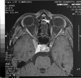
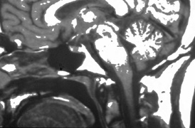
innervation in Duane's syndrome and related congenital ocular motor disorders. Am J Ophthalmol 60:443, 1965

69. Phillips WH, Dirion JK, Graves GO: Congenital bilateral palsy of abducens. Arch Ophthalmol 8:355, 1932

70. Blodi FC: Electromyographic evidence for supranuclear gaze palsies. Trans Ophthalmol See UK 90:451, 1970

71. Demer JL: The orbital pulley system: a revolution in concepts of orbital anatomy. Ann NY Acad Sci 956:17, 2002

72. Demer JL: Ocular kinematics, vergence, and orbital mechanics. Strabismus 11:49, 2003

73. Kono R, Clark RA, Demer JL: Active pulleys: magnetic resonance imaging of rectus muscle paths in tertiary gazes. Invest Ophthalmol Vis Sci 43:2179, 2002

74. Clark RA, Miller JM, Demer JL: Three-dimensional location of human rectus pulleys by path inflections in secondary gaze positions. Invest Ophthalmol Vis Sci 41:3787, 2000

75. Demer JL, Oh SY, Poukens V: Evidence for active control of rectus extraocular muscle pulleys. Invest Ophthalmol Vis Sci 41:1280, 2000

76. Clark RA, Miller JM, Demer JL: Locations and stability of rectus muscle pulleys. Muscle paths as a function of gaze. Invest Ophthalmol Vis Sci 38:227, 1997

77. Oh SY, Clark RA, Velez F, Rosenbaum AL, Demer JL: Incomitant strabismus associated with instability of rectus pulleys. Invest Ophthalmol Vis Sci 43:2169, 2002

78. Huber A: Electrophysiology of the retraction syndromes. Br J Ophthalmol 58:293, 1974

79. Matteucci P: I difetti congeniti di abduzione (“congenital abduction deficiency”) con particolare riguardo alla patogenesi. Rass Ital Ottal 15:345, 1946

80. Hotchkiss MG, Miller NR, Clark AW, Green WR: Bilateral Duane's retraction syndrome: a clinical-pathologic case report. Arch Ophthalmol 98:870, 1980

81. Miller NR, Kiel SM, Green WR, Clark AW: Unilateral Duane's retraction syndrome (type 1). 100:1468, 1982

82. Mims JL III: Describing Duane's retraction syndrome. Binoc Vis Strabismus Q 17:86, 2002

82A. Thomas R, Mathai A, Gieser SC, et al: Bilateral synergistic divergence. J Pediatr Ophthalmol Strabismus 30:122, 1993

83. Scott AB, Wong GY: Duane's syndrome: An electromyographic study. Arch Ophthalmol 87:140, 1972

84. Pressman SH, Scott WE: Surgical treatment of Duane's syndrome. Ophthalmology 93:29, 1986

85. Raab EL: Clinical features of Duane's syndrome. J Pediatr Ophthalmol Strabismus 33:64, 1986

86. Hoffman RJ: Monozygotic twins concordant for bilateral Duane's retraction syndrome. Am J Ophthalmol 99:563, 1985

87. Chew CKS, Foster P, Hurst JA, et al: Duane's retraction syndrome associated with chromosome 4g27-31 segment deletion. Am J Ophthalmol 119:807, 1995

88. Cullen P, Rodgers CS, Callen DF, et al: Association of familial Duane anomaly and urogenital abnormalities with a bisatellited marker derived from chromosome 22. Am J Med Genet 47:925, 1993

89. Tredici TD, von Noorden GK: Are anisometropia and amblyopia common in Duane's syndrome? J Pediatr Ophthalmol Strabismus 22:23, 1985

90. Osher RH, Schatz NJ, Duane TD: Acquired orbital retraction syndrome. Arch Ophthalmol 98:1798, 1980

91. Miller GR, Glaser JS: The retraction syndrome and trauma. Arch Ophthalmol 76:662, 1966

92. Kivlin JD, Lundergan MK: Acquired retraction syndrome associated with orbital metastasis. J Pediatr Ophthalmol Strabismus 22:109, 1985

93. Rubenstein AE, Lovelace RE, Behrens MM, Weisburg LA: Möbius syndrome in association with peripheral neuropathy and Kallman syndrome. Arch Neurol 32:480, 1975

94. Wishnick MM, Nelson LB, Huppert L, Reich EW: Möbius syndrome and limb abnormalities with dominant inheritance. Ophthalmic Pediatr Genet 2:77, 1983

95. Miller MT, Stromland K: The Mobius sequence: a relook. J AAPOS 3:199, 1999

96. Croemberger MF, de Castro Moreira JB, Brunoni D, et al: Ocular and clinical manifestations of Mobius syndrome. J Pediatr Ophthalmol Strabismus 38:156, 2001

97. Johansson M, Wentz E, Fernell E, et al: Autistic spectrum disorders in Mobius sequence: a comprehensive study of 25 individuals. Dev Med Child Neurol 43:338, 2001

98. Elsahy N: Möbius syndrome associated with mother taking thalidomide during gestation. Plast Reconstr Surg 51:93, 1973

99. Pedraza S, Gemez J, Rovira A, et al: MRI findings in Mobius syndrome: correlation with clinical features. Neurology 55:1058, 2000

100. Konkol RJ, D'Cruz O, Splaingard M: Central respiratory dysfunction in Möbius syndrome. J Child Neurol 6:278, 1991

101. Gilmore RL, Falace P, Kanga J, et al: Sleep-disordered breathing in Möbius syndrome. J Child Neurol 6:73, 1991

102. Szajnberg NM: Möbius syndrome: alternatives in affective communication. Dev Med Child Neurol 36:459, 1994

103. Terzis JK, Noah Em: Dynamic restoration in Mobius and Mobius-like patients. Plast Reconstr Surg 111:40, 2003

104. Terzis JK, Noah EM: Mobius and Mobius-like patients: etiology, diagnosis, and treatment options. Clin Plast Surg 29:497, 2002

105. Zuker RM, Goldberg CS, Lanktelow RT: Facial animation in children with Mobius syndrome after segmental bracilis muscle transplant. Plast Reconstr Surg 106:1, 2000

106. Goldberg C, DeLorie R, Zuker RM, et al: The effects of gracilis muscle transplantaiton on speech in children with Moebisu syndrome. J Craniofac Surg 14:687, 2003

107. Ziffer AJ, Rosenbaum AL, Demer JL, et al: Congenital double elevator palsy: vertical saccadic velocity utilizing the scleral search coil technique. J Pediatr Ophthalmol Strabismus 29:142, 1992

108. Jampel RS, Fells P: Monocular elevation paresis caused by a central nervous system lesion. Arch Ophthalmol 80:45, 1968

109. Ford CS, Schwartze GM, Weaver RG, Troost BT: Monocular elevation paresis caused by an ipsilateral lesion. Neurology 34:1264, 1984

110. Munoz M, Page LK: Acquired double elevator palsy in a child with a pineocytoma. Am J Ophthalmol 118:810, 1994

111. von Noorden GK: Burian and von Noorden's Binocular Vision and Ocular Motility: Theory and Management of Strabismus, 6th ed. St. Louis: CV Mosby, 2001

112. Jacobs L, Heffner RR, Newman RP: Selective paralysis of downward gaze caused by bilateral lesions of the mesencephalic periaqueductal gray matter. Neurology 35:516, 1985

113. Scassellati-Sforzolini G: Una sindroma motto rare: diffeto congenito monolaterale della elevazione con retrazione del globe. Riv Otoneurooftalmol 33:431, 1958

114. Khodadoust AA, von Noorden GK: Bilateral vertical retraction syndrome: a family study. Arch Ophthalmol 78:606, 1967

115. Pruksacholawit K, Ishikawa S: Atypical retraction syndrome: a case study. J Pediatr Ophthalmol 13:215, 1976

116. Wartenberg R: Inverted Marcus Gunn phenomenon. Arch Neurol Psychiatry 60:584, 1948

117. Carmicheal EA, Critchley M: The relations between eye movements and other cranial muscles. Br J Ophthalmol 9:49, 1925

118. Sano K: Trigemino-oculomotor synkineses. Neurologia 1:29, 1959

119. Wartenberg R: Associated movements in the oculomotor and facial muscles. Arch Neurol Psychiatry 55:439, 1946

120. Beyer-Machule CK, Johnson CC, Pratt SG, Smith BR: The Marcus-Gunn phenomenon. Orbit 4:15, 1985

121. Doucet TW, Crawford JS: The quantification, natural course, and surgical results in 57 eyes with Marcus Gunn (jaw-winking) syndrome. Am J Ophthalmol 92:702, 1981

122. Mrabet A, Oueslati S, Gazzah H, et al: Clinical and electrophysiological study of 2 familial cases of Marcus Gunn phenomenon [transl. French]. Rev Neurol 147:215, 1991

123. Dryden RM, Fleming JC, Quickert MH: Levator transposition and frontalis sling procedure in severe unilateral ptosis and the paradoxically innervated levator. Arch Ophthalmol 100:462, 1982

124. Dillman DB, Anderson RL: Levator myectomy in synkinetic ptosis. Arch Ophthalmol 102:422, 1984

125. Lubkin V: The inverse Marcus-Gunn phenomenon: An electromyographic contribution. Arch Neurol 35:249, 1978

126. McLead AR, Glaser JS: Deglutition-trochlear synkinesis. Arch Ophthalmol 92:171, 1974

127. Pang MP, Zweifach PH, Goodwin J: Inherited levator-medial rectus synkinesis. Arch Ophthalmol 104:1489, 1986

128. Hwang J. A case of Marcus Gunn jaw winking and speudo inferior oblique overaction. Am J Ophthalmol 131:148, 2001

129. Brown HW: True and simulated superior oblique tendon sheath syndromes. Doe Ophthalmol 34:123, 1973

130. Katz NNK, Whitmore PV, Beauchamp GR: Brown's syndrome in twins. J Pediatr Ophthalmol Strabismus 18:32, 1981

131. Catford GV, Dean-Hart JC: Superior oblique tendon sheath syndrome: an electromyographic study. Br J Ophthalmol 55:155, 1971

132. Sevel D: Brown's syndrome: a possible etiology explained embryologically. J Pediatr Ophthalmol Strabismus 18:26, 1981

133. Beck M, Hickling P: Treatment of bilateral superior oblique tendon sheath syndrome complicating rheumatoid arthritis. Br J Ophthalmol 64:358, 1980

134. Wang FM, Wertenbaker C, Behrens MM, Jacobs JC: Acquired Brown's syndrome in children with rheumatoid arthritis. Ophthalmology 91:23, 1984

135. Moore AT, Morin JD: Bilateral acquired inflammatory Brown's syndrome. J Pediatr Ophthalmol Strabismus 22:26, 1985

136. Clarke WN, Noel LP, Agapitos PJ: Traumatic Brown's syndrome. Am Orthoptic J 37:100, 1987

137. Blanchard CL, Young LA: Acquired inflammatory superior oblique tendon sheath (Brown's) syndrome. Arch Otolaryngol 110:120, 1984

138. Wesley RE, Pollard ZF, McCord CD: Superior oblique paresis after blepharoplasty. Plast Reconstr Surg 66:283, 1980

139. Hermann JS: Acquired Brown's syndrome of inflammatory origin. Arch Ophthalmol 96:1228, 1978

140. Booth-Mason S, Kyle GM, Rosser M, Bradbury P: Acquired Brown's syndrome: an unusual cause. Br J Ophthalmol 69:791, 1985

141. Hertle RW, Katowitz JA, Young TL, et al: Congenital unilateral fibrosis, blepharoptosis, and enophthalmos syndrome. Ophthalmology 99:347, 1992

142. Engle EC: The molecular basis of the congenital fibrosis syndromes. Strabismus 10:125, 2002

143. Wang SM, Zwaan J, Mullaney PB, et al: Congenital fibrosis of the extraocular muscles Type 2, an inherited exotropic strabismus fixus, maps to distal 11q13. Am J Hum Genet 63:517, 1998

144. Engle EC, McIntosh N, Yamada K, et al: CFEOM1, the classic familial form of congenitla fibrosis of the extraocular muscles, is genetically heterogeneous but does not result from mutations in ARIX. BMC Genetics 3:3, 2002

145. Pieh C, Goebel HH, Engle EC, et al: Congenital fibrosis syndrome associated with central nervous system abnormalities. Graefe Arch Clin Exp Ophthalmol 241:546, 2003

146. Good WV, Hou C, Carden SM: Transient idiopathic nystagmus in infants. Dev Ned Child Neurol 45:304, 2003

147. Dell'Osso LF, van der Steen J, Steinman RM, et al: Foveation dynamics in congenital nystagmus: I. Fixation. Doe Ophthahmol 79:1, 1992

148. Dell'Osso LF, van der Steen J, Steinman RM, et al: Foveation dynamics in congenital nystagmus: II. Smooth pursuit. Doe Ophthalmol 79:25, 1992

149. Dell'Osso LF, Weissman BM, Leigh RJ, et al: Hereditary congenital nystagmus and gaze-holding failure: the role of the neural integrator. Neurology 43:1741, 1993

150. Hertle RW, Chan CC, Galita DA, et al: Neuroanatomy of the extraocular muscle tendon enthesis in macaque, normal human, and patients with congenital nystagmus. J AAPOS 6:319, 2002

151. Abel LA, Williams IM, Levi L: Intermittent oscillopsia in a case of congenital nystagmus: dependence upon waveform. Invest Ophthalmol Vis Sci 32:3104, 1991

152. Gresty MA, Bronstein AM, Page NG, et al: Congenital-type nystagmus emerging in later life. Neurology 41:653, 1991

153. Hertle RS, FitzGibbon EJ, Avallone JM, et al: Onset of oscillopsia after visual maturation in patients with congenital nystagmus. Ophthalmology 108:2301, 2001

154. Safran AB, Gambazzi Y: Congenital nystagmus: rebound phenomenon following removal of contact lenses. Br J Ophthalmol 76:497, 1992

155. Zubcov AA, Stark N, Weber A, et al: Improvement of visual acuity after surgery for nystagmus. Ophthalmology 100:1488, 1993

156. Helveston EM, Ellis FD, Plager DA: Large recession of the horizontal recti for treatment of nystagmus. Ophthalmology 98:1302, 1991

157. Dell'Osso LF: Development of new treatments for congenital nystagmus. Ann NY Acad Sci 956:361, 2002

158. Hertle RW, Dell'Osso LF, FitzGibbon EJ, et al: Horizontal rectus tenotomy in patients with congenital nystagmus: results in 10 adults. Ophthalmology 110:2097, 2003

159. Gresty MA, Metcalfe T, Timms C, et al: Neurology of latent nystagmus. Brain 115(Pt 5):1303, 1992

160. Kommerell G, Zee DS: Latent nystagmus: release and suppression at will. Invest Ophthalmol Vis Sci 34:1785, 1993

161. Liu C, Gresty M, Lee J: Management of symptomatic latent nystagmus. Eye 7(Pt 4):550, 1993

162. Abadi RV, Pascal E: Periodic alternating nystagmus in humans with albinism. Invest Ophthalmol Vis Sci 35:4080, 1994

163. Shalloh-Hoffman J, Faldon M, Tusa RJ: The incidence and waveform characteristics of periodic alternating nystagmus in congenital nystagmus. Invest Ophthalmol Vis Sci 40:2546, 1999

164. Gradstein L, Reinecke RD, Wizov SS, et al: Congenital periodic alternating nystagmus. Diagnosis and management. Ophthalmology 104:918, 1997

165. Smith JL, Flynn JT, Spiro HJ: Monocular vertical oscillations of amblyopia: the Bielschowsky phenomenon. J Clin Neuroophthalmol 2:85, 1982

166. Pritchard C, Flynn JT, Smith JL: Wave form characteristics of vertical oscillations in long-standing vision loss. J Pediatr Ophthalmol Strabismus 25:233, 1988

167. Prasad P, Nair S: Congenital ocular motor apraxia: sporadic and familial: support for natural resolution. J Neuroophthalmol 14:102, 1994

168. Jan JE, Kearney S, Groenveld M, et al: Speech, cognition, and imaging studies in congenital ocular motor apraxia. Dev Med Child Neurol 40:95, 1998

169. Sargent MA, Poskitt KJ, Jan JE: Congenital ocular motor apraxia: imaging findings. Am J Neuroradiol 18:1915, 1997

170. Anteby I, Lee B, Noetzel M, et al: Variants of congenital ocular motor apraxia: associations with hydrocephalus, pontocerebellar tumor, and a deficit of vertical saccades. J AAPOS 1:201, 1997

171. Punal JE, Rodriguez E, Pintos E, et al: Congenital ocular motor apraxia associated with myopathy, external hydrocephalus and NADH dehydrogenase deficiency. Brain Dev 20:175, 1998

172. Churchyard A, Stell R, Mastaglia FL: Ataxia telangiectasia presenting as an extrapyramidal movement disorder and ocular motor apraxia without overt telangiectasia. Clin Exp Neurol 28:90, 1991

173. Steinlin M, Thun-Hohenstein L, Boltshauser E: Congenital oculomotor apraxia. Presentation developmental problems: differential diagnosis [transl. German]. Klin Monatsbl Augenheilkd 200:623, 1992

174. Young TL, Weis JR, Summer G, Esbert JE: The association of strabismus, amblyopia, and refractive errors in spasmus nutans. Ophthalmology 104:112, 1997

175. Gottlob IF, Zubcov AA, Wizov SS, et al: Head nodding is compensatory in spasmus nutans. Ophthalmology 99: 1024, 1992

176. Gottlob I, Wizov SS, Reinecke RD: Spasmus nutans. Invest Ophthalmol Vis Sci 36:2768, 1995

177. Wizov SS, Reinecke RD, Bocarnea M, et al: A comparative demographic and socioeconomic study of spasums nutans and intantile nystagmus. Am J Ophthalmol 133:256, 2002

178. Gottlob I, Wizov SS, Reinecke RD: Quantitative eye and head movement recordings of retinal disease mimicking spasmus nutans. Am J Ophthalmol 119:374, 1995

179. Lambert SR, Newman NJ: Retinal disease masquerading as spasmus nutans. Neurology 43:1607, 1993

180. Shawkat FS, Harris CM, Wilson J, et al: Eye movements in children with opsoclonus-polymyoclonus. Neuropediatrics 24:218, 1993

181. Wang PY: Acute cerebellitis with ocular flutter and truncal ataxia: a case report. Chin Med J (Engl) 50(2):169, 1992

182. De Graaf JH, Tamminga RY, Kamps WA: Paraneoplastic manifestations in children. Eur J Pediatr 153:784, 1994

183. Koh PS, Raffensperger JR, Berry S, et al: Long-term outcome in children with opsoclonus-myoclonus and ataxia and coincident neuroblastoma. J Pediatr 125(5 Pt 1):712, 1994

184. Pranzatelli MR, Tate ED, Wheeler A, et al: Screening for autoantibodies in children with opsoclonus-myoclonus-ataxia. Pediatr Neurol 27:384, 2002

185. Fisher PG, Wechsler DS, Singer HS: Anti-Hu antibody in a neuroblastoma-associated paraneoplastic syndrome. Pediatr Neurol 10:309, 1994

186. Wilfong AA, Parke JT, McCrary JA III: Opsoclonusmyoclonus with Beckwith-Wiedemann syndrome and hepatoblastoma. Pediatr Neurol 8:77, 1992

187. Pranzatelli MR, Kao PC, Tate ED, et al: Antibodies to ACTH in opsoclonusmyoclonus [review]. Neuropediatrics 24:131, 1993

188. Mitchell WG, Davalos-Gonzalez Y, Brumm VL, et al: Opsoclonus-ataxia caused by childhood neuroblastoma: developmental and neurolgoic sequelae. Pediatrics 109:86, 2002

189. Gambini IG, Conte M, Bernini G, et al: Neuroblastic tumors associated with opsoclonus-myoclonus syndrome: histological, immunohistochemicl and molecular features of 15 Italian cases. Virchows Arch 44w:555, 2003

190. Yee RD, Spiegel PH, Yamada T, et al: Voluntary saccadic oscillations, resembling ocular flutter and opsoclonus. J Neuroophthalmol 14:95, 1994

191. Shaywitz SE, Shaywitz BA: The science of reading and dyslexia. J AAPOS 7:158, 2003

192. Irlen H: Improving reading problems due to symptoms of scotopic sensitivity syndrome using Irlen lenses and overlays. Education 109:413, 1989

193. Menacker SJ, Breton ME, Breton ML, et al: Do tinted lenses improve the reading performance of dyslexic children? Arch Ophthalmol 111:213, 1993

194. Metsahonkala L, Sillanpaa M: Migraine in children: an evaluation of the IHS criteria. Cephalalgia 14:285, 1994

195. Katerji MA, Painter MJ: Infantile migraine presenting as colic. J Child Neurol 9:336, 1994

196. Norton G: Letter to the Editor: migraine and somnambulism. Austr NZ J Med 23:715, 1993

197. Shevell MI, Silver K, Watters GV, et al: Transient oculosympathetic paresis (group II Raeder paratrigeminal neuralgia) of childhood: migraine variant. Pediatr Neurol 9:289, 1993

198. Ottman R, Lipton RB: Comorbidity of migraine and epilepsy. Neurology 44:2105, 1994

199. Lessell S: Pediatric pseudotumor cerebri (idiopathic intracranial hypertension) [review]. Surv Ophthalmol 37:155, 1992

200. Phillips PH, Repka MX, Lambert SR: Pseudotumor cerebri in children. J AAPOS 2:33, 1998

201. Kesler A, Fattal-Valevski A: Idiopathic intracranial hypertension in the pediatric population. J Child Neurol 17:745, 2002

202. Balcer LJ, Liu GT, Forman S, Pun K, et al: Idiopathic intracranial hypertension: relation of age and obesity in children. Neurology 52:870, 1999

203. Cinciripini GS, Donahue S, Borchert MS: Idiopathic intracranial hypertension in prepubertal pediatric patients: characteristics, treatment, and outcome. Am J Ophthalmol 127:178, 1999

204. Scott IU, Siatkowski RM, Eneyni M, et al: Idiopathic intracranial hypertension in children and adolescents. Am J Ophthalmol 124:253, 1997

205. Quinn AG, Singer SB, Buncic JR: Pediatric tetracycline-induced pseudotumor cerebri. J AAPOS 3:53, 1999

206. Mochizuki K, Takahashi T, Kano M, et al: Pseudotumor cerebri induced by minocycline therapy for acne vulgaris. Jpn H Ophthalmol 46:668, 2002

207. Rogers AH, Rogers GL, Bremer DL, et al: Pseudotumor cerebri in children receiving recombinant human growth hormone. Ophthalmology 106:1186, 1999

208. Weig SG. Asymptomatic idiopathic intracranial hypertension in young children. J Child Neurol 17:239, 2002

209. Jeng MR, Rieman M, Bhakta M, et al: Pseucotumor cerebri in two adolesecnts with acquired aplastic anemia. J Pediatr Hematol Oncol 24:765, 2002

210. Nazir SA, Siatkowski RM: Pseudotumor cerebri in idiopathic aplastic anemia. J AAPOS 7:71, 2003

211. Kodsi SR, Younge BR: Acquired oculomotor, trochlear, and abducens cranial nerve palsies in pediatric patients. Am J Ophthalmol 114:568, 1992

212. Norman AA, Farris BK, Siatkowski RM: Neuroma as a chase of oculomotor palsy in infancy and early childhood. J AAPOS 5:9, 2001

213. Hamed LM: Associated neurologic and ophthalmologic findings in congenital oculomotor nerve palsy. Ophthalmology 98:708, 1991

214. Ing EB, Sullivan TJ, Clarke MP, et al: Oculomotor nerve palsies in children. J Pediatr Ophthalmol Strabismus 29:331, 1992

215. Wolin MJ, Saunders RA: Aneurysmal oculomotor nerve palsy in an 11-year-old boy. J Clin Neuroophthalmol 12:178, 1992

216. Branley MG, Wright KW, Borchert MS: Third nerve palsy due to cerebral artery aneurysm in a child. Austr NZ J Ophthalmol 20:137, 1992

217. Nazir SA, Murphy SA, Siatkowski RM: Recurrent para-infectious third nerve palsy with cisternal enhancement on MRI. J Neurol Ophthalmol 24:96, 2004

218. Hamed LM, Fang EN, Fanous MM, et al: The prevalence of neurologic dysfunction in children with strabismus who have superior oblique overaction. Ophthalmology 100:1483, 1993

219. Named LM, Maria BL, Quisling RG, et al: Alternating skew on lateral gaze. Ophthalmology 100:281, 1993

220. Davitt BD, Fenton GA, Cruz OA: Childhood myasthenia. J Ped Ophthalmol Strab 37:5, 2000

221. Mullaney P, Vajsar J, Smith R, Buncic JR: The natural history and ophthalmic involvement in childhood myasthenia gravis at the Hospital for Sick Children. Ophthalmology 107:504, 2000

222. Kim J, Hwang J, Hwang Y, et al: Childhood ocular myasthenia gravis. Ophthalmology 110:1458, 2003

223. Skelly CL, Jackson CC, Wu Y, et al: Thorascopic thymectomy in children with myasthenia gravis. Am Surg 69:1087, 2003

224. Seaber JH: The effect of supranuclear midbrain lesions on ocular motility. In Ravault AP, Lenk M (eds): Transactions of the Fifth International Orthoptic Congress. Lyon, France: LIPS, 1984:105

225. Lepore FE: Disorders of ocular motility following head trauma. Arch Neurol 52:924, 1995

226. Pratt-Johnson JA, Tillson G: Acquired central disruption of fusional amplitude. Ophthalmology 86:2140, 1979

227. Stanworth A, Mein J: Loss of fusion after removal of cerebellar tumor. Br J Orthopt 28:97, 1971

228. Pratt-Johnson JA, Tillson G: The loss of fusion in adults with intractable diplopia (central fusion disruption). Aust NZ J Ophthalmol 16:81, 1988

229. Lee MC: Acquired central fusional disruption with spontaneous recovery. Strabismus 6:175, 1998

230. Pratt-Johnson JA: Fusion ability lost and regained in visual adults. Graefes Arch Clin Exp Ophthalmol 226:111, 1988

231. MacLellan AV: A case of recovery from traumatic loss of fusion. Br J Orthopt 31:102, 1974

232. Kushner BJ: Unexpected cyclotropia simulating disruption of fusion. Arch Ophthalmol 110:1415, 1992

233. Miller MM, Guyton DL: Loss of fusion and the development of A or V patterns. J Pediatr Ophthalmol Strabismus 31:220, 1994

234. Morris RJ, Scott WE, Dickey CF: Fusion after surgical alignment of long-standing strabismus in adults. Ophthalmology 100:135, 1993

235. Kushner BJ, Morton GV: Postoperative binocularity in adults with long-standing strabismus. Ophthalmology 99:316, 1992

236. Daw NW: Mechanisms of plasticity in the visual cortex. Invest Ophthalmol Vis Sci 34:4168, 1994

237. Boyd TAS, Budd GE: The monofixation syndrome: heterophoria and asthenopia. Am J Orthopt 26:43, 1976

238. Parks MM: Monofixation syndrome. In Tasman W, Jaeger EA (eds): Duane's Clinical Ophthalmology. Philadelphia: JB Lippincott, 1990:1

239. Burgess D, Roper-Hall G, Burde RM: Binocular diplopia associated with subretinal neovascular membranes. Arch Ophthalmol 98:311, 1980

240. Bixenman WW, Joffe L: Binocular diplopia associated with retinal wrinkling. J Pediatr Ophthalmol Strabismus 21:215, 1984

241. Goldman HD, Nelson LB: Acute acquired comitant esotropia. Ann Ophthalmol 17:777, 1985

242. Burian HM, Miller JE: Comitant convergent strabismus with acute onset. Am J Ophthalmol 45:55, 1958

243. Biexnman WW, Laguna JF: Acquired esotropia as initial manifestation of Arnold-Chiari malformation. J Pediatr Ophthalmol Strabismus 24:83, 1987

244. Lewis AR, Kline LR, Sharp JA: Acquired esotropia due to Arnold Chiari I malformation. J Neuro-ophthalmol 16:49, 1996

245. Biousse V, Newman NJ, Petermann Sh, et al: Isolated comitant esotropia and Chiaria I malformation. Am J Ophthalmol 130:216, 2000

246. Deefort-Dhellemmes S, Denion E, Bouvet-Drumare I, et al: Resoluation of acute acquired comitant esotropia after suboccipital decompression for Chiara I malformation. Am J Ophthalmol 133:723, 2002

247. Pokharel D, Siatkowski RM: Progressive cerebellar tonsillar herniation with recurrent divergence insufficiency esotropia. J AAPOS 8:286, 2004

248. Stark N, Walther C: Cyclische esotropiebei einem erwaschsenen. Klin Monatsbl Augenheilkd 184:553, 1984

249. Troost BT, Abel L, Noreika J, Genovese FM: Acquired cyclic esotropia in an adult. Am J Ophthalmol 91:8, 1981

250. Friendly DS, Manson RA, Albert DG: Cyclic strabismus: a case study. Doe Ophthalmol 34:189, 1973

251. Roper-Hall MJ, Yapp JMS: Alternate day squint. In The First International Congress of Orthoptics. St. Louis: CV Mosby, 1968:262

252. Windsor CE, Berg EF: Circadian heterotropia. Am J Ophthalmol 67:656, 1969

253. Hugonnier R, Maynard P: Les desequilibres oculomoteur observes en cas de myopie forte. Ann Ocul 202:713, 1969

254. Krzizok TH, Kaulmann H, Traupe H: New approach in strabismus surgery in high myopia. Br J Ophthalmol 81:617, 1997

255. Lieppman ME: Accommodative and convergence insufficiency after decompression sickness. Arch Ophthalmol 99:453, 1981

256. von Noorden GK, Jenkins RH, Rosenbaum AL: Horizontal transposition of the vertical rectus muscles for treatment of ocular torticollis. J Pediatr Ophthalmol 30:8, 1993

257. Rubin SE, Wagner RS: Ocular torticollis. Surv Ophthalmol 30:366, 1986

258. Catalano RA, Simon JW, Krohel GB, et al: Functional visual loss in children. Ophthalmology 93:385, 1986

259. Jacobson AM, de Groot M: Psychology of visual loss. In Principles and Practice of Ophthalmology. Philadelphia: WB Saunders

Back to Top