Chapter 12
Infranuclear Disorders of Eye Movement
JOEL S. GLASER and R. MICHAEL SIATKOWSKI
Main Menu   Table Of Contents

Search

NEUROANATOMY
ABDUCENS PALSIES
TROCHLEAR PALSIES
OCULOMOTOR PALSIES
ISCHEMIC (“DIABETIC”) OCULOMOTOR PALSY
OCULOMOTOR SYNKINESIS
HERPES ZOSTER
ACUTE INFECTIOUS POLYNEUROPATHY
OPHTHALMOPLEGIC MIGRAINE
OCULOMOTOR PALSIES OF CHILDHOOD
CYCLIC OCULOMOTOR PALSY
COMBINED OCULAR MOTOR PALSIES AND PAINFUL OPHTHALMOPLEGIAS
ORBITAL LESIONS
TOLOSA-HUNT SYNDROME
PARASELLAR SYNDROMES
OTHER OCULAR POLYNEUROPATHIES
DISORDERS OF THE NEUROMUSCULAR JUNCTION AND OCULAR MYOPATHIES
CONDITIONS SIMULATING PROGRESSIVE EXTERNAL OPHTHALMOPLEGIA
TOLOSA-HUNT SYNDROME
PARASELLAR SYNDROMES
OTHER OCULAR POLYNEUROPATHIES
DISORDERS OF THE NEUROMUSCULAR JUNCTION AND OCULAR MYOPATHIES
CONDITIONS SIMULATING PROGRESSIVE EXTERNAL OPHTHALMOPLEGIA
MYOTONIC DYSTROPHY
OCULAR NEUROMYOTONIA
DORSAL MIDBRAIN SYNDROME
DIPLOPIA AFTER OCULAR SURGERY
MISCELLANEOUS OCULAR MUSCLE CONDITIONS
REFERENCES

NEUROANATOMY
The supranuclear control of eye movements is discussed in detail in Chapters 9 and 10. However, for purposes of review it should be recalled that supranuclear ocular motor pathways descend from the cerebral hemispheres, decussate in the caudal midbrain, and terminate in the pontine horizontal gaze complex (Fig. 1). From here, the motor nuclei of the ocular muscles are integrated by way of the medial longitudinal fasciculus.

Fig. 1. Supranuclear oculomotor system. Conjugate horizontal gaze to left originates in right frontal optomotor cortex (R). Pathway descends in area of internal capsule, decussates at level of trochlear nucleus (IV) to synapse in left pontine paramedian reticular formation (PPRF). Via direct connection to abducens nucleus (VI; labeled on right side), left lateral rectus (LR is innervated. From abducens nucleus via contralateral medial longitudinal fasciculus (MLF) impulse is directed to contralateral oculomotor nuclear complex (III; labeled on left side), then to right medial rectus (MR). (VN, vestibular nuclear complex; SCC, semicircular canals)

The nuclear complex of the third (oculomotor) nerve lies beneath the aqueductal gray matter of the rostral midbrain at the level of the superior colliculus (Fig. 2). The medial longitudinal fasciculus (MLF) passes just lateral to the oculomotor nuclei, and the fourth (trochlear) cranial nerve nucleus is contiguous caudally. The organization of the oculomotor nuclear complex into distinct motor cell pools subserving individual extraocular muscles was investigated by Warwick,1 from whose schema (Fig. 3) the following points are especially noteworthy: (1) both lid levators are served by a single dorsal-caudal midline nucleus; (2) the motor cell pool of the superior rectus muscle sends fibers across to the contralateral oculomotor nerve; (3) a nucleus for convergence (Perlia) has not been consistently demonstrated in primates, including humans; and (4) at the caudal aspect of the oculomotor complex is the trochlear nucleus, whose axons turn dorsally to cross in the anterior medullary velum and innervate the contralateral superior oblique. Therefore, the nuclear motor pools of the superior rectus and superior oblique muscles are contralateral to the eye that they move. Warwick proposed that each muscle (intrinsic and extrinsic) innervated by the oculomotor nerve is subserved by a single, circumscribed mass of cells called a subnucleus. Modern tracer techniques have, to date, confirmed this concept for all the extrinsic muscles except the medial rectus, which has three definable subnuclei2,3; there are also afferent fibers to the ipsilateral trigeminal ganglion. Results of elegant axonal tracer studies mapping the intricate connections of the visceral nuclei of the oculomotor complex have been summarized by Burde.4

Fig. 2. Diagrammatic section of brain stem. A. Cross-section at level of superior colliculus. (N-III, oculomotor nucleus; MLF, medial longitudinal fasciculus; LEM, medial lemniscus; RN, red nucleus; SN, substantia nigra; CS, corticospinal tract) B. Cross-section at level of abducens nucleus (N-VI). (PPRF, pontine paramedian reticular formation [note that reticular formation continues rostrally and caudally (arrows)]; N-VII [FN], facial nucleus; VN, vestibular nuclear complex; SNT, spinal nucleus of trigeminal; STT, spinal tract of trigeminal; STH, spinothalamic tract)

Fig. 3. Organization of oculomotor nuclear complex viewed from above, left posterior. E-W, Edinger-Westphal parasympathetic subnucleus; subnuclei IR, inferior rectus; IO, inferior oblique; MR, medial rectus. Note that SR, superior rectus motor pool, is crossed, as is SO, superior oblique; CCN, caudal nucleus to both lid levators; LR, abducens nucleus for lateral rectus (Adapted Warwick R: Representation of the extra-ocular muscles in the oculomotor complex. J Comp Neurol 98:449, 1953)

From the oculomotor complex the efferent fibers exit ventrally and pass through the red nucleus and medial aspect of the cerebral peduncles. The fascicles emerge in the interpeduncular space anterior to the midbrain as the paired oculomotor nerves. The oculomotor nuclei obtain their vascular supply from the terminal bifurcation of the basilar artery. Multiple arteries perforate the median mesencephalon in the interpeduncular space.

The sixth (abducens) nucleus is situated in the caudal portion of the paramedian pontine tegmentum, beneath the floor of the fourth ventricle (see Fig. 2). Facial nerve fibers loop around the abducens nucleus before exiting in the cerebellopontine angle. This intimate relationship accounts for frequent concurrent damage seen clinically. Although lateral to the third nuclear complex in the rostral mesencephalon, the MLF passes medial to the abducens nucleus. There are two populations of neurons with cell bodies within the abducens nucleus.5,6 One group forms the sixth nerve; these abducens fibers pass ventrally to exit at the pontomedullary junction. The other internuclear neurons send fibers to join the contralateral MLF, with projection to the medial rectus subnucleus. Therefore, lesions involving the sixth nucleus produce an ipsilateral conjugate gaze palsy (i.e., ipsilateral lateral rectus and contralateral medial rectus).7

The abducens, trochlear, and oculomotor nuclei are integrated via the MLF, which also has major connections with the vestibular nuclear complex (see Fig. 1). Lesions involving the MLF typically result in internuclear ophthalmoplegia (see Chapter 10), which consists of faulty adduction of the ipsilateral eye (observed especially with attempted saccades) and dissociated nystagmus greater in the abducting eye on attempted lateral horizontal gaze (Fig. 4). In addition, some degree of vertical nystagmus in upward gaze is often present, and skew deviation may account for vertical diplopia.8 Internuclear ophthalmoplegia, usually bilateral, is by far the most common ocular motor disturbance of demyelinative origin. As a rule, unilateral ophthalmoplegia is usually due to a vascular incident, but bilateral involvement should not be considered rare in brain stem infarction.9

Fig. 4. Left internuclear ophthalmoplegia. On attempted right gaze, the left eye fails to adduct due to lesion of left medial longitudinal fasciculus.

The peripheral course of the third nerve is as follows (Fig. 5): as the nerve exits ventrally from the midbrain into the interpeduncular space, it passes beneath the origin of the posterior cerebral artery and lies parallel and lateral to the posterior communicating artery. Klintworth10 pointed out that, in instances where the basilar artery bifurcates at a low level, the oculomotor nerve may be angled downward at the point where the posterior cerebral artery crosses the nerve, and vascular grooving of the superior aspect of the nerve is present in approximately one third of normal brains. The nerve runs between the free edge of the tentorium and the lateral aspect of the posterior clinoid, where it pierces the dura to enter the cavernous sinus. The oculomotor trunk occupies the superior aspect of the cavernous sinus (see ahead to Fig. 17) and separates into superior and inferior divisions at 4 to 5 mm before the superior orbital fissure. Fiber count of the superior division is about one-third that of the inferior,11 and it has been suggested that relative pupil sparing in cavernous sinus or orbital apex lesions may reflect preservation of the inferior branch. The inferior branch supplies the medial and inferior recti, inferior oblique, and parasympathetic root to the ciliary ganglion (pupil sphincter); the superior branch innervates the superior rectus and levator palpebrae.

Fig. 5. Representation of cranial nerves and related basal structures in the area of the midbrain, middle fossa, cavernous sinus, and orbital apex. The roof of the right orbit, sphenoid wing, floor of middle fossa, and petrous ridge have been sectioned; a section of the tentorial edge is removed to demonstrate the course of trochlear nerve (4). The cerebellum is retracted to show structures in the posterior fossa. Cross-section of the midbrain is at level of superior colliculi and red nuclei. (2, optic nerves and chiasm; 3, oculomotor nerves [note relationship to posterior cerebral and posterior communicating arteries]; 4, trochlear nerve in edge of tentorium; 5, trigeminal nerve [51, ophthalmic division; 52, maxillary division; 53, mandibular division]; 6, abducens nerve [note course up clivus and passage under petroclinoid ligament into posterior aspect of cavernous sinus]; 7, facial nerve; 8, acoustic nerve)

Fig. 17. Bulbar polyneuritis (Fisher syndrome) in a 16-year-old boy with rapidly progressive facial diplegia and total ophthalmoplegia, including pupillary and accommodative paresis. A. Acute phase. B. Complete resolution 3 months later.

According to Kerr,12 the pupillomotor fibers are superficial in the oculomotor nerve trunk, lying just internal to the epineurium. It is thought that this superficial position makes the pupillomotor fibers especially vulnerable to compression. In more anterior segments (e.g., cavernous sinus), however, pupillomotor fibers may be preferentially spared even in the presence of total oculomotor palsy. It is likely that involvement or “sparing” of the pupil reflects the nature and acuteness of the lesion, rather than which specific segment of the oculomotor trunk is compromised.

The trochlear fascicles pass dorsally, lateral to the aqueduct, and the nerve exits the midbrain and crosses the contralateral fourth nerve in the anterior medullary velum, just caudal to the inferior colliculi. The fourth nerve is the only cranial nerve to exit the brain stem dorsally. The nerve continues laterally around the midbrain tectum, crosses the superior cerebellar artery, and reaches the free edge of the tentorium, where it enters the dura and runs forward into the cavernous sinus (see Fig. 5). The fourth nerve enters the orbit through the superior orbital fissure, but above the annulus formed by the origin of the recti muscles, and innervates only the superior oblique muscle.

The abducens nerve emerges from the brain stem at the lower border of the pons in the pontomedullary sulcus, approximately 1 cm from the midline. The nerve ascends the ventral face of the pons for a short distance, is crossed by the anterior inferior cerebellar artery, and pierces the dura of the clivus approximately 2 cm below the posterior clinoids (see Fig. 5). The sixth nerve traverses or passes above the inferior petrosal sinus, runs beneath the petroclinoid ligament (Gruber), and enters the cavernous sinus. The sixth nerve lies freely within the body of the cavernous sinus, unlike the oculomotor and trochlear nerves that are supported in the lateral wall of the sinus. Some sympathetic fibers are briefly attached to the abducens, passing onward to the ophthalmic trigeminal.13 From the cavernous sinus, the nerve passes through the annular segment of the superior orbital fissure and innervates only the lateral rectus.

Milisavljevi and associates14 demonstrated penetration of oculomotor trunks by circumflex mesencephalic arteries or branches of the posterior cerebral perforating vessels, but clinical implications of such anatomic anomalies are not clear.

According to the anatomic review by Lapresle and Lasjaunias,15 there are three arterial “systems” that vascularize the cranial nerves: the inferolateral trunk arises from the intracavernous siphon of the internal carotid artery and nourishes cranial nerves III, IV, VI, and V-1; the middle meningeal system supplies nerves VII and V-2,3; and the ascending pharyngeal system supplies nerves IX through XII. The oculomotor nerve is also vascularized by the basilar artery system in the region of the posterior perforated substance, and in the supracavernous region by the artery of the free tentorial margin (Bernasconi). The clinical correlation of ischemic cranial neuropathies with these arterial territories is not entirely clear.

Back to Top
ABDUCENS PALSIES
It is not proper to equate all lateral rectus malfunction with “sixth nerve palsy”: to do so confuses the issue and leads to inappropriate diagnostic procedures. For example, myasthenia, Graves' (dysthyroid) myopathy, or orbital inflammation may all produce deficits of abduction, none of which is due to sixth nerve lesions. The “neural” etiology of lateral rectus weakness must be established or, at least, other causes excluded when possible (Table 1). Along with details provided by a thorough medical history, special diagnostic techniques should always be employed, including intravenous edrophonium (Tensilon), forced duction (see Chapter 3, Fig. 6), and forced generation tests. Until local orbital diseases have been excluded, it is premature for the ophthalmologist to refer a patient for a “neurologic workup.”


TABLE 1. Causes of Abduction Deficits

  Sixth nerve palsies
  Graves' myopathy (fibrotic medial rectus)
  Myasthenia gravis
  Orbital pseudotumor/myositis
  Orbital trauma (medial rectus entrapment)
  Congenital defects (Duane, Möbius syndromes)
  “Convergence spasm” (spasm of the near reflex)


ETIOLOGY

The causes of actual sixth nerve palsy are legion (Table 2). As noted previously, the peripheral course of the abducens nerve is a lengthy one that predisposes this cranial nerve to involvement at all levels, from the brain stem and base of the skull, through the petrous tip and cavernous sinus, to the superior orbital fissure and orbit.


TABLE 2. Causes of Sixth Nerve Palsies

  Nonlocalizing
  Increased intracranial pressure
  Intracranial hypotension
  Head trauma
  Lumbar puncture or spinal anesthesia
  Vascular, hypertension
  Diabetes/microvascular
  Parainfectious processes (postviral; middle ear infections in children)
  Basal meningitis
  Localizing
  Pontine syndromes (infarction, demyelination, tumor); contralateral hemiplegia; ipsilateral facial palsy, ipsilateral horizontal gaze palsy (± ipsilateral internuclear ophthalmoplegia); ipsilateral facial analgesia
  Cerebellopontine angle lesions (acoustic neuroma, meningioma): in combination with disorders of the eighth, seventh, and ophthalmic-trigeminal nerves (especially corneal hypoesthesia), nystagmus, and cerebellar signs
  Clivus lesions (nasopharyngeal carcinoma, clivus chordoma)
  Middle fossa disorders (tumor, inflammation of medial aspect of petrous): facial pain/numbness, ± facial palsy
  Cavernous sinus or superior orbital fissure (tumor, inflammation, aneurysm): in combination with disorders of the third, fourth, and ophthalmic-trigeminal nerves (pain/numbness)
  Carotid-cavernous or dural arteriovenous fistula


Lesions in the area of the abducens nucleus produce an ipsilateral horizontal gaze paresis, since both the internuclear neurons and the motor neurons of the sixth nerve originate in this nucleus. Although it is not certain whether the horizontal gaze palsy that occurs from damage to the abducens nucleus is always symmetric, it is clear that isolated lateral rectus pareses should never be considered nuclear in origin.

The fascicular (intrapontine) portion of the abducens nerve may be involved along with adjacent structures (see Fig. 2) to produce (1) ipsilateral paralysis of abduction (ipsilateral gaze palsy if sixth nucleus and/or pontine paramedian reticular formation [PPRF] is affected); (2) ipsilateral facial palsy; (3) ipsilateral Horner syndrome; (4) ipsilateral facial analgesia; (5) ipsilateral peripheral deafness; and (6) contralateral hemiparesis. These signs constitute the dorsolateral and ventral pontine syndromes (Foville, Millard-Gubler) at the level of the abducens fasciculus in the distribution of the anterior inferior cerebellar artery or its paramedian perforating arteries.

As the sixth nerve ascends the clivus in the subarachnoid space it is vulnerable to various insults, including neoplasms in the prepontine basal cistern (e.g., clivus chordoma, intraforaminal extension of nasopharyngeal carcinoma), compression by downward or forward movement of the brain stem (e.g., transtentorial herniation from supratentorial space-occupying lesions, head trauma, posterior fossa masses or structural anomalies, intracranial hypotension from cerebrospinal fluid leaks), and meningitis. It is here also that the nerve is probably affected by changes in intracranial pressure. Unilateral or bilateral abducens palsies can develop in association with pseudotumor cerebri or the syndrome of spontaneous intracranial hypotension,16 as well as after the following procedures: lumbar puncture, shunting for hydrocephalus, contrast myelography, spinal anesthesia,17 and treatment of cervical fractures.18

Before entering the cavernous sinus, the abducens nerve lies in relationship to the medial aspect of the petrous bone. Inflammation of the petrous bone and its dura may occur secondary to middle-ear infections, with involvement of the facial nerve (facial palsy), the trigeminal ganglion (pain in the eye or face), and the abducens nerve (lateral rectus palsy). These signs and symptoms constitute the now rare Gradenigo syndrome. The combination of sixth and seventh nerve palsies, even bilateral, is not uncommon in closed-head trauma, especially when the skull is compressed in its horizontal diameter (Fig. 6); this results in transverse fractures of the temporal bone. Leakage of blood or spinal fluid from the external ear canal, hemotympanum, or mastoid ecchymosis (Battle's sign) may be further evidence of basal fracture.

Fig. 6. Bilateral sixth and seventh nerve palsies due to basal skull fracture. A. Left gaze. B. Right gaze. C. Attempted lid closure demonstrates intact Bell's phenomenon and lagophthalmos.

In the cavernous sinus, the abducens nerve may be involved in combination with the ophthalmic-trigeminal, third, or fourth nerves. Abducens monoparesis is frequent with cavernous sinus lesions, perhaps related to the nerve's location within the sinus, inferolateral to the carotid artery and unsupported by the dural wall of the sinus.19,20 Isolated abducens palsy occurs with carotid-cavernous fistulas (especially with spontaneous dural shunts21) and intracavernous aneurysms20 (Fig. 7), and is the earliest indication of contralateral spread of cavernous sinus thrombosis. Sixth nerve palsy accompanied only by ipsilateral Horner's syndrome also points to the cavernous sinus, since the ocular sympathetics from the carotid plexus may be simultaneously involved.22

Fig. 7. A. Chronic isolated sixth nerve palsy. B. Coronal and Axial (C) MRI sections showing large intracavernous internal carotid aneurysm (arrows).

Lesions involving abducens nerve function at the superior orbital fissure or orbital apex regularly involve other motor nerves to the eye, or produce proptosis and/or visual compromise.

Keane23 provided an analysis of 125 cases of bilateral sixth nerve palsies. Unlike the many isolated, unilateral abducens palsies that wander in the limbo of “vascular disease,” more accurate pathoanatomic diagnoses were made where bilateral palsies existed. One fourth of the patients in this series demonstrated deficits of other cranial nerves, and many had additional neurologic signs and symptoms, as well as spinal fluid abnormalities.

ISOLATED ABDUCENS PALSY

As noted previously, all patients with abduction defects do not have sixth nerve lesions per se, and additional diagnostic techniques such as forced ductions/generations and intravenous Tensilon tests are mandatory. If the onset of diplopia is associated with acute eye, orbital, or head pain, then neither Graves' disease nor myasthenia is a likely cause. In addition to the ocular motor (III, IV, VI) nerves, the function of cranial nerves V (especially corneal sensation), VII, and VIII should be examined.

The question now arises as to how one should proceed in the workup of the patient with a truly isolated sixth nerve palsy. In the elderly, as well as in younger adults (see later discussion), vascular disease (hypertensive or otherwise) has been considered a cause of sixth nerve palsies, and remitting abducens palsies are regularly encountered in the adult diabetic population. Therefore, a history of hypertension or diabetes should be sought. Considering the latter, a fasting and 2-hour postprandial glucose level or glycosylated hemoglobin should be obtained. There is no correlation between severity of glucose metabolism defect and occurrence of cranial nerve palsies; thus, an isolated sixth, fourth, or pupil-sparing third nerve palsy may signal the presence of otherwise occult diabetes. Diabetic retinopathy is frequently absent in these cases.

Although it would be rather extraordinary for cranial arteritis to present as an isolated sixth nerve palsy, an erythrocyte sedimentation rate is a reasonable laboratory study in patients 65 years of age or older. Of the neurologic complications of systemic lupus erythematosus, both sixth and third nerve palsies, and brain stem motility disorders, are recognized.24 Because of the propensity for nasopharyngeal carcinoma to spread through the extradural space via basal skull foramina, basisphenoidal sections should be included in gadolinium-enhanced magnetic resonance imaging (MRI) (Table 3).


TABLE 3. Diagnostic Studies for Isolated Abduction Palsy

  Tensilon test
  Forced ductions/forced generations
  Serum glucose: fasting or after glucose load, or glycosylated hemoglobin
  Erythrocyte sedimentation rate
  Imaging studies*

  Contrast-enhanced CT: axial and coronal views, especially of orbital apex, cavernous sinus, sella, and clivus
  Contrast-enhanced MRI: cavernous sinus, clivus


* Pain or mild ocular discomfort may occur with onset of ischemic-diabetic abducens palsy. If pain persists or worsens, imaging studies should be performed.


When associated orbital and cranial signs and symptoms are absent, and when laboratory and appropriate radiologic studies are normal, the most prudent and practical course to follow is continued observation. An examination by a neurologist is comforting, but even Tensilon testing may be postponed. In this specific clinical situation, neuroimaging is infrequently productive, and its application in the elucidation of acute or subacute isolated sixth nerve palsies is moot. However, further radiologic evaluation must be undertaken if pain persists or develops, if other cranial nerves become involved, or if the palsy does not begin to improve over a 3- to 4-month period. Although it is true that some isolated “chronic” sixth nerve pareses last longer than 6 months, yet follow a completely benign course,25 others are indeed caused by potentially treatable basal tumors.26 Volpe and Lessell27 reported seven patients with relapsing or remitting sixth nerve palsies, which were ultimately identified to be secondary to extramedullary compression of the abducens nerve by skull-based tumors. No patients had diabetes or vascular disease, and all recovered completely at least once (and in one case in five separate episodes) without surgical intervention, radiotherapy, or chemotherapy before a definitive diagnosis was made. Lesions included chordomas (Fig. 8), chondrosarcomas, and presumed meningiomas.

Fig. 8. MRI sagittal section, gadolinium enhanced, shows clivus chordoma (arrows). Patient presented with chronic bilateral sixth nerve palsies.

When pain persists and all radiologic and orbital investigations are unrevealing, a trial of corticosteroids (e.g., 60 mg prednisone orally for 5 days) may result in prompt and dramatic relief, in which case a tentative diagnosis of nonspecific inflammation of the superior orbital fissure or cavernous sinus may be made. However, the “response to steroid trial” may produce relief of pain with neoplasms as well as inflammation (see below, Combined Ocular Motor Palsies and Painful Ophthalmoplegia). Certainly, unrelenting eye or orbital or facial pain is an indication for exquisite visualization of the cavernous sinus and parasellar area by thin-section MRI scanning and either magnetic resonance or conventional angiography, if vascular abnormalities are suspected.

Isolated sixth nerve palsies in children often resolve spontaneously. Newborns may rarely manifest a transient lateral rectus weakness, with resolution occurring by 6 weeks.28 Knox and colleagues29 called attention to transient isolated abducens palsy of presumed postviral origin developing in children 1 to 3 weeks after nonspecific febrile or respiratory illness. The age range of the patients reviewed was 18 months to the early teens; recovery of abducens function occurred within 10 weeks (one case continued for 9 months). A similar pattern may be seen with specific viral illnesses such as varicella,30 after immunization,31 or in association with Epstein-Barr viremia.32 A spate of reports indicates that some patients have multiple recurrences of these isolated “benign” sixth nerve palsies and that these recurrences have no serious implications.31,33–36 A 35-year-old with cluster headaches also experienced three consecutive right lateral rectus palsies, on each occasion beginning a day or two after onset of pain, with recovery within 2 weeks37; radiologic studies were unremarkable, suggesting the possibility of migrainous abducens palsy.

Robertson and co-workers38 found a high incidence of brain tumor in his Mayo Clinic series of children with sixth nerve paresis. Although referred to as cases of “isolated” sixth nerve paresis, this definition signifies only that no other ocular motor nerves were defective. Indeed, of the children with neoplasms, one-third had papilledema and one-half demonstrated nystagmus at the time of initial examination. Of the patients with tumor who truly presented with only isolated abducens palsy, additional signs appeared within a few weeks or, rarely, within 2 to 3 months. Harley,39 in his series of children with sixth nerve palsies of all causes, whether isolated or not, noted that approximately one third had suffered trauma and one fourth had underlying tumors; the total clinical picture in these cases is not described.

In the child with an isolated sixth nerve palsy without other neurologic signs, including papilledema, headaches, or ataxia, the following approach is suggested: (1) rule out middle-ear infection; and (2) obtain a peripheral blood count (lymphocytosis may be considered an indication of recent viral infection). The child must be reexamined at regular intervals until the paresis clears, and the parents must be advised to observe for new signs or symptoms. If the paresis persists or worsens, MRI is mandated. A persistent sixth nerve paresis in childhood may be the first clinical sign of a pontine astrocytoma or other posterior fossa mass lesion.

In a 10-year series of 49 “younger adults” (age range, 15 to 50 years) with isolated sixth nerve palsy, Moster and colleagues40 reported that about one third had diabetes and/or hypertension; 8 had basal tumors; 6 had isolated abducens palsies as the initial sign of multiple sclerosis (all with subsequent clinical or spinal fluid abnormalities), usually with spontaneous resolution; and 11 had no specific etiology disclosed, but did have a sanguine outcome. This series excluded patients with a positive Tensilon test, abnormal forced ductions, other known neurologic disease, or with bilateral palsies.

Larger series of sixth nerve palsies reported41–43 are helpful in reviewing the causes, although these patients were somewhat preselected. The largest number of cases fell into the nonspecific “vascular” category (37%, 7%, and 18%, respectively) and the “undetermined” category (24%, 21%, and 29%, respectively). All three series included a modest number of patients with multiple sclerosis (13%, 7%, and 4%, respectively; also 12% in the Moster40 series), but abducens palsy in demyelinative disease has been exceedingly rare in our personal experience. Nevertheless, with the advent of MRI, acute sixth nerve palsies with multiple sclerosis may be more frequently discovered, with abnormal signals in the pons.44

Minimal abduction paresis may herald increased intracranial pressure, such that the patient complains of horizontal diplopia when viewing distant objects. Clinically, these symptoms and findings may mimic divergence insufficiency. Kirkham and co-workers,45 using electro-oculographic techniques, have demonstrated reduced abduction saccadic velocities, which suggest bilateral minimal sixth nerve dysfunction. Although neurons that discharge with divergence (and convergence) have been found in the mesencephalic reticular formation of the monkey,46 the existence of a “divergence center” in the brain stem still remains controversial.

Symptomatic treatment of an acute sixth nerve palsy includes the use of Fresnel prisms, or alternate patching to avoid diplopia and possible medial rectus contracture. The injection of botulinum toxin type A (Botox) into the ipsilateral medial rectus is quite effective at preventing muscle contracture and improving fusion in the primary position of gaze. However, this procedure carries the disadvantage of significant crossed diplopia (exotropia) in contralateral gaze. Definitive surgical correction of a sixth nerve paresis should be employed when the deviation has been stable for at least 6 months. The primary consideration in these cases is the amount of residual abducting power of the lateral rectus, since moderate to good abduction is associated with successful ipsilateral recess/resect procedures (perhaps combined with contralateral medial rectus recession). However, if residual abduction is poor or absent, ipsilateral medial rectus recession may be combined with either a full- or partial-thickness tendon transplant procedure of the superior and inferior recti, or a Jensen procedure. The latter may have a decreased risk of anterior segment ischemia in susceptible patients.

Divergence paralysis is characterized by acute comitant esotropia at distance, normal fusion at near, and full ocular ductions. Of 11 patients with nontraumatic cause, Krohel and associates47 found 3 with neurologic disease and other findings. Otherwise, there was no tendency to late evolution of neurologic sequelae. Stern and Tomsak48 reported a young adult with a lower pontine lesion and also provided a useful overview of “divergence paralysis.” Divergence paralysis has been reported in association with the Arnold-Chiari malformation; in some cases, fusion returns to normal after neurosurgical correction.49 In general, however, the process is idiopathic and benign, and neuroimaging is neither useful nor necessary. When surgical correction is indicated in these cases, lateral rectus resection, without medial rectus recession, effectively eliminates diplopia.

Back to Top
TROCHLEAR PALSIES
Palsy of the superior oblique, as in the case of isolated lateral rectus weakness, may be due to local orbital processes that should be distinguished from a neurogenic lesion per se. As always, myasthenia and Graves myopathy (an especially frequent cause of incomitant vertical strabismus) must be suspected and appropriate tests performed. The pattern of muscle imbalance and ocular versions can be quite similar in ipsilateral inferior rectus fibrosis (e.g., in Graves disease) and contralateral superior oblique paresis50; however, worsening of the vertical deviation in upgaze is seen in inferior rectus fibrosis, and in downgaze in superior oblique palsy. Also, intraocular pressure may increase in attempted upgaze in restrictive (Graves) disease. Excyclodeviations are more prominent in superior oblique paresis than in thyroid myopathy, which may show significant excyclotorsion only in abduction.

Superior oblique palsies may present spontaneously in late childhood and possibly represent “decompensated” congenital fourth nerve palsies. This diagnostic concept (i.e., that fusional mechanisms decompensate in later life) has also been applied to otherwise healthy adults with spontaneously acquired, unremitting superior oblique palsies of unknown origin (Fig. 9). A similar, often transient, occurrence of superior oblique palsy has been reported during pregnancy.51 These adults complain of reading difficulties, principally momentary diplopia, and show all of the attributes of superior oblique palsy. Old photographs may document tilting of the head, and further inquiry may uncover a forgotten history of childhood squint. Patients with congenital trochlear palsies commonly show increased amplitudes of vertical vergence.52 Normally, only 2 to 4 prism diopters (D) of vertical fusional amplitude are found, but patients with congenital superior oblique pareses may be able to fuse a 10-D or even a 30-D deviation. However, the presence of large vertical fusional amplitudes does not necessarily imply a congenital etiology, because vertical fusional vergence may increase in adults within weeks or months after an acquired vertical strabismus.

Fig. 9. An 18-year-old with head tilt complained of intermittent diplopia for several months. Muscle balance measured 8Δ of right hypertropia in primary position and 16Δ in left gaze. Snapshots at ages 12 and 2 demonstrate habitual head tilt consistent with right superior oblique palsy.

The cause of most congenital superior oblique palsies is unknown; however, agenesis of the trochlear nucleus has been described in association with agenesis of other cranial-nerve nuclei,53 but never in the situation of an isolated congenital fourth nerve palsy. Dysplasia (aplasia) of cranial nuclei may occur after perinatal peripheral injuries to nerves, with secondary “dying back.” Also, axonal death, with selective elimination and preservation, is an established phenomenon during neurogenesis of all cranial nerves.54 Absence of the superior oblique tendon has been observed during surgery to correct putative isolated congenital superior oblique palsies. This phenomenon may be more common in patients with craniofacial dysostoses.55 Indeed, Helveston et al56 reported congenital absence of the superior oblique tendon in 18% of patients with congenital superior oblique palsy, in whom a tuck of the superior oblique had been contemplated, and in another subgroup of patients with congenital superior oblique pareses, abnormally lax superior oblique tendons have been described. Bilateral congenital superior oblique palsies (particularly asymmetric pareses), as with bilateral acquired superior oblique palsies, may initially appear to be unilateral until corrective surgery “unmasks” the contralateral palsy.

Other than in the context of trauma, acquired isolated fourth nerve palsy occurs far less frequently than abducens or oculomotor palsies. In a retrospective study of 412 patients,57 third and sixth nerve palsies were seven times more common than fourth nerve palsy. As with isolated abducens palsy, many spontaneous trochlear palsies are classified as “unknown” or “vascular.” In the older age group, isolated fourth nerve palsy is frequently associated with diabetes. Keane58 provided an excellent overview of fourth nerve palsy among 215 patients, with head trauma representing the cause in more than 50%; no tumors showed isolated palsies, but were accompanied by other defects related to lesions in the cavernous sinus. In comparison, 149 patients with ocular myasthenia did not show isolated superior oblique palsies. Bilateral fourth palsies occurred in 19%; again, the majority of these cases were due to head trauma. Herpes zoster ophthalmicus may be associated with isolated trochlear palsy,59 with variable recovery, but meningitis produces other signs and symptoms.58 Although extremely rare, intracranial aneurysms (e.g., superior cerebellar artery) have been documented to cause superior oblique palsy as well.60 Autosomal-dominant inheritance of superior oblique palsy, some bilateral, is also documented.61 The causes of fourth nerve palsies are listed in Table 4.


TABLE 4. Causes of Superior Oblique Paresis

  Fourth nerve palsy

  Traumatic (may be bilateral)
  Vascular mononeuropathy
  Diabetic
  Decompensated congenital paresis
  Posterior fossa tumor (rare)
  Cavernous sinus/superior orbital fissure syndromes
  Neurosurgical procedures
  Herpes zoster


  Myasthenia gravis
  Graves' myopathy (fibrotic inferior oblique, superior rectus)*
  Orbital inflammatory pseudotumor
  Orbital injury to trochlea


* Note that a fibrotic inferior rectus may mimic a contralateral superior oblique palsy.


Susceptibility of the trochlear nerve to injury in closed head traumas has been attributed to the position of the nerves with respect to the tentorial edge. According to Lindenberg,62 the tectum of the midbrain is subject to contrecoup contusion at the tentorial notch when the forehead or skull vertex strikes a stationary object, with the impact force directed toward the tentorium. The fourth nerves may be injured as they sweep laterally around the midbrain or dorsally in the anterior medullary velum, or in the substance of the lower midbrain. In these situations, bilateral fourth nerve palsies are common. Lindenberg also pointed out that, in blows to the base of the occiput or even in falls on the buttocks, forces are transmitted such that the cerebellum is thrust against the tentorium from below. In contrast to Lindenberg's neuropathologic material, fourth nerve palsies may result from minimal, if not insignificant, trauma. Radiographic documentation of the site of damage to the fourth nerve is unusual after trauma, but MRI of the posterior fossa has greatly increased the possibility of identifying minor intra- and extra-axial lesions in these cases.

As a rule, the diagnosis of an acute superior oblique palsy is not difficult (Fig. 10). Many patients rapidly learn to tilt the head toward the side opposite the defective eye. Such head tilting is not an exclusive sign of trochlear palsy, but it is seen more consistently here than with other vertical muscle pareses.63 Nevertheless, it must be cautioned that a number of patients will have no consistent head tilt, or may even show a tilt toward the palsied side, possibly to achieve greater subjective image separation so that the more peripheral image can be ignored.

Fig. 10. Ocular deviation with right superior oblique palsy. Left. Defective duction of right eye on gaze down and left. Right. Bielschowsky head-tilt test: (A) Tilt to left; (B) Tilt to right (note increased right hyperdeviation.

According to the data accumulated by Khawam et al,63 all patients with unilateral superior oblique palsies show vertical deviation in the primary position, with an increase in deviation on adduction of the involved eye. A common pattern wherein the vertical deviation is greatest in adduction and elevation is seen with secondary inferior oblique overaction. With time, the vertical deviation often becomes comitant. In patients with bilateral palsies, almost all due to head trauma, the vertical deviation in primary position tends to be smaller. Anomalous head positions were present in half of the unilateral palsies and 70% of the bilateral palsies, but more than 90% of all patients demonstrated greater vertical deviation with the head tilted toward the side of the paretic muscle (i.e., a positive Bielschowsky forced head-tilt test; see Fig. 10). Sydnor and colleagues64 concurred that unilateral fourth nerve palsies have large hypertropias and more vertical than torsional diplopia, whereas bilateral palsies show small hypertropias in primary gaze, large V-pattern esotropias, a compensating chin-down position that permits fusion in upgaze, and excyclotorsion greater than 10° to 12°. Lee and Flynn65 noted that bilateral superior oblique palsies should be suspected when the Bielschowsky test is positive with head tilt in both directions, especially after a relatively severe head trauma.

In the clinical dilemma of acquired verticle deviations, Trobe66 provided practical data regarding cyclotorsional defects: excyclodeviation occurred in 30 of 33 patients with trochlear palsies, 8 of 15 patients with Graves ophthalmopathy, and 1 of 13 patients with myasthenia, but did not occur in any case of skew deviation. Keane67 provided an excellent summary of the vertical diplopia syndromes seen during a 15-year inpatient hospital experience, including oculomotor palsy (579 cases), trochlear palsy (133 cases), skew deviation (434 cases), myasthenia (94 cases), and others, such as Guillain-Barré syndrome, orbital floor fracture, and Graves ophthalmopathy.

In the patient with an isolated superior oblique palsy, without antecedent trauma and with a negative Tensilon test, forced duction, and serum glucose tests, observation is the rule. Radiologic studies are of minimal value. Most of these patients spontaneously recover in less than 6 months.43 If the deviation is stable, prisms can be incorporated into spectacles, but this may fail because of torsional defects or persistent incomitance. After measurements remain stable for at least 6 months, surgical correction may be undertaken with a high rate of success. In general, ductional defects when present should be addressed: for example, if the superior oblique is underactive, a tendon-tucking procedure is preferred; with significant inferior oblique overaction, inferior oblique myectomy or recession is quite effective. Depending on the degree and pattern of the vertical misalignment, recessions of the ipsilateral superior rectus and/or contralateral inferior rectus using the adjustable suture technique may be employed. These procedures have worked well in cases of congenital absence of the superior oblique tendon68 or in cases of bilateral superior oblique palsies. For bilateral cases, surgery should be commensurate with the relative degree of symmetry between the two eyes. In cases of persistent V-pattern esotropia and excyclodeviation, after bilateral superior oblique tucks, bilateral inferior rectus recessions often yield significant improvement.69 Finally, in cases where there is minimal vertical deviation but excyclodiplopia is the chief complaint, advancement of the anterior superior oblique tendon fibers (Harada-Ito procedure) may improve the torsional diplopia without disturbing vertical ocular alignment.

Acting as a pseudopalsy of the inferior oblique, Brown's syndrome refers to an anomalous articulation between the trochlea and superior oblique tendon sheath, resulting in a restrictive strabismus showing lack of elevation in adduction. This is most often a congenital disorder, in which diplopia is infrequent and good fusion is the rule. Many patients enjoy spontaneous improvement by late childhood or early adulthood.70 When surgery is required for large hypotropia in the primary position, or for disfiguring chin-up posture in order to promote fusion, superior oblique tenectomy with or without ipsilateral inferior oblique weakening is the most commonly employed procedure. Other surgical techniques include introduction of a silicone tendon expander and superior oblique and trochlear luxation.71 A form of secondary Brown's syndrome may also be acquired after orbital trauma72 or with orbital metastasis.73 It has been described in association with systemic collagen vascular disease, such as juvenile rheumatoid arthritis,74 systemic lupus erythematosus,75 and hypogammaglobulinemia. Such cases typically respond well to oral steroids or nonsteroidal agents. Local injection of depot steroids in the region of the trochlea can produce improvement as well, but may cause secondary scarring and fibrosis. Brown's syndrome has also been described as a transient phenomenon in the postpartum period.76

Superior oblique myokymia (see also Chapter 11) consists of spasms of cyclotorsional and vertical eye movements. These are often difficult to appreciate on gross examination, but are easily seen with the slit lamp or ophthalmoscope. There is an initial intorsion and depression of the affected eye, followed by irregular torsional oscillations of minor amplitude. This phenomenon is strictly unilateral, and generally occurs in the absence of neurologic disease. Brazis et al77 have reported a long-term follow-up of 16 patients. Therapeutic options include carbamazepine or propranolol. If medical treatment is ineffective or intolerable, superior oblique tenectomy combined with inferior oblique myectomy to avoid iatrogenic trochlear paresis has been documented to be an effective treatment.

Back to Top
OCULOMOTOR PALSIES
Oculomotor nerve function may be affected at a nuclear level, in the fascicular portion within the midbrain, in the interpeduncular space, in its course forward alongside the posterior communicating artery, at its entrance into the dura lateral and anterior to the dorsum sellae, in the cavernous sinus, in the superior orbital fissure, and in the orbit itself. The combination of oculomotor palsy with other cranial nerve (II, IV, VI, V) deficits, or with corticospinal or cerebellar-system signs, permits accurate localizing diagnoses (Table 5).


TABLE 5. Causes of Oculomotor Palsies

  Nuclear
  Infarction
  Demyelination
  Metastatic tumor
  Fascicular
  Infarction
  Demyelination (rare)
  Tumor
  Interpeduncular
  Aneurysm
  Trauma
  Meningitis
  Cavernous Sinus
  Carotid-cavernous fistula
  Granulomatous inflammation (Tolosa-Hunt syndrome)
  Intracavernous aneurysm
  Extrasellar extension of pituitary tumor
  Meningioma
  Sphenoid sinus carcinoma
  Metastatic tumor
  Mucormycosis (other fungus)
  Herpes zoster
  Orbit
  Nonspecific inflammation (pseudotumor)
  Trauma
  Tumor
  Ischemic (diabetic) Ophthalmoplegia
  Miscellaneous
  Polyneuritis (Guillain-Barré-Fisher syndrome)
  Cyclic oculomotor palsy (Bielschowsky)
  Migraine
  Arteritis


NUCLEAR LESIONS

Disorders of the oculomotor nerve at its nuclear source may be encountered in rare instances. Based on Warwick's anatomic configuration of the third nerve nuclear complex (see Fig. 3), the following clinical rules are applicable:

  1. Conditions that cannot represent nuclear lesions
    1. Unilateral external ophthalmoplegia (with or without pupil involvement) associated with normal contralateral superior rectus function
    2. Unilateral internal ophthalmoplegia
    3. Unilateral ptosis

  2. Conditions that may be nuclear
    1. Bilateral total third nerve palsy
    2. Bilateral ptosis
    3. Bilateral internal ophthalmoplegia
    4. Bilateral medial rectus palsy
    5. Isolated single muscle involvement (except levator and superior rectus)

  3. Obligatory nuclear lesions
    1. Unilateral third nerve palsy with contralateral superior rectus and bilateral partial ptosis
    2. Bilateral third nerve palsy (with or without internal ophthalmoplegia) associated with spared levator function

Because ocular myasthenia, and in some instances, Graves myopathy, may mimic various patterns of oculomotor nerve dysfunction, Tensilon testing and forced ductions should be considered in cases of painless ophthalmoplegia where pupils are normal. Supranuclear lesions involving midbrain structures near the third nerve nucleus may closely simulate direct damage to the nucleus. If ocular motility improves with vestibular stimulation (oculocephalic reflex, calorics) or with Bell's phenomenon, a supranuclear lesion is present. Failure of ocular motility to improve with any of these stimuli indicates the presence of a nuclear or infranuclear lesion, but does not exclude additional supranuclear abnormalities.

Several clinicopathologic studies have supported Warwick's schema. There are documented instances of isolated bilateral ptosis, with discrete foci in the nuclear complex of the third nerve, in the midline at the level of the central caudal nucleus.78 Keane et al79 described a case of unilateral oculomotor palsy, complete except for near-normal levator function; at autopsy, a solitary midbrain metastasis was seen involving the rostral ipsilateral third nerve nucleus, but sparing the central caudal levator nucleus. The MRI findings in two instances of levator-sparing nuclear oculomotor palsies, with contralateral elevator palsy, defined focal rostral midbrain infarcts.80 In several patients, isolated extraocular muscle palsies (e.g., isolated inferior rectus palsy) have been ascribed on clinical grounds to small lesions of the oculomotor nuclear complex.81

Acquired bi-nuclear total ophthalmoplegia is occasionally seen (Fig. 11), as reported by Masucci.82 These findings are the result of thrombotic or embolic processes at the level of the basilar bifurcation, with occlusion of the median mesencephalic perforating arteries. Congenital bilateral total ophthalmoplegia with or without levator and pupil sparing has been reported and may be associated with dysplasia of the corpus callosum.83

Fig. 11. Bilateral oculomotor palsies (nuclear?) associated with abrupt onset of vertigo and mild left hemiparesis.

FASCICULAR LESIONS

Deficits of the oculomotor fasciculus are usually identified by the accompanying brain stem signs. Oculomotor palsy with contralateral hemiplegia (Weber syndrome) indicates involvement of the corticospinal tracts. Contralateral ataxia and intention tremor (Benedikt syndrome) indicates involvement of the red nucleus (see Fig. 2). Nothnagel syndrome is an eponym given when signs of both Weber and Benedikt syndromes are present. Midbrain vascular accidents account for most fascicular defects.

Ksiazek84 shed some light on the fascicular arrangement of the oculomotor nerve based on two patients with partial oculomotor paresis, each with pupillary mydriasis, significant inferior rectus paresis, and medial rectus paresis. Neuroimaging revealed a lesion in the fascicular portion of the nerve, thus indicating the proximity of these fibers in the fasciculus. Monocular elevator paresis (superior rectus and inferior oblique) in mass compression of the oculomotor fasciculus has also been reported.85 In this regard, Castro and associates86 proposed the mediolateral somatotopy of the oculomotor fascicular fibers within the mesencephalon with the inferior oblique and superior rectus muscles being most lateral, and the pupilloconstrictor fibers and inferior rectus being most medial. The levator palpebrae fascicles are in an intermediate location between the superior rectus and medial rectus fascicles.

INTERPEDUNCULAR LESIONS

Basal lesions, including the rare rostral basilar artery aneurysm, may encroach on the oculomotor nerves as they exit in the interpeduncular space. Such slow-growing aneurysms, either saccular or fusiform, may present as partial oculomotor palsies with or without involvement of pyramidal tracts, and without subarachnoid hemorrhage.87 Aneurysms of the posterior communicating artery, on the other hand, are probably the most common lesions causing acute spontaneous oculomotor palsies (Fig. 12). According to Hyland and Barnett,88 the oculomotor palsy that occurs with posterior communicating aneurysm is not necessarily due to mass effect per se, but rather is attributed to hemorrhage that suddenly enlarges the aneurysmal sac to which the oculomotor nerve is adherent, or to hemorrhage into the substance of the nerve itself. Most patients present, therefore, with an intensely painful, complete unilateral oculomotor palsy in association with other signs and symptoms of subarachnoid hemorrhage. Few patients with symptomatic posterior communicating aneurysms are found in office waiting rooms: they are usually obtunded or comatose in emergency rooms.

Fig. 12. Sudden total right ophthalmoplegia accompanied by orbital pain, due to posterior communicating artery aneurysm. A. Complete right ptosis. B. Right eye in abducted position, with dilated pupil, fixed to light. C. Failure of adduction on left gaze. D. Right eye intorts (arrow) on downward gaze, indicating retained function of fourth nerve. E. Contrast-enhanced T-1 weighted MRI axial section shows aneurysm (arrows). Confirmed by angiography.

Involvement of pupillary fibers is such a consistent finding in third nerve palsies due to bleeding aneurysms that most clinicians concur in this useful dictum: a pupil-sparing, but otherwise complete, third nerve palsy is very unlikely to be due to posterior communicating aneurysms. Careful pupil evaluation may disclose subtle abnormalities in “apparent pupil-sparing,” especially in cases of aberrant regeneration or with chronic cavernous sinus lesions. Generally, in patients at least 50 years of age or older, an acute, isolated, painful oculomotor palsy that spares the pupil is caused by intraneural ischemia; nevertheless, these patients must be carefully observed for further evolution. In our opinion, an acute complete oculomotor palsy with moderate to major mydriasis, even when diabetes is present, is an indication for cerebral arteriography. It should be emphasized that magnetic resonance angiography may not detect aneurysms smaller than 3 to 4 mm.89

The clinical management of patients with relative pupil-sparing third nerve palsies remains in debate. Observation alone arguably is appropriate management of such patients; however, since practically every conceivable combination of partial ophthalmoplegia and pupillary abnormality has been reported in aneurysmal compression of the third nerve, it is better to err on the side of caution and perform angiography more frequently. It is incumbent upon the physician to evaluate carefully the proportion of ophthalmoplegia and ptosis in relation to the degree of pupillary abnormality when deciding appropriate workup of these patients. Again, the increasing sensitivity of magnetic resonance angiography has not yet entirely replaced formal angiography. Certainly, neurosurgical intervention requires conventional cerebral arteriography before surgical treatment. Capó and colleagues90 pointed out that the interval from onset to maximal ophthalmoplegia does not differentiate between microvascular (3.3 days) and aneurysm (3 days), but that failure to recover within 4 to 8 weeks requires further evaluation.

Other partial oculomotor palsies occur regularly with cavernous sinus masses and parasellar syndromes (see below), accompanied by variable pupillary findings. Furthermore, both acute and chronic lesions may produce incomplete palsy of the superior division (supplying levator palpebrae and superior rectus muscles) or of the inferior division (medial and inferior recti, inferior oblique and pupillomotor fibers). If pain or first trigeminal division numbness are absent, and if the pupil is uninvolved, such fractional oculomotor pareses are regularly misinterpreted as myasthenia or local orbital inflammations. Guy et al91 described five patients with isolated ptosis and elevator paresis in abduction, consistent with selective “superior division” involvement. They also discussed five previously reported cases with the following respective diagnoses: (1) intracavernous aneurysm (usually with associated Horner's syndrome) and basilar artery aneurysm; (2) diabetic ophthalmoplegia; (3) meningitis; (4) dural lymphoma; and (5) postsurgical manipulation of parasellar structures. In essence, there was little anatomic correlation with the physical separation into superior and inferior oculomotor trunks that occurs in the cavernous sinus. Moreover, two patients sustained superior division palsies during surgical manipulation of the subarachnoidal portion of the oculomotor nerve trunk. A number of cases of inferior rectus paresis, isolated or in combination with ipsilateral or contralateral superior rectus paresis, have been construed as focal lesions involving the rostral portion of the oculomotor nuclear complex.80–82

Oculomotor palsy following head trauma is not rare, but probably occurs less frequently than traumatic fourth nerve palsies. As a rule, such closed-head injury causes loss of consciousness and is accompanied by skull fracture, but this is not invariable. Injury to the ocular motor nerves in road accidents was studied by Heinze,92 who dissected the cadavers of 21 fatal cases. He found that the relationship of frontal or temporal fractures to neural damage was unpredictable. In fact, intact nerves were encountered adjacent to gross fracture sites. The oculomotor nerve was damaged at three locations: (1) avulsion of the rootlets at their ventral exit from the brain stem; (2) contusion necrosis of the most proximal portion of the nerve trunk; and (3) intraneural and perineural hemorrhage of the nerve trunks at the level of the superior orbital fissure. Of great interest are Heinze's findings of focal hemorrhages in extraocular muscles, usually associated with fractured orbital bones.

Eyster et al93 reported three patients with large basicranial tumors, who presented with oculomotor palsies precipitated by mild blows to the head that were insufficient to cause fracture or loss of consciousness. The oculomotor nerves were encased and stretched by tumor, which apparently rendered these tethered nerves vulnerable when innocent head blows abruptly shifted the brain. The authors pointed out that such atypical presentations of intracranial tumors may further mimic aneurysms, since subarachnoid hemorrhage does occasionally occur with tumors. Neetens94 reported an additional three cases of oculomotor nerve palsies after minor trauma in the presence of basal intracranial tumors; the trochlear nerve was involved in all three cases, and in two cases the oculomotor nerve was partially affected. Walter et al95 reported two instances of minor head trauma resulting in complete third nerve palsies attributed to occult posterior communicating artery aneurysms. We have seen a 45-year-old school teacher who experienced an immediate right abducens palsy when playfully slapped on the back of the head; within weeks, other cranial nerve palsies announced the presence of diffuse meningeal spread of carcinoma.

In the United States, basilar meningitis is rare, but was formerly encountered with tuberculosis and syphilis. When the third nerve is involved in such cases, progressive defects are the rule and other cranial nerve palsies are commonly found. Oculomotor palsy may especially occur with meningitides in infants, including instances of viral and bacterial (e.g., Streptococcus pneumoniae, Haemophilus influenzae) infections.96

Oculomotor nerve compression by the proximal segment of the posterior cerebral artery, or by the uncus against the petroclinoid ligament, can be seen with increasing cerebral edema or with an ipsilateral expanding supratentorial mass, and it is often heralded by unilateral pupillary dilation (Hutchinson pupil). Progression rapidly leads to complete ocular motor nerve palsy. Keane97 reviewed the ocular motor signs of tentorial herniation, which include anisocoria and parasympathetic pupillary abnormalities, unilateral or bilateral ptosis, internuclear ophthalmoplegia, vertical gaze paresis, and partial third nerve palsies.

CAVERNOUS SINUS LESIONS

The oculomotor nerve may be involved by inflammatory disease, tumor, aneurysm, arteriovenous fistula, or thrombosis at the level of the cavernous sinus. The third nerve is usually involved in combination with the fourth, sixth, and ophthalmictrigeminal nerves, and accompanying sympathetic paresis may minimize pupillary dilation. The syndrome of the cavernous sinus, therefore, includes multiple ocular motor nerve palsies and pain or numbness in the first trigeminal division. In practice, lesions involving primarily the superior orbital fissure produce signs and symptoms that, with the possible exception of proptosis, cannot be distinguished from those of the anterior cavernous sinus. In particular, dural carotid cavernous fistulas that drain primarily into the inferior petrosal sinus may cause third nerve pareses without significant orbital congestion.98

Third nerve palsies due to lesions in the cavernous sinus tend to be partial in that all muscles innervated by the oculomotor branches need not be involved. This is especially true of pupillomotor fibers, such that the pupil may be normal or minimally involved. This “pupil- sparing” is offhandedly attributed to the superimposition of sympathetic paresis (Horner syndrome), but appropriate pharmacologic tests rarely substantiate this explanation (see below, Parasellar Syndrome). More likely, slowly expanding masses (e.g., infraclinoid aneurysm, meningioma) functionally spare the pupilloconstrictor fibers in the intracavernous portion of the oculomotor nerve. In addition, the levator, superior, inferior, and medial recti may be involved in unequal degrees, but progressive paresis evolves. (Once again, myasthenia must be suspected in any nonpainful, pupil-sparing, nonproptotic ophthalmoplegia, with or without ptosis.) Cavernous sinus lesions are further discussed below.

Primary neurinoma of the oculomotor nerve is a relatively rare lesion that should be considered in children or young adults with insidious third nerve palsy. These may occur in the cavernous or interpeduncular portion of the nerve (Fig. 13).99,100

Fig. 13. Insidiously progressive third nerve palsy due to oculomotor neurinoma (arrows) in 16-year-old girl. MRI T-1, enhanced axial (top) and coronal (bottom) sections.

ORBITAL LESIONS

Oculomotor nerve palsies with orbital lesions are usually accompanied by abducens weakness and proptosis. In the absence of proptosis, anterior cavernous sinus lesions may not be distinguishable from those involving the superior orbital fissure or orbital apex. Nonspecific inflammations of orbital tissues (orbital pseudotumor) may produce palsies of extraocular muscles in variable combinations, but other manifestations (e.g., pain, chemosis, ocular inflammation, proptosis) are usually present. Orbital trauma, unless overlooked or forgotten, usually presents no difficulty in diagnosis. Forced duction testing and orbital ultrasonography and/or enhanced CT imaging are indicated.

Back to Top
ISCHEMIC (“DIABETIC”) OCULOMOTOR PALSY
As noted in the discussion of fourth and sixth nerve palsies, self-limited vasculopathic extraocular muscle pareses may be associated with diabetes, although the incidence of ophthalmoplegia in diabetes is relatively rare. Jacobson et al101 reported that diabetes and left ventricular hypertrophy are associated risk factors, but often enough the patient is an adult with latent diabetes first brought to light during the evaluation of the acute ophthalmoplegia. Surprisingly, very few of these patients have significant diabetic retinopathy at the time of their ophthalmoplegia. Although recurrences, on either the same or opposite side and involving either the same or other nerves (III, IV, VI), are not infrequent, simultaneous involvement of two or more ocular motor nerves is a rare occurrence in diabetes and should be viewed with caution.102 We may enunciate a useful clinical dictum: a patient with diabetes is permitted one cranial neuropathy at a time; exceptions to this rule, that is, either more than one motor nerve per eye, or simultaneous bilateral cranial palsies, make investigation mandatory. Nevertheless, Eshbaugh et al103 reported three patients with multiple simultaneous bilateral cranial neuropathies secondary to diabetes mellitus, all of which spontaneously resolved without sequelae. Their paper also reviewed the literature regarding reports of such patients, as well as those with multiple recurrent episodes.

Clinically, patients with ischemic oculomotor palsy present with acute pain in and about the involved eye. The pain may precede actual ophthalmoplegia and can be so severe as to suggest a bleeding aneurysm (but without neck stiffness, photophobia, obtundation, etc.), or it may accompany the onset of ptosis or diplopia as a mild brow pain or headache. The pupil is almost always spared, in contradistinction to posterior communicating artery aneurysm (see above). Iridoplegia, when present, is moderate. Ophthalmoplegia may be partial, with some muscles spared entirely (Fig. 14). Recovery is predictable, occurring after several weeks and usually before 4 months.

Fig. 14. Ischemic (diabetic) right oculomotor ophthalmoplegia of mild degree. A. Note failure of adduction on left gaze and unusual lack of ptosis. B. Pupil is spared, with intact light reaction. C. Superior rectus weakness. D. Inferior rectus weakness. Mild pain accompanied onset of diplopia, with complete resolution in 3 weeks. This was the second episode in a 62-year-old patient with known diabetes.

Studies by Weber et al104 and Asbury et al105 have indicated the following: (1) the lesion is primarily a focal demyelinization with minimal axonal degeneration; (2) remyelinization is thought to be responsible for recovery without aberrant regeneration; and (3) ischemia due to closure of intraneural arterioles is thought to cause the demyelinative lesion, which may occur in the intracavernous105 or subarachnoid104 segment of the third nerve. However, the advent of MRI has clearly demonstrated that intraparenchymal midbrain lesions may be responsible for pupil-sparing ophthalmoplegia.106,107

The key to diagnosis is threefold: a high level of suspicion should be entertained when a middle-aged or elderly person (1) presents with a pupil-sparing ophthalmoplegia involving all or most of the ocular muscles innervated by the third nerve only (with or without accompanying pain); (2) has elevated serum glucose levels either during fasting or after a measured glucose load; most recently, the American Diabetes Association has recommended108 a more reliable assay of hyperglycemia, namely, glycosylated hemoglobin (GHb or hemoglobin A1c), reflecting mean glycemia over the preceding 2 to 3 months; and (3) has a documented recovery within 3 to 4 months. The significant proportion of cranial nerve pareses due to diabetes or another microvascular disease cannot be overemphasized. The Mayo Clinic has reviewed more than 4000 cases of third, fourth, or sixth cranial nerve palsy.43 The order of frequency was as follows: abducens pareses (43.9%), oculomotor pareses (28%), trochlear pareses (15%), and multiple simultaneous oculomotor pareses (13.1%). Slightly more than 15% of all cranial neuropathies were attributed to vascular causes (e.g., diabetes, hypertension, atherosclerosis, vasculitis). Note, however, that these figures pertain to adults only: microvascular cranial neuropathies do not occur in the pediatric population.

Back to Top
OCULOMOTOR SYNKINESIS
Since the era of Ramon y Cajal, it has been appreciated that, following trauma, regenerating axons of peripheral motor nerves may randomly access structures other than those originally innervated. This neurophysiologic phenomenon accounts for intrafacial dyskinesias following seventh nerve palsies, or for paradoxic gustatory facial sweating (Frey syndrome) and gustatory tearing (“crocodile tears”) with misdirection of regenerated autonomic axons. Since the oculomotor nerve innervates multiple extraocular muscles, the levator palpebrae, and the pupil sphincter, aberrant innervation patterns may be produced by misdirected axonal sprouting after injury, producing variable patterns of dyskinetic ocular movement.

The precise mechanism whereby oculomotor synkinesis occurs is controversial and unsettled. Sibony et al109 reviewed the various hypotheses proposed to explain oculomotor synkinesis. Random misdirection of regenerating motor axons within an injured oculomotor nerve trunk, with erroneous muscle re-innervation, is the traditional explanation for oculomotor synkinesis, and indeed histopathologic, clinical, and experimental evidence may be cited in support of this hypothesis.110 But transient oculomotor synkinesis,111–113 spontaneous “primary” oculomotor synkinesis,111,114–117 and synkinesis involving superior and inferior divisions of the third nerve after damage to only the superior division112 have been observed and are difficult, but not impossible,109 to explain by the hypothesis of indiscriminate axonal regeneration. Such clinical oddities prompted Lepore and Glaser111 to challenge the concept that misdirection of regenerating oculomotor fibers is responsible for all oculomotor synkinesis, and to invoke two alternative mechanisms: ephaptic transmission (axo-axonal “cross-talk”), and synaptic reorganization of the oculomotor nucleus following retrograde axonal degeneration.

After acute ophthalmoplegia, clinical signs of dyskinesia begin at about 8 to 10 weeks, accompanying variable degrees of recovered function (Fig. 15). When recovery begins earlier, synkinesia is less frequent—and, of course, when there is no recovery, there is no synkinesia. Several paradoxic patterns may evolve: elevation of the upper lid on attempted use of the inferior (pseudo-von Graefe sign) or medial rectus muscles (“reverse Duane” syndrome) (see Fig. 13); adduction or retraction of the globe on attempted downward or upward gaze; and a light-near (or lateral gaze) pupillary dissociation with constriction of a larger than normal pupil occurring on attempted adduction. According to Hepler and Cantu,118 of 25 patients with aberrant oculomotor regeneration after posterior communicating artery aneurysms, 20 demonstrated the pseudo-von Graefe phenomenon of lid elevation on attempted downgaze. This critical sign is best appreciated by directing the patient's gaze first downward, and then slowly from side to side. Czarnecki and Thompson119 reported other pupillary anomalies, including asynchronous sectoral contractions with both light stimulation and eye movement.

Fig. 15. Aberrant regeneration of left oculomotor nerve following head trauma. Note modest limitation of medial rectus function and marked deficit of superior rectus. On attempted downward gaze (inferior rectus) the left lid elevates, that is, the pseudo-Graefe phenomenon.

Oculomotor synkinesis is frequently observed in congenital third nerve palsies.39,120–122 The spontaneous and progressive development of oculomotor synkinesis in patients with no prior acute oculomotor palsy111,114–117 is seen most commonly with slowly growing lesions in the cavernous sinus, usually meningiomas or aneurysms, and has been termed primary oculomotor synkinesis. Varma and Miller123 pointed out that although aneurysms typically present with acute, painful oculomotor pareses, they may produce painless third nerve palsies with primary aberrant regeneration. Secondary oculomotor synkinesis, that is, synkinesis following recognized acquired oculomotor palsy, most commonly occurs in association with trauma or aneurysms, but may rarely be seen with neoplasia or even ophthalmoplegic migraine.124 Misdirection has not been documented after ischemic (diabetic) ophthalmoplegia or demyelinative syndromes.

Bilateral misdirection produces the extraordinary picture of gaze-dependent (adduction and/or depression) alternating lid retraction: that is, right lid retraction on left gaze, and left lid retraction on right gaze. An instance of apparent combined oculomotor-abducens synkinesis has been reported125 in one unusual case of posttraumatic “total ophthalmoplegia”: on attempted adduction, the right eye slightly abducted, but adducted upon attempted abduction; in addition, attempted abduction produced pupillary constriction. An extraordinary case of trigeminal-abducens synkinesis has been reported126 following head trauma. In a 29 year old woman, left eye abduction was enhanced by 30° when accompanied by tight jaw closure, attributed to misdirection of mandibular division motor fibers accessing the abducens sheath at the petrous bone.

Pallini and colleagues,127 in their work in adult guinea pigs, noted frequent aberrant regeneration following oculomotor nerve repair after transection proximally at the tentorial edge, versus full recovery without aberrant phenomena when the nerve was severed distally at the orbital fissure. The presence of aberrant regeneration correlated inversely with reestablishment of normal topographic bias in the regenerated nerves.

Back to Top
HERPES ZOSTER
Ocular motor nerve palsies are frequently enough encountered in herpes zoster ophthalmicus, but their presence neither reflects special severity nor predicts a complicated outcome. Since lid swelling with complete ptosis is so common a finding, many instances of ocular motor deficits may be muted. Of such deficits, partial or complete oculomotor palsy is the most common (Fig. 16), but isolated internal ophthalmoplegia, independent of iritis, is also frequent. Pupillary involvement, with or without oculomotor palsy, may resolve as a “pseudo-Argyll Robertson pupil” (i.e., a mid-dilated pupil with defective light reaction, but constriction on near effort). Marsh et al128 reviewed 58 patients with herpes zoster pareses: 34 had involvement ipsilateral to the cutaneous eruption (16 oculomotor, 11 abducens) and 9 had contralateral involvement (2 oculomotor, 8 abducens); 6 had evolving contralateral paresis; 5 had bilateral involvement; and 4 had complete ophthalmoplegia. Prognosis for full recovery of motor function is excellent, although an interval of several months may be required. The mechanism whereby other cranial nerves are involved in herpes zoster ophthalmicus is not known, but in ipsilateral cases it is likely that inflammation extends from the trigeminal ganglion to the ocular motor nerves as they traverse the cavernous sinus and superior orbital fissure (see also Trochlear Palsies). Other cranial neuropathies include those of the facial, glossopharyngeal, and vagus nerves.

Fig. 16. Herpes zoster ophthalmicus with right oculomotor palsy. Double vision was noted when lid swelling abated and patient held up lid. Pupil is dilated and fixed.

Back to Top
ACUTE INFECTIOUS POLYNEUROPATHY
The bulbar variant of the Guillain-Barré syndrome (Landry ascending paralysis) often presents as a painless, rapidly progressive bilateral ophthalmoplegia. As it evolves, this cranial polyneuropathy may mimic unilateral or bilateral oculomotor palsies, but it usually progresses to a more or less total symmetric ophthalmoplegia that may include the pupils and accommodation. Lid elevators can be normal or minimally involved. The presence of acute or subacute facial diplegia confirms the diagnosis and practically excludes other considerations (Fig. 17).

Commonly, the disorder follows a febrile or “viral” illnesses, or is seen in association with infectious mononucleosis. Although the well-known cerebrospinal fluid protein elevation, in the absence of cellular response, is a sine qua non of this disorder, by no means is this dissociation a constant finding. Detailed nerve conduction studies in one patient with generalized Guillain-Barré syndrome129 revealed that demyelination occurred first in the most distal nerve and progressed to the spinal root; during recovery, remyelination occurred initially at the spinal root level. This sequence may explain the typical interval of days to weeks between onset of symptoms and rise in cerebrospinal fluid protein, which probably increases only when the spinal roots become involved.

In 1956, Fisher130 documented the ophthalmoplegic variant of acute idiopathic polyneuritis, characterized by oculomotor palsies, areflexia, and ataxia. Pathologic material published by Asbury et al131 demonstrated inflammatory infiltration of nerve roots, including peripheral and cranial nerves; these authors proposed that their findings suggest that the polyneuritis syndrome is related to a lymphocyte-mediated autoimmune reaction. The patient studied by Grunnet and Lubow,132 however, showed central chromatolysis in the nuclei of the third, fourth, fifth, and twelfth nerves, and of the anterior horn cells, with only sparse lymphocytic infiltration. Additional clinical evidence that Fisher's syndrome can indeed affect the central nervous system (CNS) includes the following: loss of voluntary saccades with preservation of pursuit; upgaze paresis with intact Bell's phenomenon; internuclear ophthalmoplegia; cerebellar ataxia; hemiparesis; extrapyramidal signs; and disorders of consciousness, including obtundation and electroencephalographic abnormalities.133 Radiologic abnormalities include enhancing midbrain tegmental lesions in a case of bilateral internuclear ophthalmoplegia, vertical gaze palsy, ataxia, and hyporeflexia.134

Although it is estimated that ophthalmoplegia occurs in 15% of patients with Guillain-Barré syndrome,135 nonetheless, on the basis of both clinical and neuropathologic findings, Fisher's syndrome (ophthalmoplegia, ataxia, areflexia) may not always represent a variant of the Guillain-Barré syndrome, but could represent several disease states. In fact, ophthalmoplegia may accompany a third type of idiopathic demyelinative polyneuropathy characterized by a prolonged course of symmetric sensory and motor limb symptoms.136 Acute ophthalmoparesis may be associated with high serum IgG antibody to GQ1b ganglioside, with or without ataxia.137 (See also Other Ocular Polyneuropathies, below).

In an extensive review of patients with Fisher's syndrome, Berlit and Rakicky133 provided the following data:

  Male:female ratio: 2:1
  Mean age of onset: 43.6 years
  Percentage with a preceding viral prodrome: 72%, with a 10-day interval
  Symptoms: diplopia (39%), ataxia (21%), and areflexia (82%)
  Non-ocular nerve involvement included:
  seventh (46%), ninth and tenth (40%), and twelfth (13%) nerves
  Percentage with elevated CSF protein: 64%

Prognosis was considered good, with a mean recovery time of 10 weeks, but residual symptoms were present in 33% of cases. Therapy with corticosteroids or plasmaphoresis seemed without benefit.

Back to Top
OPHTHALMOPLEGIC MIGRAINE
Although rarely encountered, migrainous ophthalmoplegia is a distinct clinical entity; at times it is confused with carotid aneurysm, even though the victims are usually in the pediatric age group (Fig. 18). Migraine in its various forms occurs in at least 5% of school-age children, but may actually begin in infancy. In the early years, head pain is less frequent than cyclic vomiting or vertigo, and the typical episode in infancy is characterized by crying, irritability, photophobia, pallor, and vomiting, followed by sleep. In older children, cyclic vomiting may be the only sign of migraine, but more frequently the child experiences either classic recurrent migraine or “cluster” episodes. A family history of migraine is obtained in nearly 90% of the childhood cases of migraine (see also Chapter 16). Many clinicians believe that childhood “motion sickness” is also a migraine variant.

Fig. 18. Ophthalmoplegic migraine. This 3-year-old experienced an episode of nausea, vomiting, and headache of several hours' duration. After 12 hours of sleep, child awoke with left ptosis but felt absolutely well. A and B. Examination revealed complete left oculomotor palsy. The pupil was slightly dilated but reactive. C. Five weeks later all deficits had cleared. No diagnostic studies beyond skull x-rays were performed. One year later a second episode occurred exactly duplicating the first. D. A 60-year-old man with history of multiple episodes of left hemicrania in childhood, relieved by onset of left ptosis. At age 18 years, the left oculomotor palsy became fixed following typical attack. Residua include mild ptosis, left exotropia, elevation deficit, and small nonreactive pupil (fixed to all pharmacologic agents except strong mydriatics).

Friedman et al138 analyzed eight cases of ophthalmoplegic migraine culled from 5000 migraine patients. Ages at onset of the first ophthalmoplegic episode were as follows: 2, 2, 3, 3, 5, 8, 17, and 30 years; that is, six of eight patients experienced ocular palsy at age 8 years or younger. All patients had oculomotor nerve involvement, the majority of which involved the pupil. A typical episode consisted of pain in and about the involved eye, nausea, and vomiting. With the onset of ophthalmoplegia, the head pain often resolved. As a rule, paresis clears completely within 1 month, but residua may persist (Fig. 16D). Although patients can suffer multiple attacks, many years may intervene.

The mechanism of ophthalmoplegic migraine is obscure, but surely is peripheral in nature. Vijayan139 observed sparing of the pupil in a single patient during three episodes of ophthalmoplegic migraine and, on reviewing the literature, reiterated that pupil sparing is not uncommon in this entity. He argued that this makes compressive neuropathy an unlikely mechanism, but proposed instead that “swelling of the walls of the carotid or basilar arteries leads to occlusion of the smaller vessels which supply the involved cranial nerves.” However, MRI has shown enhancement of the oculomotor nerve in acute ophthalmoplegic migraine, with normal appearance on repeat imaging after the attack.140–142

The question of cerebral angiography is an important one. In the typical case of a young child with nausea, vomiting, and headache followed by third nerve palsy upon resolution of the premonitory symptoms, as well as a positive family history of migraine, perhaps no angiography is indicated. It is extremely unlikely than an intracranial tumor would present as a sudden painful third nerve palsy in a child, and aneurysms are quite rare among pediatric patients. A small perimesencephalic vascular anomaly was found angiographically in a 31-year-old patient with a partial oculomotor palsy and a 25-year history of recurrent ophthalmoplegic migraine.143 The point that migrainous ophthalmoplegia and Tolosa-Hunt syndrome (see below) share many features is well taken,141,144 but the argument that steroids should be used seems superfluous given the rapid resolution of pain and spontaneous recovery.

Osuntokun and Osuntokun145 reported an extraordinarily high incidence of ophthalmoplegic migraine in Nigerians. Of practical importance is their finding of concurrent hemoglobin AS, and they suggested that hemoglobin electrophoresis be obtained in any black patient presenting with complicated migraine, including the ophthalmoplegic form.

Back to Top
OCULOMOTOR PALSIES OF CHILDHOOD
Isolated, nontraumatic oculomotor palsy is a rare event in childhood, but as noted above, ophthalmoplegic migraine occurs almost exclusively in the first decade of life, and may be suspected as a cause of recurrent, benign oculomotor palsies in this age group. Miller120 reviewed 30 cases of isolated third nerve palsy occurring in childhood (23 patients younger than 10 years of age). Thirteen children (43%) had congenital palsies, but in only 4 was the palsy associated with birth trauma or forceps delivery. Aberrant regeneration was a common sequela, and cyclic phenomena developed in 3 patients (see below). Six patients had palsies secondary to trauma, most commonly orbital fractures, and 4 had palsies secondary to established or presumed (possibly viral) meningitis. Only 3 cases of tumor-related palsies were recorded: 1 case each of leptomeningeal sarcoma, leptomeningeal lymphoma, and orbital apex tumor. Migraine ophthalmoplegia occurred in 2 children, ages 3 and 6. Posterior communicating artery aneurysms occurred in 2 adolescents, ages 16 and 17, both with signs and symptoms of subarachnoid hemorrhage. Likewise, Ing et al96 reviewed 54 cases of childhood third nerve palsy, 31 of which were related to trauma, and 11 were evident at birth; of the remaining cases, 4 were associated with meningitis, 3 were “viral,” 2 were migrainous, and only 1 brain stem glioma (late in the course with accompanying sixth and seventh nerve palsies), and 1 due to an orbital hemangioma.

Balkan and Hoyt122 cautioned that congenital third nerve palsies may not be monosymptomatic. Of their 10 patients with congenital third nerve palsies, 4 had associated focal neurologic deficits and 2 had generalized developmental delay. In children, isolated third nerve palsy may follow viral illness123,146 or immunization,147 but seems to occur much less frequently in this setting than sixth nerve palsy. Recall also that “cryptogenic,” progressive oculomotor pareses may be secondary to a neurinoma of the peripheral nerve or masses in the cavernous sinus, for which thin-section MRI is required.

Harley123 provided a useful overview of “paralytic strabismus” in children, a retrospective analysis of the Wills Eye Hospital experience of 121 patients ranging in age from birth to 16 years. The common causes of isolated oculomotor palsy included 15 congenital cases (including 4 with double elevator palsies), frequently showing aberrant regeneration signs; 4 traumatic cases; and 3 migrainous cases. Of the abducens palsy cases, 21 were traumatic; 17 were associated with neoplasms, including brain stem gliomas, and posterior fossa astrocytomas and medulloblastomas. Although no further clinical data are supplied, in the tumor group surely other neurologic signs and symptoms were present, although the abducens may have been the predominant malfunctioning motor nerve to the eye. Of 18 trochlear palsy cases, 12 were congenital (including 5 bilateral cases) and 5 were the result of head trauma. Other patients in this series showed eye movement defects that were due to local orbital inflammation or tumor, but not strictly due to focal ocular motor palsies per se.

In the situation of nontraumatic, acquired, isolated oculomotor palsy in childhood, where the diagnosis is in doubt, it is reasonable to proceed with MRI scanning of basal structures including the orbits. In cases where meningitis or intracranial hemorrhage is suspected, cerebrospinal fluid examination is also mandatory.

Back to Top
CYCLIC OCULOMOTOR PALSY
Alternating paresis and spasm of the extraocular and intraocular muscles supplied by the oculomotor nerve is an extraordinary cyclical phenomenon that usually occurs in early childhood or may be noted at birth. With few exceptions, once the cycles begin, they persist throughout life. The relationship to birth trauma is unsettled, and other mechanistic concepts are speculative. Loewenfeld and Thompson148 reviewed 57 cases, and their analysis is authoritative. During the paretic phase, the eyelid is ptotic, the pupil is dilated but not necessarily fixed to light, accommodation is impaired, and the eye is usually in an exotropic position with paresis of abduction and vertical movement. The shorter spastic phase may be initiated by attempts at adduction and begins with lid twitching, which progresses to lid elevation. The pupil constricts, accommodation increases, and the globe may turn to the midline or beyond. As a rule, cycles continue during sleep but may be abolished during deep anesthesia.

One extraordinary case149 showed involvement of the pupil and accommodation only, with no evidence of levator or extraocular muscle involvement. In another unusual case,150 cyclic oculomotor palsy accompanied by oculomotor synkinesis was presumably caused by a supraclinoid aneurysm in an elderly woman.

Back to Top
COMBINED OCULAR MOTOR PALSIES AND PAINFUL OPHTHALMOPLEGIAS
It is imperative to distinguish simultaneous palsies of the oculomotor, trochlear, and abducens nerves (Table 6) from oculoparesis due to diffuse orbital inflammatory disease (Graves ophthalmopathy, myositis), slowly progressive myopathy (chronic progressive external ophthalmoplegia), and ocular myasthenia.


TABLE 6. Combined Oculopareses


 Graves OphthalmopathyMyastheniaOcular MyopathyCombined III,IV, VI
Course*Chronic/rarely acute; ± history of dysthyroidismAcute/chronic; intermittentChronicAcute/chronic
BilateralUsuallyUsually; may alternateAlwaysRarely
Pain± Foreign body sensationNoNoVariable
PupilsNormalNormalNormalVariable
Tensilon testNegativePositiveNegativeNegative
Forced duction†PositiveNegativeVariableNegative
Other signsLid retraction,scleral injection, proptosis,lid edema; classic echographic, CT or MRI changesPtosis, lid fatigability, orbicularis weakness, Cogan's lid twitchPtosis,orbicularis weakness, ±temporalis wasting± Trigeminal hypoesthesia

* Regardless of etiology, diplopia is considered acute by most patients.
† Any chronic oculoparesis may show positive forced ductions.


Combined ocular neural palsies are most commonly unilateral because lesions involve the superior orbital fissure or cavernous sinus. Orbital diseases producing oculoparesis usually also result in some degree of proptosis and congestion of the lids and conjunctiva. Minimal proptosis (2 to 3 mm) has been attributed to lack of tone of the extraocular muscle cone in the presence of profound oculomotor nerve palsy, but with such “neurogenic proptosis,” the globe may be easily retropulsed into the orbit by applying firm digital pressure through the closed lids. In the presence of an orbital mass lesion or Graves disease, increased orbital resistance prevents retropulsion of the globe.

Bilateral combined palsies may be seen in a variety of etiologic settings (see Table 6), including the following: acute infectious polyneuropathy (Fisher syndrome), enterovirus 70 infections, brain stem toxoplasmosis, infiltrative brain stem tumors, basicranial extension of nasopharyngeal carcinoma, basal sarcoidosis, carcinomatous seeding of basal arachnoid, clivus chordoma, extrasellar extension of pituitary tumor into both cavernous sinuses (may occur acutely with hemorrhage [i.e., “pituitary apoplexy”;]), sphenoid carcinoma, cavernous sinus thrombosis, or arteriovenous fistula. In a review of 4278 cases of ocular palsies encountered at the Mayo Clinic,108 the abducens nerve was affected in 1918, oculomotor in 1225, trochlear in 657, and multiple cranial nerve palsies in 573. According to this series, in patients with simultaneous multiple ocular cranial neuropathies, neoplasm was by far the most common cause (35.3%). However, 4.1% of such patients had microvascular disease as the underlying etiology. These diverse clinical situations are usually differentiated by temporal course, associated signs and symptoms (including involvement of other cranial nerves), examination of the cerebrospinal fluid, and neuroimaging techniques that include CT, arteriography, and MRI, all performed with special regard to orbital and basicranial areas.

The combination of single or multiple ocular motor nerve palsies with pain in and about the eye, usually unilateral, constitutes the syndrome of painful ophthalmoplegia. This syndrome has a wide variety of underlying pathophysiologic mechanisms, ranging from benign ischemic oculomotor palsy to carcinoma involving the cavernous sinus (Table 7).


TABLE 7. Painful Ophthalmoplegia Syndromes

  Orbit
  Inflammatory pseudotumor
  Contiguous sinusitis
  Mucormycosis or other fungal infections
  Metastatic tumor
  Lymphoma
  Superior Orbital Fissure/Anterior Cavernous Sinus
  Nonspecific granulomatous inflammation (Tolosa-Hunt syndrome)
  Metastatic tumor
  Nasopharyngeal carcinoma
  Lymphoma
  Herpes zoster
  Carotid-cavernous fistula
  Cavernous sinus thrombosis
  Parasellar Area
  Pituitary adenoma
  Intracavernous aneurysm
  Metastatic tumor
  Nasopharyngeal carcinoma
  Sphenoid sinus mucocele
  Meningioma, chordoma
  Petrositis (Gradenigo's syndrome)
  Posterior Fossa
  Posterior communicating artery aneurysm
  Basilar artery aneurysm (rare)
  Miscellaneous
  Diabetic ophthalmoplegia
  Migrainous ophthalmoplegia
  Cranial arteritis


Back to Top
ORBITAL LESIONS
In the orbit (also see Chapter 14), acute painful ophthalmoplegia may be associated with inflammation in the adjacent sinuses, in children151 as well as adults. Orbital cellulitis per se is not a requisite, but thrombosis of orbital veins, demonstrable by echography, may account for congestive signs and symptoms. Bergin and Wright152 found abnormal sinuses radiographically in 61% of 49 cases of orbital cellulitis, with an average age presentation of 31 years. Some degree of proptosis and lid swelling is invariably present, but fever is more common in infants and children.

Of great clinical importance is the early recognition of an acute orbital inflammatory syndrome in the diabetic patient, which should immediately suggest an opportunistic fungal infection such as mucormycosis (phycomycosis). Contrary to popular opinion, uncontrolled acidosis need not be present, and in fact, orbitocerebral phycomycosis can occur in otherwise healthy patients.153 Although the exception, mucormycosis may run a subacute or even chronic course. Retinal artery occlusion is highly suggestive, and classically a progressive and often fatal picture of cavernous sinus thrombosis rapidly evolves.

The basic pathophysiologic process by which mucormycosis produces orbital signs and symptoms is ischemic necrosis due to arterial thrombosis. All orbital structures may be involved, and there may also be occlusion of the cavernous sinus, other cerebral venous sinuses, the arterial circle, internal carotid artery, and central retinal artery. Rarely, orbital disease is minimal, but more commonly a full-blown orbital apex syndrome is observed. Premorbid diagnosis is dependent on a high index of suspicion and immediate sinus exploration with mucosal biopsy. Survival depends on the combined effort of the ophthalmologist, rhinologist, mycologist, and internist. Amphotericin B is the drug of choice, but adequate therapy also requires surgical débridement of necrotic tissue.

A major review by Yohai et al154 analyzed survival factors in mucormycosis. Criteria related to a lower survival rate included delayed diagnosis and treatment, the presence of hemiparesis or hemiplegia, bilateral sinus involvement, leukemia, renal disease, and prior treatment with deferoxamine. Facial necrosis also played an important role in the clinical prognosis of these patients. Newer adjunctive therapies include local irrigation with amphotericin B and alteration of immunosuppressive regimens in patients being treated for transplants or malignancies. In addition, hyperbaric oxygen therapy had a favorable effect on prognosis.

Inflammatory orbital pseudotumor is a distinct, if somewhat difficult to define, clinical entity that may run an acute or indolent course. Usually associated with proptosis, orbital pseudotumor is a source of variable degrees of ophthalmoplegia and pain, but visual loss is infrequent. It occurs in all age groups; recurrences are common, and both orbits may be involved.155 Orbital pseudotumor is an infrequent manifestation of the collagen vasculitides,156 and in rare cases may be part of a syndrome of multifocal idiopathic fibrosclerosis.157

Rootman and Nugent,158 in their series of 17 patients with acute orbital pseudotumor, described five patterns determined by clinical and CT criteria: anterior, diffuse, posterior, lacrimal, and myositic. Of particular interest here is the myositis form of orbital pseudotumor (orbital myositis). This entity, like other forms of orbital pseudotumor, is characterized by inflammation of unknown etiology; may be associated with systemic collagen vascular disease; may be recurrent or chronic; occurs in all age groups; and shows a dramatic response to steroids. The patient typically presents with symptoms of acute periorbital pain, eyelid swelling, and diplopia with proptosis. Ocular echography and CT or MRI quickly exclude a number of diagnostic possibilities, such as carotid-cavernous fistula, infectious cellulitis, metastatic neoplasm, and cavernous sinus thrombosis.

Siatkowski et al159 performed a large retrospective review of 75 patients (age range, 9 to 84 years) with orbital myositis. Female patients were affected more than twice as frequently as male patients; 68% of patients had single muscle involvement, the lateral and medial recti being affected most commonly. In almost half of these patients, affected muscles functioned normally, but in the other patients muscle function was equally distributed between paretic or restrictive, or combined “paretic-restrictive” myopathies. Early treatment with systemic corticosteroids was advocated to avoid permanent restrictive myopathies. Interestingly, in 9% of patients with typical unimuscular orbital myositis, classic thyroid eye disease later developed. Usually, the clinical distinction between these two entities is straightforward: myositis presents with pain, muscle enlargement, and involvement of the tendon insertion on CT or orbital echography, with low muscle reflectivity seen on standardized A-scan; thyroid myopathy is typically painless and spares the tendon, with notably high reflectivity echographically.

On relatively infrequent occasions, the clinical symptoms of cranial arteritis include defects of ocular motility. The incidence of subjective diplopia is uncertain, but it is estimated at 10% to 17%.160 Ophthalmoplegia of any degree, however, may be obscured by the more dramatic symptoms of visual loss. In the only complete pathologic study of ophthalmoplegia occurring in cranial arteritis,161 the ocular motor system was unremarkable except for the extraocular muscles, which showed variable degrees of ischemic necrosis. It is also possible that some instances of diplopia may be due to cranial nerve infarctions, but pathologic confirmation of this mechanism is lacking.

Slowly progressive ophthalmoplegia, usually without pain, may occur rarely in amyloidosis, and intraductal (sclerosing) breast carcinoma produces a syndrome of insidious fibrous fixation (desmoplasia) of orbital soft tissues with atrophy and enophthalmos, despite infiltrative metastases.162

Combined unilateral palsies of the third, sixth, or fourth nerves, or involvement of the trigeminal nerve (producing pain or hypoesthesia), suggest a lesion at the skull base, especially in the cavernous sinus or parasellar area. Keane163 reviewed case material accrued at the Los Angeles County Medical Center and documented that 30% of such cases were due to tumors, two thirds of which were malignant.

Back to Top
TOLOSA-HUNT SYNDROME
It is probable that the same nonspecific inflammatory reaction that characterizes orbital pseudotumor also accounts for acute inflammatory syndromes that involve the superior orbital fissure or anterior cavernous sinus (i.e., the so-called Tolosa-Hunt syndrome, or “painful ophthalmoplegia”). In 1954, Tolosa164 described a 47-year-old man with recurrent orbital pain and ophthalmoplegia, who died after exploratory craniotomy. At autopsy, the intracavernous carotid was surrounded by granulomatous tissue that invested cranial nerves and partially filled the cavernous sinus. In 1961, Hunt et al165 described six similar clinical cases and concluded that the process was self-limited and responded to corticosteroid therapy, and that Tolosa's case did not represent an actual arteritis. The use of corticosteroids as a “diagnostic test,” often with dramatic therapeutic response, should be viewed with caution because both spontaneous and steroid-induced remissions of symptoms and signs, in both tumorous and nontumorous lesions, have been recorded.166 Therefore, prompt clinical response to corticosteroid therapy does not confirm the nature of the disease process.167

The lesions responsible for the Tolosa-Hunt form of painful ophthalmoplegia have been pathologically confirmed in very few instances; these descriptions include nonspecific granulation tissue in the cavernous sinus164 and pachymeningitis of the superior orbital fissure. Kline168 provided a subject review with additional cases cited, and a patient with necrotizing inflammation of the intracavernous and intracranial portions of the internal carotid artery is documented.169

The following criteria are suggested for the diagnosis of the Tolosa-Hunt (painful ophthalmoplegia) syndrome:

  1. The patient has steady, boring pain in and about the eye (ophthalmic division of the trigeminal nerve).
  2. There is ophthalmoplegia with partial or total palsy of the extraocular muscles innervated by nerves III, IV, or VI, in any combination.
  3. The pupil may be partially dilated and sluggish, dilated and fixed, spared entirely, or small (because of involvement of the sympathetic nerves).
  4. Sensory defects may be found in the distribution of the ophthalmic-trigeminal nerve (rarely the second division).
  5. The optic nerve may rarely be involved.
  6. Symptoms are acute or subacute and respond dramatically to large doses of corticosteroids (e.g., 60 to 100 mg prednisone).
  7. Spontaneous remissions may occur with complete or partial regression of deficits.
  8. Episodes may recur at intervals of months or years.
  9. Diagnostic studies (CT, MRI, arteriography, rhinologic examination) show no evidence of involvement of structures outside of the cavernous sinus).

Radiologic findings may be relatively meager, but include soft tissue densities (Fig. 19) in the cavernous sinus,170,171 some with resolution following corticosteroid therapy; sellar erosion,172 and, in a patient with painful ophthalmoplegia associated with diabetes and hypoadrenalism, enlargement of the hypophysis and infundibulum173, biospy of which demonstrated chronic inflammation. Other various mechanisms that mimic Tolosa-Hunt include dural arteriovenous fistula,174 ophthalmoplegic migraine with enhanced oculomotor nerves,141,142 and lymphoma.175

Fig. 19. A 42-year-old woman presented with severe pain in left orbit and brow, and diplopia due to partial oculomotor paresis. MRI with enhancement showed soft tissue densities (arrows) in left cavernous sinus. Coronal (top) and axial (bottom) sections. All laboratory studies and CSF results were normal. There was complete clinical resolution with corticosteroid therapy. Follow-up neuroimages were declined.

The clinician should be mindful that a diagnosis of Tolosa-Hunt syndrome is one made by default when exhaustive examination has seemingly ruled out other causes of painful ophthalmoplegia (see Table 7). More specific underlying processes may surface with the passage of time, and radiologic studies may bear repeating. If the preceding criteria are met, it is reasonable to begin corticosteroid therapy to alleviate severe pain while radiologic studies are being completed. If mucormycosis or other fungal infection is suspected, the use of corticosteroids is strictly contraindicated, and may indeed cause or hasten a fatal course.

The relationship between nonspecific inflammation involving the cavernous sinus or superior orbital fissure and idiopathic orbital pseudotumor is of interest. One may consider that these syndromes are indeed caused by the same process, in different locations. It is speculated that, on the basis of antineutrophil cytoplasmic antibodies, Tolosa-Hunt may represent a limited form of Wegener granulomatosis176 in some instances. Also, idiopathic cranial pachymeningitis or fibrosclerosis may be related.177 Concurrent autoimmune diseases such as Hashimoto's thyroiditis may be likewise incriminated.178

Back to Top
PARASELLAR SYNDROMES
Lesions in and about the sella turcica may involve the ocular motor nerves in their course through the cavernous sinus (Figs. 20 and 21). Pituitary tumors, or even supposedly normal glands, may suddenly enlarge and expand laterally into the cavernous sinus. Such extrasellar extension produces a clinical picture of multiple ocular motor palsies (often bilateral), severe headache, and variable disturbances of vision, including abrupt bilateral blindness. This constellation is highly suggestive of spontaneous infarction of a pituitary adenoma (i.e., so-called “pituitary apoplexy”). An enlarged sella confirms the diagnosis, and enhanced CT or MRI can discriminate among densities related to blood, infarction, and necrosis. Chronically progressive cavernous sinus syndromes due to pituitary tumors are relatively rare; rather, an acute or rapidly progressive ophthalmoplegia is the rule. Bills et al179 noted oculomotor pareses in 78% of patients with pituitary apoplexy, and Vidal et al180 in 67%. As Weinberger et al181 noted, pituitary tumors that produce visual symptoms due to insidious chiasmal compression do not, as a rule, cause ophthalmoplegias. They quoted Schaeffer's observation that anatomic variations in the size of the aperture in the diaphragma sellae, and the strength of the diaphragma itself, may influence the growth pattern and actually determine the direction of extrasellar extension of pituitary adenomas. Pituitary apoplexy is further considered in Chapter 6.

Fig. 20. An exposed view of the cavernous sinus. The neurovascular anatomy of the cavernous sinus is shown in detail. (BAS, basilar artery; SCA, superior cerebellar artery; PCA, posterior cerebral artery; Tent. (cut), tentorium cut to demonstrate underlying arteries and nerves; Post Clin., posterior clinoid; MCA, middle cerebral artery; Post. Comm., posterior communicating artery; Pit., pituitary gland; SCCA, supraclinoid carotid artery; ACA, anterior cerebral artery; Ant. Comm., anterior communicating artery; Dura (cut), dura cut for exposure of cavernous sinus; Oph., ophthalmic artery; Ant. Clin., anterior clinoid; MMA (from f. spinosum), middle meningeal arterial branch coming from foramen spinosum; AMA, accessory meningeal artery at level of foramen ovale; ICSA, artery to the inferior cavernous sinus; Sup. br., superior branch of ICSA; Ant. br., anterior branch of ICSA; Med. ra., medial ramus of anterior branch of ICSA; Lat. ra., lateral ramus of anterior branch of medial ICSA; Post. br., posterior branch of ICSA; Med. ra p., medial ramus of posterior branch of ICSA, Cap., capsular arteries; CCA, cavernous carotid artery; MHT, meningohypophyseal trunk; Inf. Hyp., inferior hypophyseal artery of MHT; Brain stem, anterior portion of mesencephalon exposed; II, optic nerve; III (Cut), oculomotor nerve cut to allow visualization of intracavernous carotid artery and its branches; IV (cut), trochlear nerve cut; VI, abducens nerve cut; V1, first division of fifth cranial nerve ophthalmic; III, oculomotor nerve seen posteriorly and anteriorly (not cut) on reader's right; V, fifth cranial nerve seen (not cut) on reader's right. V (cut) on left. Dor. Men., dorsal meningeal artery; Ten, tentorial artery.

Fig. 21. Microtomic preparation, coronal section through cavernous sinuses. SS, sphenoid sinus; P, pituitary gland; AC, anterior clinoids; OC, optic chiasm; 3, 4, 6, 5 1 (ophthalmic), and 5 2 (maxillary, cranial nerves). Siphons of intracavernous carotid arteries (A) cut in cross-section. White arrowheads indicate dura of lateral wall of cavernous sinuses. (Courtesy of Dr. David Daniels, Medical College of Wisconsin, Milwaukee)

Intracavernous aneurysms (see Fig. 7B and C) constitute only 2% to 3% of all intracranial aneurysms, but they may represent up to 15% of symptomatic unruptured aneurysms,19 and 20% to 25% of lesions producing a cavernous sinus syndrome.166 The only other entities that are as frequently responsible for this syndrome are nasopharyngeal and metastatic neoplasms.163,166,182 As Meadows183 noted, intracavernous aneurysms “behave differently from aneurysms arising elsewhere in the skull by virtue of their position … and [they] tend to present themselves to ophthalmologists on account of ocular features.” Although Meadows wrote that “rupture may certainly occur,” with subsequent formation of an intracavernous arteriovenous fistula, this complication must be exceedingly rare. Fatal subarachnoid hemorrhage has been reported.184 On occasion, uncontrollable nasal bleeding may be caused by erosion into the sphenoid sinus, but this phenomenon pertains to the post-traumatic, acquired intracavernous aneurysm.185

The clinical features of intracavernous aneurysms may be summarized as follows186:

  1. There is usually slowly progressive diplopia first noted on eccentric gaze, due to variable involvement of the ocular motor nerves.
  2. There is usually an abduction defect (abducens palsy) coupled with partial oculomotor palsy; pure abduction defects have been reported.186,187
  3. Less frequently, patients present with an abrupt and simultaneous onset of diplopia, unilateral ptosis, and severe ipsilateral periocular pain or trigeminal dysesthesia; however, pain may be minimal or absent, even in the presence of profound ocular motor palsies.
  4. Ptosis may be minimal to complete.
  5. The pupil may show sympathoparesis, parasympathoparesis, or (rarely) a combination that renders the pupil smaller (pharmacologic testing for Horner syndrome confirms the presence of sympathoparesis, whereas sluggish light reaction suggests parasympathoparesis).
  6. “Primary misdirection” (see above) may be observed.
  7. There is an unexplained higher frequency of these aneurysms in middle-aged and elderly women (Fig. 22).
  8. Involvement of the optic nerve (visual loss and optic atrophy) is rare, and indicates encroachment of the aneurysm superiorly toward the ipsilateral anterior clinoid.
  9. Thin-section, enhanced CT scanning of basicranial structures, or MRI, demonstrates the lesion, which may be confirmed by angiography or radionuclide dynamic flow studies.
  10. Longstanding, unruptured aneurysms are compatible with long life, and indications for surgical intervention are indistinct, although intervention would seem reasonable to treat intractable trigeminal pain.
  11. Cardiovascular disease, including hypertension, is commonly associated with these aneurysms.

Fig. 22. A. A 72-year-old man with slowly progressive left ophthalmoplegia. Note paresis of left medial, lateral, superior, and inferior recti. Left pupil showed inextensive motor function. Left lid retraction on downward gaze (pseudo-vonGraefe phenomenon) indicates oculomotor “misdirection.” B. Left carotid arteriogram demonstrates large intracavernous aneurysm (A). C. Similar left intracavernous aneurysm (A) displayed on coronal MRI; note partial thrombosis (T) surrounded by signal void. (P, pituitary gland)

Of 59 cavernous aneurysms reported with ocular motor involvement,188 17 involved the sixth nerve only, 5 involved the third nerve, and 37 involved multiple nerves, including 13 with complete unilateral ophthalmoplegia. The onset of oculomotor involvement was painful in all but three patients, and the conditions of all nine patients with sudden ophthalmoplegia improved spontaneously within 6 weeks.188

Barr et al19 documented the pathologic changes of intracavernous aneurysms, including the disposition of the ocular motor nerves displaced on the medial convexity of the aneurysmal sac.

It is clinically valid to separate from the general category of middle fossa or sphenoid ridge meningiomas, a distinctive type whose center of growth, symptomatically and radiologically, is the cavernous sinus. Although meningiomas represent 15% to 20% of intracranial tumors, origin in the dura of the cavernous sinus is not acknowledged in most comprehensive series.189 In all probability, these tumors derive from the meninges covering the floor of the middle fossa, but they are clinically unlike the tumors designated as “middle fossa meningiomas.” Typical middle fossa tumors, which constitute 2% to 15% of meningiomas, evidently originate at some distance lateral to the cavernous sinus, for they produce headaches, seizures, memory disturbances, hemiparesis, homonymous hemianopia, and papilledema before producing ophthalmoplegia. Meningiomas of the more medial sphenoid ridge frequently produce ophthalmoplegia, proptosis, and possible compromise of the optic nerve.

As with intracavernous aneurysms, “intracavernous” meningiomas may masquerade for years as slowly progressive unilateral ophthalmoplegia without pain, but commonly with proptosis, moderate ptosis, and occasionally primary aberrant regeneration phenomena.186 Pupillary abnormality is usually of the parasympathoparetic type (i.e., somewhat dilated and with a sluggish light reflex). CT or MRI of the sellar area is typical, if not pathognomonic (Fig. 23).

Fig. 23. A. A 68-year-old man with progressive left ptosis and diplopia. Left pupil slightly larger than right, with sluggish light reaction. Note pseudo-vonGraefe lid retraction in downgaze. B. CT shows enhancing soft tissue mass involving left cavernous sinus, petrous ridge, dorsum, and sella, compatible with meningioma. C. Enhanced T-1 weighted MRI. Axial (top) and coronal (bottom) sections show medial sphenoidal (“cavernous”) meningioma (large arrows). Note tentorial extension (small arrows).

Aside from histopathologic confirmation, surgery generally affords no relief of diplopia; the effectiveness of fractionated or stereotactic radiation therapy is moot.190

In the Thomas and Yoss166 series of 102 patients with parasellar syndrome, nasopharyngeal carcinoma was the most common cause, accounting for approximately 1 in every 5 patients. Also, according to Godtfredsen and Lederman,182 20% of nasopharyngeal tumors present as a cavernous sinus syndrome and, conversely, 20% of cavernous sinus syndromes are due to malignant nasopharyngeal tumors. Although nasopharyngeal tumors may occur at any age, they most frequently do so in the seventh and eight decades and occur more frequently in males. Tumorous growth usually begins in the roof of the nasopharynx or in the lateral region about the ostium of the eustachian tube. Therefore, symptoms of tubal occlusion, including recurrent serous otitis, may be the initial sign of a nasopharyngeal tumor. Tumor extension commonly involves the basal foramina of the middle cranial fossa such that trigeminal involvement, especially maxillary division (e.g., pain or numbness in the cheek or side of the face), is common. According to Godtfredsen and Lederman,182 among patients with neuro-ophthalmic signs of nasopharyngeal tumors, the following frequencies of involvement are found: in 70%, neuralgias of the first and second trigeminal divisions; in 65%, ophthalmoplegia (most often affecting the abducens, then the oculomotor and trochlear nerves); in 17%, exophthalmos; in 12%, optic nerve defect; and in 16%, Horner syndrome. Although strictly ocular signs occurred alone in 25% of the patients, they were associated either with first- or second-division trigeminal defects or with varying lower cranial nerve palsies in 50% of the patients.

Because of the high rate of ophthalmologic signs and symptoms in nasopharyngeal tumors, and the large percentage of cavernous sinus syndromes due to such lesions, competent nasopharyngeal examination is mandatory in multiple ocular motor palsies or painful ophthalmoplegia. Since subtle submucosal extension of tumor occurs, “blind” nasopharyngeal biopsy may be positive in the absence of visible tumor mass, but CT and MRI studies of the paranasal sinuses often disclose evidence of soft tissue masses or bone erosion, in which case biopsy is performed at the indicated site.

Although associated with a more chronic variety of frontal and periocular headaches, sphenoidal sinus mucoceles (pyoceles) may also produce ophthalmoplegia. The review by Nugent et al191 indicates that approximately one in three patients with a sphenoidal mucocele evidences palsy of the oculomotor or abducens nerves. However, in the series by Valvassori and Putterman,192 no patient showed oculoparesis. One or both optic nerves are commonly involved, with a picture of chronically progressive visual loss and optic atrophy, but visual loss may be abrupt.193 Occasionally, patients demonstrate chiasmal defects, usually with severe visual loss in one eye and a temporal hemianopic field defect in the other. It should be recalled that the sphenoidal sinus shares a common bony wall with the optic canal, tuberculum sellae, cavernous sinus, and superior orbital fissure (See Chapter 4, Fig. 5; Chapter 5, Fig. 32). With large expansion, exophthalmos may occur, as well as disc edema.194

Patients harboring sphenoidal mucoceles may have a history of otolaryngologic disease, but many do not. The diagnosis rests on typical radiologic features best seen by CT scanning, and recovery of visual and ocular motor function is dependent on chronicity of symptoms.

Metastatic tumors to the cavernous sinus comprised 23% of the 102 parasellar lesions reviewed by Thomas and Yoss,166 and 33% of all neoplastic disease. Other than the nasopharynx, common primary sites include lung, breast, prostate, and systemic lymphomas. In the series of 17 patients with cavernous sinus metastases reported by Post et al,195 unilateral, rather severe periorbital pain was the initial symptom in 12; 9 patients, including most of those with pain, had decreased sensation in trigeminal distribution, the ophthalmic division alone or with the maxillary in six cases, with the mandibular division in two cases, and isolated maxillary division in one case; four patients also had ipsilateral optic nerve involvement. High-resolution, contrast CT scanning demonstrated an enhancing soft tissue mass that bulged the wall of the cavernous sinus laterally, and secondary bone invasion was often present (see also Kline et al196). In 6 of 17 patients, the cavernous sinus syndrome was the initial presentation of occult malignancy, and it represented the first sign of metastases in 5 patients with known disease. Median survival was 4.5 months from onset of parasellar symptoms, but focal radiation therapy was useful in pain control.

Skin carcinoma, especially squamous cell, may spread centripetally from the face or neck via perineural routes to the orbital apex or superior fissure, and may present years after dermatologic excisions.197 Patients may present with diplopia due to minimal ocular duction deficits with few orbital signs and can also have trigeminal hypoesthesia or pain, or facial neuropathies.198 tenHove et al199 documented nine such patients, noting that enhanced MRI of infraorbital and orbital nerves was helpful for confirmatory biopsy; in addition, radiation therapy may stabilize the diplopia in these patients, allowing for the possibility of strabismus surgery. Contiguous perineural and endoneural extension may also lead to meningeal carcinomatosis.200

Identification of specific pathologic entities causing the cavernous sinus syndrome is rarely possible by clinical criteria alone. The mode of onset, frequency of remissions, rate of progression, presence or absence of pain, pattern of neurologic deficit, and response to steroid therapy cannot reliably predict the precise nature of a cavernous sinus lesion. Nonetheless, it is often possible, on the basis of history, physical findings, radiography, and other clinical factors, at least to limit etiologic considerations and manage accordingly. Although it is true that ultimate diagnosis rests with biopsy, either transnasal or transcranial, in some instances even without histologic confirmation radiation therapy may be a better choice than craniotomy, especially if abnormal tissue is not encountered outside of the cavernous sinus. Surgery indeed may further jeopardize visual function or intensify general morbidity.

Back to Top
OTHER OCULAR POLYNEUROPATHIES
Diffuse or multifocal seeding of the leptomeninges by carcinoma, so-called meningeal carcinomatosis, often presents as simultaneous or rapidly sequential cranial nerve disorders, with or without headache, altered mentation, or signs of meningeal irritation. Although such neurologic complications are usually a late manifestation of systemic cancer, on occasion these signs may be the first evidence of occult carcinoma. According to the reviews by Olson et al,201 Little et al,202 and Wasserstrom et al,203 ophthalmoplegia due to oculomotor and/or abducens involvement is strikingly common; also frequently involved are the facial, trigeminal, and acoustic nerves. Cerebrospinal fluid findings variably include raised pressure, elevated protein, depressed glucose, lymphocytic pleocytosis, and atypical (malignant) cells on cytologic preparations. Filling defects of the basal cisterns, subarachnoid space, and enhancement of leptomeninges, fissures, and cerebral sulci are best seen with gadolinium-MRI studies; hydrocephalus may be an indirect sign.204

Although the nervous system is involved relatively infrequently in patients with systemic sarcoidosis, multiple cranial neuropathies, aseptic meningitis, hydrocephalus, disease of CNS parenchyma, peripheral neuropathies, and myopathies may all occur with neurosarcoidosis. Of cranial palsies, peripheral facial nerve weakness is most frequent,205 but the ocular motor nerves may also be involved as well as the optic nerves and chiasm (see Chapter 5). Of 50 cases of neurosarcoidosis, Oksamen206 found that the angiotensin-converting enzyme (ACE) level in the cerebrospinal fluid was elevated in 18 of 31 patients.

Ocular motor nerve palsies may be the initial presenting sign of CNS toxoplasmosis, a potentially treatable disorder. Toxoplasma gondii is an opportunistic, neurotropic organism that usually causes multifocal CNS lesions and frequently involves the thalamus and brain stem.207 We have observed several immunodeficient or immunosuppressed patients who presented with ptosis or diplopia due to toxoplasmosis involving the brain stem. Asymptomatic lesions are often found elsewhere in the brain. Other cranial nerve and brain stem functions may become progressively involved.

The acquired immunodeficiency syndrome (AIDS) provides the clinical substrate for cranial neuropathies secondary to infections and lymphomas (Table 8). Third and sixth nerve palsies may herald CNS infection with cryptococcosis and toxoplasmosis,207 as well as with large cell lymphoma.208 Other ocular motility disorders include conjugate gaze palsies and internuclear ophthalmoplegia.209


TABLE 8. The Potential Etiologies of Cranial Nerve Palsies with HIV Infection

  Infectious Meningitis
  Fungal

  Cryptococcus
  Histoplasmosis
  Mucormycosis


  Bacterial

  Mycobacteria tuberculosis
  Listeria monocytogenes
  Treponema pallidum


  Viral

  Herpes zoster/varicella
  Cytomegalovirus
  HIV meningitis


  Neoplastic Meningitis
  Lymphoma
  Other malignancies
  Compression From Intracranial Mass Lesions
  Neoplastic

  Brain lymphoma
  Other malignancies


  Infectious

  Toxoplasmosis
  Cryptococcoma
  Tuberculomas and tuberculous abscess


  Vasculitis Complicating HIV Infection
  Inflammatory
  Guillain-Barré syndrome
  Chronic inflammatory polyradiculoneuropathy
  Miscellaneous
  Malignant otitis externa
  Idiopathic Bell's palsy
  Other

(Berger JR, Flaster M, Schatz N et al: Cranial neuropathy heralding otherwise occult AIDS-related large cell lymphoma. J Clin Neuro Ophthalmol 13:113, 1993)


Another infectious agent, enterovirus 70, causes acute hemorrhagic conjunctivitis that, in a number of cases, is associated with dysfunction of any of the spinal cord and/or cranial motor nerves.210,211 One series211 yielded the following results: 50% of the patients showed cranial nerve disturbances; sole involvement of the seventh or fifth cranial nerves was most common, and when multiple cranial nerves were involved, these same two nerves were again most frequently affected; prognosis was related to both severity and type of cranial nerve dysfunction; and patients with mild initial weakness and involvement of cranial nerves VII, IX, and X showed complete recovery, whereas patients with severe weakness or involvement of nerves III, IV, VI, and V did not show significant improvement.

The spirochete Borrelia burgdorferi produces Lyme disease, which may manifest a variety of acute, subacute, and chronic ocular and neurologic symptoms, including conjunctivitis, scleritis, uveitis, panophthalmitis, neuroretinitis, fluctuating meningoencephalitis, peripheral radiculopathies, and cranial nerve palsies.212 By far, the facial nerve is most commonly affected, but other cranial nerve involvement, including the third, sixth, or optic nerve, is relatively rare even in endemic areas. Elevated serum IgM or IgG antibody titers are variably present, but false-negative results are common. The Western blot is a helpful confirmatory test, but cases of seronegative neuroborreliosis are well documented in the literature. Lyme disease is also a common cause of false-positive FTA-absorbed tests. Polymerase chain reaction may provide a sensitive tool for organism detection to complement immunologic techniques. The circular cutaneous lesion of erythema chronicum migrans is pathognomonic, as is the characteristic tick bite, but these phenomena may be unnoticed by the patient. The optimal treatment regimen for Lyme disease has not been defined, but a course of ceftriaxone (2 g/day) or cefotaxime (6 g/day) for 3 to 4 weeks is commonly prescribed. Intravenous penicillin and oral doxycycline (200 mg/day) for 2 weeks have been used successfully to treat Lyme meningitis, but these results require confirmation.

Back to Top
DISORDERS OF THE NEUROMUSCULAR JUNCTION AND OCULAR MYOPATHIES
Although of diverse nosology, disorders that primarily involve the neuromuscular junction of the extraocular muscles, or the muscles themselves, will be considered together in this section. Myasthenia and related myasthenic syndromes represent neuromuscular conduction defects. Graves ocular myopathy represents a more or less specific immunologically mediated inflammatory reaction, especially of the extraocular muscles. What constitutes a primary ocular muscle dystrophy versus a neural abiotrophy is not precisely understood, but the presentation here nonetheless provides a useful clinical classification (Table 9).


TABLE 9. Disorders of the Neuromuscular Junction and Ocular Muscles

  Myasthenia
  Myasthenia-like syndromes

  Eaton-Lambert syndrome (distant effect of neoplasia)
  Drug-induced myopathies


  Thyroid-related myopathy (Graves disease)
  Progressive external ophthalmoplegia

  Congenital
  Mitochondrial cytopathies

  Isolated ocular
  Oculofacial
  With pigmentary retinopathy
  Kearns-Sayre syndrome


  Oculopharyngeal (oculofacial pharyngeal, oculopharyn-godistal)
  Familial ophthalmoplegia with intestinal pseudo-obstruction
  Associated with neurodegenerative disorders

  Spinocerebellar degenerations, heredoataxias
  Juvenile spinal muscular atrophy (Wohlfart-Kugelberg-Welander syndrome)
  Infantile spinal muscular atrophy (Werdnig-Hoffman syndrome)
  Abetalipoproteinemia (Bassen-Kornzweig syndrome)


  Myotonic dystrophy
  Ocular neuromyotonia
  Conditions simulating progressive external ophthalmoplegia

  Progressive supranuclear (bulbar) palsy (Steele-Richardson-Olszewsky)
  Parkinsonism
  Rostral-dorsal midbrain syndrome


MYASTHENIA

The diagnosis of myasthenia is at times simple and straightforward, and on other occasions frustrating and elusive. Despite the well-publicized ocular signs, myasthenia ranks high on the list of “missed diagnoses,” simply because many physicians are unaware of the variations in presentation, do not know how to examine for subtle signs, and often do not think of this possibility, at any rate. Any puzzling acquired ocular motility disturbance—with or without ptosis, but with clinically normal pupils—should raise the question of myasthenia.

Myasthenia may be characterized as follows: weakness without other signs of neurologic deficit (no reflex changes, sensory loss, or muscle atrophy); variability of muscle function within minutes, hours or weeks; remissions and exacerbations (sometimes triggered by infection, fever, or trauma); and tendency to affect extraocular, facial, and oropharyngeal muscles. In addition, there is usually reversal or improvement of muscle function with cholinergic drugs. The onset of myasthenia may occur at any age, but before age 40, the disease is more common in women. Neonatal forms are rarely encountered, and the clinical course in children and infants differs from adults, demonstrating a wider spectrum of myasthenic syndromes.213 (See also Chapter 13.) The association with thymoma is well known (approximately 10% of myasthenia patients), and in such patients morbidity tends to be more severe and mortality rates are higher. In addition, dysthyroidism is found in approximately 5% of myasthenic patients, such that ocular signs may be admixed (e.g., exophthalmos and ptosis, paretic and restrictive motility defects). There is also a distinct relationship between collagen vascular disorders, thymoma, and myasthenia, and a familial incidence of myasthenia has been reported.

Myasthenia gravis is an autoimmune disorder characterized by a reduction of available postsynaptic acetylcholine receptors on the end plates of the neuromuscular junctions of skeletal muscle. Antibody-receptor interactions block neuromuscular transmission and subsequently destroy the receptor complex. The humoral immune response (e.g., polyclonal IgG produced by B lymphocytes) apparently plays a critical role in producing this disease. Antiacetylcholine receptor antibody is said to be present in 85% to 90% of patients with generalized myasthenia gravis (GMG) but less in patients with myasthenia “restricted” to ocular muscles (OMG).214 Indeed, given the “embryonic” type of acetylcholine receptor in ocular muscles, there is evidence for considerable immunologic heterogeneity between GMG and OMG.215 This phenomenon may partially account for cases of “sero-negative” OMG. Although actual antibody titers correlate poorly with the severity of the disease, Drachman et al216 demonstrated that the antibodies do accelerate degradation of acetylcholine receptors and increase the extent of receptor blockade. In turn, increased receptor degradation and blockade correspond closely with clinical status, and thus confirm the relevance of antiacetylcholine receptor antibodies in the pathogenesis of myasthenia. Thus, the concept of a “safety margin” is important in the pathophysiology of myasthenia. Normally, both acetylcholine receptors and acetylcholine molecules at the neuromuscular junction are in significant excess. Any aberration that decreases the likelihood of molecular interaction between these two entities reduces this safety margin and produces clinical symptoms. The cause of the autoimmune attack on acetylcholine receptors is not known, but the thymus regularly shows prominent germinal centers (presumably the source of antibody-forming cells), if not actual tumoral growth. Epithelial (“myoid”) cells normally present in the thymus do indeed histologically resemble skeletal muscle, complete with acetylcholine receptors; these cells may become antigenic.

Ocular muscle involvement eventually occurs in 90% of all myasthenia and accounts for the initial complaint in approximately 75%.217 In a study involving 1487 patients,218 more than 50% presented with manifestations limited solely to extraocular muscles and levator palpebrae; of those patients with strictly ocular involvement during the first month after onset, 34% continued to have clinically “ocular” myasthenia over a four-decade follow-up period. That is, in 14% of this series, clinical manifestations were limited to the ocular muscles during the entire observation interval (mean, 17 years). Generalized myasthenia evolved in 68% of the initially “ocular” group, 78% of whom had clinical evidence within 12 months. Bever et al,219 in a similar study, reported that 49% of “ocular” myasthenia remains “ocular,” and that 82% of those patients who later developed generalized myasthenia did so within 2 years.

Since muscle fatigability and remissions are the hallmarks of the disease, it is not surprising that ocular signs may vary, lasting from a period of hours to a course of weeks or months. Although some degree of ptosis is almost invariable, it may at first be unilateral and noted only as the day or the fatigue progresses, or the ptosis may “shift” from eye to eye. Clinically, ptosis may be made more apparent by repeated eyelid closure or during sustained upward gaze (Fig. 24). In cases of unilateral ptosis, the contralateral upper lid may be retracted, but will assume a normal position if the eye with ptosis is occluded or if the ptotic lid is lifted with a finger (“curtaining” sign or “enhanced ptosis”). This represents an example of Hering's law of equivalent innervation to the lid levators, the intact lid responding to increased innervation evoked by the effort to raise the ptotic lid. Cogan220 described a “lid twitch” sign that is elicited by having the patient rapidly redirect gaze from the downward to the primary position. The lid will be seen to twitch upward and then resettle to its original ptotic position. Occasionally, fine fluttering vibrations of the lash margins are observed in myasthenic lids. Although this is not a pathognomonic sign, Cogan's lid twitch is only rarely associated with other causes of ptosis.

Fig. 24. A. Ocular myasthenia presenting as unilateral ptosis in an 8-year-old child. B. Levator can be fatigued by sustained upward gaze. C. Increasing ptosis. D. With ptosis accentuated, intravenous edrophonium (Tensilon) relieves fatigued left lid. E. Paradoxically, right lid is paralyzed.

Authoritative consensus dictates that pupillary and accommodative musculature is clinically uninvolved by myasthenia, sporadic reports and laboratory data to the contrary, notwithstanding.217,221 If pupillary signs are present, another diagnosis must be entertained.

Extraocular muscle involvement does not follow any set pattern, although some have suggested that upward movements may be involved earliest. In our experience, medial rectus weakness is quite common, but essentially any ocular movement pattern may develop, such that isolated muscle palsies, or even total external ophthalmoplegia, may evolve. The motility pattern can mimic central gaze palsies or even internuclear ophthalmoplegia, complete with nystagmoid movements in the abducting eye (Fig. 25).222 Myasthenic “nystagmus” has been otherwise documented.223 Eye movement recordings may be helpful in the diagnosis of myasthenia, which tends to demonstrate hypometric large saccades, hypermetric smaller saccades, and intrasaccadic fatigue (resolved with edrophonium).224 Intersaccadic fatigue may result in the appearance of a bimodal saccade or very rapid small saccades (“lightning eye movements”) terminating in an apparent, small “quiver.” These movements are quite characteristic and may be observed clinically. That is, supernormal saccadic velocities are the rule in myasthenia, even when significant ductional defects are present. Weakness of the orbicularis oculi is a very consistent sign in ocular myasthenia (Fig. 26) and serves as a further clue in diagnosis. Rarely does corneal exposure occur, but lower lid ectropion is occasionally seen. The “peek” sign results from orbicularis fatigue during eyelid closure, resulting in one or both eyes slightly opening spontaneously as the patient appears to peek at the examiner.225

Fig. 25. Ocular myasthenia with motility pattern mimicking bilateral internuclear ophthalmoplegia. Lag of adducting eye on right (A) and left (C) gaze, relieved with edrophonium (Tensilon) (B and D). Convergence before (E) and after (F) edrophonium administration. Note relief of ptosis (B and D). (Glaser JS: Myasthenic pseudointernuclear ophthalmoplegia. Arch Ophthalmol 75:363, 1966. Copyright © 1966, American Medical Association)

Fig. 26. A. Ocular myasthenia with ptosis. B. Marked orbicularis weakness. C. After administration of edrophonium, ptosis is relieved. D. Orbicularis strength increased.

Although it is true that the diagnosis of myasthenia is made on the basis of history and careful physical observations, one of the most helpful and dramatic (if positive) tests in medicine is the Tensilon test (edrophonium infusion). However, Tensilon testing is complicated by the problem of interpretation of response. When deficits in lid elevation or ocular motility are moderate or marked, evaluation of response to anticholinesterase drugs is usually a simple matter. However, when signs are minimal or inconstant, edrophonium or neostigmine (Prostigmin) response is more difficult to assess. The patient's interpretation of change in diplopia pattern may be made easier by placing a red filter before one eye, but this technique shares the inadequacies of all subjective examinations. In fact, patients have reported “improvement” after intravenous saline placebo. Such artificial reactions may indicate a functional disorder, or “neurasthenia.” Before edrophonium is administered, lid weakness may be accentuated by prolonged upward gaze, but extraocular muscles are rarely weakened by exercise, with uncommon exceptions such as fatigue of sustained lateral gaze.226 The orbicularis strength should also be noted before and after administration of edrophonium (see Fig. 26).

False-positive reactions occur with both neostigmine and edrophonium, but are fortunately infrequent. False-negative tests are not uncommon. This is in part due to the physician's often rough evaluation of subtle and inconstant oculoparesis and ptosis, dependency on the patient's subjective impressions, day-to-day variability of signs, and inadequate observations of “end points.” Paradoxical edrophonium responses are recognized, including producing paresis of previously nonparetic muscles in myasthenics. Retzlaff et al227 believe such paradoxical reactions to be present in half of myasthenics subjected to Tensilon testing, using red-green glasses for diplopia assessment.

In the chronic or “fixed” form, ocular myasthenia may be confused with chronic progressive external ophthalmoplegia (CPEO), because either entity can demonstrate symmetric total external ophthalmoplegia, ptosis, facial weakness, edrophonium resistance, and pharyngeal symptoms. A slow, progressive symmetric course, without fluctuations or remissions and little or no complaints of diplopia, speaks strongly in favor of CPEO, as does familial incidence. The “fixed” type of chronic ocular myasthenia may also show mechanical resistance to attempted forced duction testing.

For the most part, intravenous edrophonium has replaced neostigmine in diagnostic testing for myasthenia, although neostigmine is useful in children or in adults with poor intravenous access. The Tensilon test is performed in the following manner: (1) ptosis and motility defects are evaluated as objectively as possible; (2) 1 mL (10 mg) edrophonium is drawn into a tuberculin or other small syringe and, after venipuncture, 0.2 mL of test dose is injected, but the needle is left in the vein; (3) tearing and fasciculations in the lids are indicators of cholinergic effect; and (4) if lid or extraocular muscle function is not improved within 30 to 60 seconds, the remaining 0.8 mL is slowly injected over similar intervals until a positive test result is observed, systemic effects (e.g., lid myokymia, tearing) occur, or the entire vial is injected, and the lid positions and eye movements assessed again.228 We have had no problem with the use of intravenous edrophonium, even in children. The effect of edrophonium is short lived, and all evaluations must be completed within 5 minutes. Many myasthenics show improvement within 30 to 45 seconds after doses as low as 0.3 mL. Rarely is the entire vial necessary to provoke a response, and in fact, large doses of edrophonium may paradoxically cause worsening of ocular motility in myasthenia.

The Hess diplopia screen (red-green glasses and matching projector lights) combined with intravenous edrophonium was used to test 10 normal control subjects, 12 nonmyasthenic patients with acquired strabismus, and 10 patients with acquired strabismus caused by OMG. A positive response to the edrophonium-Hess screen test was defined as a 50% or greater reduction in the strabismic deviation at the fixation point associated with maximum deviation within 1 minute of edrophonium infusion. All myasthenic patients had a 50% or greater reduction in the initial deviation within 1 minute of edrophonium infusion. Myasthenic patients had a statistically significant reduction in the average deviation up to 150 seconds after edrophonium infusion. In contrast, with or without edrophonium infusion, control subjects had a purely horizontal fluctuation in binocular alignment of less than or equal to 2° for the entire 4-minute period after edrophonium infusion. None of the 12 nonmyasthenic patients tested positive to the edrophonium-Hess screen test. These results suggest that clearly defined endpoint criteria make the edrophonium-Hess screen test a sensitive and specific quantitative study.229

Siatkowski et al230 performed Tensilon tests on 30 normal subjects and 14 patients with nonmyasthenic strabismus. There were no clinically significant changes in muscle balance after Tensilon injection in any of the subjects, although the normal subjects had a slight increase in their near exophorias (mean, 2 prism diopters). The strabismic patients tended to have a slight change in their vertical deviation (mean change, 1.7 prism diopters; maximum change, <5 prism diopters) which was neither clinically nor statistically significant. The mean dose of edrophonium required for systemic response was 7.1 mg.

Just as the Tensilon test may on occasion be positive when the Prostigmin test is negative, the Prostigmin test may be positive when the Tensilon test is negative. Miller et al231 suggested that neostigmine be used in patients whose signs are minimal, particularly in those with diplopia but no ptosis. Arguably, the longer duration of neostigmine's effect allows more time for quantitative measurements of ocular motility. Such patients are usually pretreated with intramuscular atropine (approximately 0.6 mg) before receiving intramuscular injection of neostigmine (approximately 1.5 mg). Ocular motility is reassessed 30 to 45 minutes thereafter.

It should be emphasized that neurasthenia may masquerade as myasthenia, especially where the chronically fatigued or tired patient shows no real eye muscle involvement, the so-called findings being limited to variable limb weakness. A placebo injection of, for example, physiologic saline that produces increased muscle strength and/or rapid alleviation of fatigue will quickly provide a useful diagnostic distinction. A small pediatric scalp-vein needle permits alternate connection of syringes first with saline and then with edrophonium.

Diagnostic procedures that complement Tensilon and Prostigmin testing, particularly in generalized myasthenia, include (1) electromyography (EMG) of muscle action potentials evoked by repetitive supramaximal nerve stimulation (approximately 3 to 5 Hz), and for the presence of the jitter phenomenon on single muscle fiber studies; and (2) antiacetylcholine receptor antibody titer (see above). In OMG, testing of the orbicularis muscles may be helpful. The relative importance of several methods—stimulated single fiber EMG (stimulated SFEMG), repetitive nerve stimulation test (RNS) of orbicularis oculi muscle, and infrared reflection oculography (IROG)—was investigated.232 Based on the results of the three neurophysiologic tests, the patients can be divided into three groups:

  Group 1: Those with an abnormal stimulated SFEMG, abnormal RNS, and/or abnormal IROG
  Group 2: Those with only a slightly abnormal stimulated SFEMG
  Group 3: Those with normal results in all three tests

The clinical diagnosis of OMG was made in all 11 patients in the first group; in 6 of 7 patients (86%) in the second group; and in 1 of 14 patients (7%) in the third group. This study emphasizes that the orbicularis oculi muscle is a suitable muscle for stimulated SFEMG in patients with suspected OMG. A seemingly simple “sleep test” was proposed,233 based on the phenomenon of myasthenic symptoms and signs improving after rest: patients are kept in a quiet room in a restful state for 30 minutes, and levator and extraocular muscle function is assessed before and after rest. In this series, the sleep test was positive in cases of known OMG.

A diagnosis or firm suspicion of myasthenia on the part of the ophthalmologist is an indication for thorough examination by a neurologist. Thin-section, contrast-enhanced CT scan or MRI of the mediastinum should be performed to search for occult thymoma, but in a significant number of patients with only hyperplastic or normal thymus glands,234 CT scan may suggest thymoma. Ideally, tests to determine thyroid function and the presence of collagen vascular disease should be performed.

The pharmacologic treatment of myasthenia, ocular or otherwise, is beyond the purlieu of even the interested ophthalmologist and is strictly the domain of an experienced neurologist, who would be more familiar with the response of myasthenics to medications, with the minor and major complications of the primary disorder, and with the difficulties of dose variations and medication schedules. The ophthalmologist should collaborate by reevaluating ocular motility and using press-on prisms and lid crutches when indicated. Large, variable, or incomitant deviations are best treated with an opaque lens. Ptosis surgery is dangerous because defective ocular motility can lead to problems of corneal exposure.

Therapy for myasthenia at present is somewhat individualized,235 but is based on one of the following options: (1) increasing the amount of acetylcholine available with cholinesterase inhibitors such as pyridostigmine (Mestinon); or (2) blunting the autoimmune response with corticosteroids, especially for the often-resistant ocular symptoms, or less frequently with immunosuppressive agents (e.g., azathioprine, cyclosporine), plasmapheresis, and/or thymectomy. Gamma-globulin therapy and plasmapheresis are rarely, if ever, indicated for purely ocular myasthenia. Other pharmacologic agents include ambenonium, a biquaternary compound that binds irreversibly to acetylcholinesterase, with a duration of approximately 8 hours.

Thymectomy is rarely used in OMG but often used in GMG. Remission postoperatively is well documented, although the benefit of surgery may be delayed from 1 to 3 years. For OMG, we have found that a combination of pyridostigmine bromide and oral corticosteroid provides salutary results, but some authors have reported improvement with the use of steroids alone.236

MYASTHENIA-LIKE SYNDROMES

The Eaton-Lambert syndrome is a paraneoplastic disorder of the neuromuscular junction that produces proximal limb weakness and fatigability resembling myasthenia in some aspects. In contrast to true myasthenia, ocular, facial, and oropharyngeal musculature is preferentially spared, a temporary increase in muscle power is seen after brief exercise, and deep tendon reflexes are diminished or absent.237 EMG diagnosis entails demonstrating a characteristic incremental response to repetitive nerve stimulation, which is precisely the opposite of myasthenia. Although patients with this disorder are sensitive to small doses of curare, as in true myasthenia the weakness is due to a presynaptic mechanism that causes impaired release of acetylcholine238 at both nicotinic and muscarinic nerve terminals. Specifically, antibodies to voltage-gated calcium channels in motor and autonomic nerve terminals disrupt calcium influx and reduce acetylcholine release. Approximately 70% of patients with Eaton-Lambert syndrome harbor malignant neoplasms, usually small-cell bronchogenic carcinoma; other instances are associated with autoimmune disorders, such as Sjögren's syndrome or discoid lupus,239 but in some cases no other primary disease can be discovered.

Ocular involvement is distinctly rare and, if present (particularly if there is isolated ocular involvement), practically excludes the diagnosis of Eaton-Lambert syndrome. Patients have been reported with ptosis and/or ocular motility disorders,240 as well as with documented abnormal eye movement recordings.241 Breen et al242 reported transient improvement of ptosis after sustained upgaze as a clinically useful sign in distinguishing Eaton-Lambert syndrome from myasthenia gravis. Grisold et al243 provided a general review of paraneoplastic neurologic syndromes in which detection of autoantibodies directed against central and peripheral nervous system structures has suggested an autoimmune etiology. The therapeutic results of 258 patients with paraneoplastic neurologic disease (e.g., paraneoplastic encephalomyelitis, sensory neuronopathy, cerebellar degeneration, motor neuron disease, stiff-man syndrome) were summarized. The results showed that in some entities, such as Lambert-Eaton syndrome, successful treatment can be expected. In other syndromes, such as subacute sensory neuronopathy or paraneoplastic cerebellar degeneration, therapeutic success varies from 5% to 10%.

Some pharmacologic agents may induce a clinical picture closely mimicking myasthenia. For example, D-penicillamine, given for rheumatoid arthritis, can produce isolated ocular signs and symptoms or generalized muscle involvement, and affected patients have elevated antiacetylcholine receptor antibodies, along with the same HLA antigens seen in true myasthenia.244 A number of antibiotics, including the polypeptides (Colistin, polymyxin B) and aminoglycosides (neomycin, streptomycin, kanamycin, azithromycin) can also induce weakness resembling myasthenia gravis.245 Diplopia, accommodative insufficiency, and bulbar muscle weakness may be encountered. The antineoplastic agents vincristine and vinblastine have special neurotoxic propensity, including ocular signs such as ptosis, external ophthalmoplegia, isolated muscle paresis, facial palsy, and lagophthalmos.246

Numerous other pharmacologic agents can decrease transmission at the neuromuscular junction,247 such as neuromuscular blockers, anticholinesterase agents, antiarrhythmics (procainamide and quinidine), anticonvulsants (phenytoin), β-blockers (propranolol, timolol), corticosteroids, cisplatin, lithium, and magnesium. Obviously, great care must be taken when patients with myasthenia or other disorders of neuromuscular transmission are exposed to or treated with these agents. Corticosteroids, for example, are commonly used to treat myasthenia and may exacerbate muscle weakness, in some instances to the point where respiratory support is necessary.

Toxins elaborated by scorpions, ticks, wasps, spiders, and bacteria (Clostridium botulinum, Clostridium tetani) also affect the neuromuscular junction. Botulinum toxin acts presynaptically to prevent release of acetylcholine and also destroys nerve endings, which require several months for regeneration. Ophthalmologic findings in botulism include ptosis, ophthalmoparesis, and dilated, poorly reactive pupils.248,249 Of course, botulinum toxin is commonly used therapeutically to produce isolated transient paresis of the extraocular muscles and of the facial and neck muscles in treating strabismus, blepharospasm, hemifacial spasm (including Meige's syndrome) and torticollis.

THYROID-RELATED MYOPATHY (GRAVES DISEASE)

Restricted eye movement caused by pathologic changes in extraocular muscles commonly, but not exclusively, associated with dysthyroidism is an extraordinarily frequent cause of diplopia. Encountered in all age groups (but rarely occurring in those less than 20 years old), thyroid-related restrictive myopathy (TRM) is the most common cause of spontaneous double vision in middle age and early senescence. Like myasthenia, TRM ranks high on the list of frequently missed diagnoses; patients are constantly subjected to inappropriate, invasive, and expensive radiodiagnostic studies. The ophthalmologist and neurologist should learn well the subtle ocular signs that usually accompany TRM and should know how to perform the single most important office maneuver, the forced duction test, to establish the presence of mechanical resistance (Chapter 3, Fig. 6). Comments here are limited to those aspects of TRM pertinent to neuro-ophthalmology: that is, those findings that permit a clinical diagnosis and obviate further uncomfortable and costly studies.

In subtle cases of TRM, the striking clinical signs of congestive proptosis are absent, but spontaneous lid retraction (stare) or lag on downward gaze is observable (see Chapter 3, Fig. 11; Chapter 14, Fig. 4). These lid signs may be elicited by having the patient perform pursuit eye movements while fixating some object moved vertically at a moderately fast speed. As the eyes turn downward, one or both lids are noted to lag (or “hang up”). A peculiar “jelly roll” edema is often evident in the upper or lower lids, but at times is difficult to distinguish from the redundancy of lid tissue that accompanies aging.

Careful inspection of the globe itself may reveal the conjunctival vessels overlying the anterior aspect of the horizontal recti muscles to be dilated and tortuous. The hypertrophied extraocular muscles themselves are occasionally visible (Fig. 27).

Fig. 27. Graves disease. Note chemotic caruncle (small arrow) in right eye and clearly visible insertion of hypertrophied left lateral rectus (large arrow). The overlying conjunctival vessels are engorged.

The single most common ocular motility abnormality encountered in TRM is unilateral “elevator palsy” (Fig. 28), or a hypodeviation that increases on upward gaze. While mimicking a superior rectus palsy, the actual problem is fibrotic shortening of the inferior rectus, which restricts upward rotations. That the globe is tethered by a taut muscle is established by the palpable resistance to mechanical elevation (i.e., a positive forced duction test). If both inferior recti are involved, the patient shows an upward gaze palsy somewhat mimicking midbrain syndromes (Fig. 29). Similar fibrotic contraction of the medial rectus produces an abduction deficit that mimics a sixth nerve palsy, but the globe resists outward rotation when the insertion of the medical rectus is grasped. Downward gaze is limited when restrictive fibrosis of the superior rectus occurs. Isolated lateral rectus involvement, with abduction deficits, is uncommon, but all gaze functions may be reduced. In addition to the myopathy itself, impaired orbital venous outflow may play a role in the exophthalmos and strabismus of this disorder.250

Fig. 28. Graves disease. Typical uniocular elevator palsy (A) due to enlarged inferior rectus (see also Fig. 30). Off-axial (B) and coronal (C) CT sections show selective enlargement of right inferior rectus muscle (IR).

Fig. 29. Graves disease. Marked lid retraction (A) is evident. Small solid arrow marks vessel loop that indicates intorsion when right eye adducts (C). Small open arrow marks vessel loop that indicates extorsion when right eye abducts (B). Note that abducting eye (B and C) depresses in lateral gaze (? tight inferior rectus). Attempted upward gaze (D) results in minimal convergence and increased lid retraction. Full downgaze (E) is achieved. A misdiagnosis of pinealoma had been made.

Commonly, torsional movements of the globe are observed on attempted horizontal or vertical versions (see Fig. 29A and B). For example, on attempted right gaze, the right eye abducts incompletely and extorts slightly as it reaches the position of maximum abduction. This excyclotorsion is seen especially in the company of elevation deficits (tight inferior rectus).

In the early stages of the disease, an affected muscle may in rare cases appear paretic rather than restricted. Hermann251 reported on two patients with diplopia with negative forced ductions, but with saccadic velocities consistent with inferior rectus paresis. We also have observed a patient with obvious Graves disease who presented with an inferior rectus paresis, including a positive three-step test and negative forced ductions; echography revealed a greatly enlarged inferior rectus muscle. Several months later the hypertropia converted to a hypotropia, as a typical restrictive pattern evolved. Such paresis is distinctly unusual, and may represent thyroid “myositis” as a precursor to muscle fibrosis. However, mechanical restriction by the involved muscle with positive forced duction testing is the rule.

An additional clue to the restrictive nature of TRM is the finding of elevation of intraocular pressure on attempted upward gaze.252 This phenomenon, attributed to a fibrotic and taut inferior rectus muscle, may aid in establishing the cause of otherwise puzzling spontaneously acquired diplopia.

The diagnosis of TRM is not so difficult a task on clinical grounds, if the physician bears in mind the characteristic lid and orbital congestive signs, the typical patterns of motility disturbance, and the use of the forced duction test. Increase in extraocular muscle bulk, the hallmark of Graves orbitopathy, may be assessed by standardized A-scan ultrasonography,253 CT scanning, or MRI (Fig. 30). Usually multiple muscles in both orbits are enlarged, but asymmetry may be striking. To affirm the diagnosis in terms of biochemical tests of thyroid function is another problem altogether. Ocular manifestations may antedate laboratory or clinical evidence of dysthyroidism, but many patients present with ophthalmologic complications occurring months to years after surgery or radioactive iodine therapy for hyperthyroid states. At the time of ocular diagnosis, many of these patients are euthyroid by all parameters of multiple laboratory tests of thyroid function. However, hypothyroidism with elevated thyrotropin (thyroid-stimulating hormone, TSH) levels after radioactive iodine treatment appears to be an important adverse risk factor for development or exacerbation of ophthalmopathy.254 In addition to a history and physical examination, laboratory procedures to determine triiodothyronine (T3), thyroxine (T4), TSH, and T3 resin uptake have assumed a fundamental role. T4 and T3 are the major circulating hormones produced by the thyroid. By increasing the sensitivity of analysis, radioimmunoassay (RIA) techniques have dramatically simplified thyroid hormone assessment. Furthermore, T3 (RIA) and T4 (RIA) levels are not influenced by inorganic iodine contamination from outside sources. Hence, the administration of iodinated radiologic contrast dye does not confound the evaluation of thyroid function when RIA methods are employed. Both the T4 (RIA) and the previously widely used T4 measurement by the Murphy-Pattee competitive protein-binding assay require simultaneous T3 resin uptake determination. The T3 resin uptake analyzes the unbound thyroxine-binding globulin. Numerous conditions (e.g., pregnancy, hepatitis, recent surgery, renal failure) and pharmacologic agents (e.g., estrogens, corticosteroids, phenytoin) may alter thyroxine-binding globulin affinity; therefore a computed value, the free T4 index, is often used to estimate the amount of free thyroid hormone available to the tissues. The free T4 index is calculated from the serum T4 concentration and the T3 resin uptake.

Fig. 30. Graves ophthalmopathy. A. CT axial section through superior aspect of orbits shows symmetrically enlarged superior rectus muscles (S). B. Midorbital plane shows enlarged medial rectus muscles (M, and arrows). C. Coronal MRI shows hypertrophied ocular muscles (arrowheads) in both orbits. D. Axial section of MRI reveals enlarged medial and horizontal recti (arrows). E. Orbital ultrasonography (A-scan) provides sensitive measurement of muscle belly diameters. Enlarged right medial rectus shows interspike interval (small arrows) corresponding to 8.9 mm. F. Left medial rectus, 7.3 mm. G. Normal muscle diameter, 4.5 mm.

Although T3 thyrotoxicosis is well recognized by internists and endocrinologists as a distinct clinical entity, ophthalmologists must be aware that T4 determination alone does not completely evaluate peripheral blood thyroid hormone status. In addition, T3 is often elevated out of proportion to T4 in Graves disease. Long-acting thyroid stimulation determinations, although still available, have no currently recognized value in diagnosis or patient care.

Since approximately two thirds of patients with euthyroid Graves disease demonstrate an autonomously functioning pituitary-thyroid axis, this homeostatic system must be evaluated. An autonomous pituitary-thyroid axis indicates that the thyroid gland has escaped the normal feedback, regulatory control of circulating TSH; it is thus known as the autonomous thyroid gland. For many years, the Werner suppression test was employed to assess any “escape” of the thyroid from normal TSH control. This test involved the administration of oral T3 (Cytomel) for 7 days with prior and follow-up determination of radioactive iodine uptake detected over the thyroid gland. With disruption of the pituitary-thyroid balance, the administration of oral T3 fails to suppress radioactive iodine uptake below 50% of the initial level, thereby indicating autonomous thyroid activity (provided that the initial uptake is at least 15%).

The intravenous thyrotropin-releasing hormone (TRH) test has now virtually supplanted the more cumbersome, time-consuming, and costly Werner suppression test. TRH is the hypothalamic regulatory factor controlling the release of TSH from the anterior pituitary gland. Normally, intravenous administration of TRH causes a four- to five-fold peak rise in blood TSH level within 20 to 30 minutes after injection. The TSH response to TRH is fairly constant, except in patients with Cushing's syndrome from either endogenous or exogenous excess corticosteroids. In addition, patients with an autonomous pituitary-thyroid axis will fail to show the expected increase in TSH response. A blunted or absent pituitary TSH response to TRH injection signifies a positive test, as found in approximately 50% of patients with Graves ophthalmopathy considered “euthyroid” by conventional testing.255 A normal TRH test is therefore seen in the remaining 50% of these patients and designates them truly euthyroid by currently available laboratory criteria.

Autoimmune abnormalities, such as thyroid-stimulating antibodies and thyroid-displacing activity, may be used in conjunction with the TRH test to detect occult thyroid disease. The collaboration of a competent endocrinologist is of obvious advantage. In addition, elevated serum IgE256 and anti-eye-muscle antibodies257 have been reported in patients with thyroid-related myopathy.

For the internist, a medical approach to therapy should be tempered by knowledge that thyroid-related orbitopathy is more or less a self-limited disease, and that emphasis should be placed on the prevention of serious ocular complications. It seems reasonable that rendering hyperthyroid patients euthyroid, and preventing them from becoming hypothyroid, is advantageous. If the euthyroid state is preferable, then no specific medical manipulation of euthyroid ophthalmopathic patients is indicated.255

Treatment of optic neuropathy in Graves disease is considered in Chapter 5. The strabismus in acute and subacute orbitopathy has been demonstrated to improve with both corticosteroid and radiation therapy,258–260 but definitive surgical treatment should be postponed until deviations have been stable for a minimum of 6 months. Because of the incomitance of the strabismus, prisms are often inadequate to control patients' symptoms, necessitating extraocular muscle surgery. As a general rule, muscle resections should not be employed in this disease, because such procedures do not address the underlying mechanism of the restrictive strabismus and may, in fact, worsen it. Long-term results of rectus muscle recessions employing the adjustable suture technique are quite favorable,261 and adjustable suspensions of the lower eyelid retractors may be employed concurrently to minimize lower eyelid retraction after inferior rectus surgery.262 The surgeon should attempt initially to undercorrect the deviation, since late overcorrections are common in Graves' ophthalmopathy.263 Botulinum toxin has a limited role, if any, in Graves' ophthalmopathy, but may be used in those rare patients with extraocular muscle paresis in order to minimize or delay contracture of the antagonist muscle.

PROGRESSIVE (CHRONIC) EXTERNAL OPHTHALMOPLEGIA

There is considerable controversy regarding the precise nosologic classification of the “muscular dystrophies,” including the chronic external ophthalmoplegias (i.e., those not involving the internal ocular muscles of the pupil). Although they were once traditionally considered primary myopathies, later contributions have challenged this etiologic concept and have argued strongly in favor of another: primary neurogenic disorders or abnormal proliferation of mitochondria are believed to cause “ragged-red fibers” (so called because of their dark red color on modified Gomori trichrome stain), which are a hallmark of the severe biochemical defects in oxidative phosphorylation characteristic of many mitochondrial encephalomyopathies.264 Recent literature has focused on the importance of diffuse, systemic mitochondrial abnormalities (Table 10). In patients with progressive external ophthalmoplegia (PEO) or CPEO, light microscopic examination of extraocular muscle, orbicularis oculi, and at times other skeletal muscle reveals ragged-red muscle fibers admixed within a population of relatively normal muscle fibers. On electron microscopic examination, these same ragged-red fibers demonstrate strikingly abnormal mitochondria. Ragged-red fibers may be found in other diseases, and small numbers are indeed seen in the orbicularis and extraocular muscles of normal persons, but not in their limb muscles. Also, mitochondria are morphologically abnormal in skeletal muscle biopsies from PEO patients with or without ragged-red fibers.265 In the Kearns-Sayre variant, such abnormal mitochondria have also been observed in liver cells, sweat glands, and granular and Purkinje cells of the cerebellum.266 All recent studies have suggested that mitochondrial dysfunction plays an essential role in producing the multisystem involvement in many PEO patients. Mitochondrial dysfunction is associated with a variety of genetic defects due to nucleotide mutations.264,267,268 Muscle mitochondrial DNA (mtDNA) deletions may be detected, localized, and quantitated by Southern blot analysis of transfer RNA (tRNA) genes in the mtDNA (+ RNA leucine, glutamine, isoleucine, and formylmethionine).269 Such techniques confirm the high frequency of mtDNA deletions or point mutations in PEO. At the onset of the disease, no clinical, morphologic, or molecular features can predict whether PEO will remain isolated or become part of a more severe multisystem disease. However, patients with mtDNA deletions are characterized by more severe ophthalmoplegia of earlier onset. Muscle alterations are roughly parallel in severity to the proportion of deleted mtDNA molecules in muscle. There are sporadic cases of patients with multitissue disease and mtDNA deletions; their clinical presentation usually closely resembles Kearns-Sayre syndrome.


TABLE 10. Manifestations of the Kearns-Sayre-Daroff Syndrome (“Ophthalmoplegia Plus”)

  Cardinal manifestations
  External ophthalmoplegia with onset in childhood
  Retinal pigmentary degeneration
  Cardiac conduction defects
  Elevated cerebrospinal fluid protein
  Abnormal muscle mitochondria
  Spongiform encephalopathy, including brain stem
  Negative family history
  Associated Manifestations
  Short stature
  Neurologic

  Deafness
  Cerebellar ataxia
  Mild Corticospinal tract signs
  “Descending” myopathy of face and limbs
  Subnormal intelligence
  Slowed electroencephalogram
  Aseptic meningitis (by history)
  Demyelinating radiculopathy
  Decreased ventilatory drive
  Hyperglycemic acidotic coma/death


  Endocrine

  Diabetes mellitus
  Hypogonadism
  Hypoparathyroidism
  Growth hormone deficiency
  Adrenal dysfunction


  Skeletal and dental anomalies
  Corneal edema


Clinically, PEO is characterized by insidiously progressive, symmetric immobility of the eyes, which are fixed to oculocephalic or caloric stimulation. There is no pain and the pupils are spared, but the lids typically are ptotic and the orbicularis oculi weak (Figs. 31 and 32). Unlike Graves' ophthalmopathy, there is no lid retraction, proptosis, or congestive conjunctival signs. In longstanding PEO, however, fibrotic changes in extraocular muscles may produce mechanical resistance and a positive forced duction test. Chronic or “fixed” ocular myasthenia may be confused with PEO, because both disorders tend to demonstrate the following: symmetric total or subtotal external ophthalmoplegia; ptosis; normal pupils; orbicularis oculi, facial, and bulbar weakness; and resistance to Tensilon or other cholinergic agents. A slowly progressive symmetric ophthalmoplegia, without fluctuations or remissions, speaks strongly in favor of PEO (see below, hereditary form). Otherwise, electrophysiologic testing and muscle biopsy may be required to distinguish between chronic myasthenia and PEO.

Fig. 31. Progressive external ophthalmoplegia. A. Marked ptosis and facial wasting in 18-year-old woman (see motility and fundus in the same patient, Fig. 32). B. Adult-onset familial oculopharyngeal dystrophy. Note marked temporalis wasting. C. Patient with lid crutches attached to glass frames (arrow). The bifocal segments are useless.

Fig. 32. Progressive external ophthalmoplegia (A). Almost complete absence of eye movements in all fields of gaze. B. Peripheral fundus of patient shows mottled degeneration of retinal pigment epithelium with minimal pigment clumping.

Although not always an easy task, it is usually possible clinically to distinguish ophthalmoplegia due to CNS lesions from the PEO syndromes. Patients with PEO, however, do not demonstrate an increase in ocular motility when the doll's head maneuver is employed. On the other hand, cerebral gaze palsies are acute, usually asymmetric, and show retention of reflex eye movements, including oculocephalic deviations. Pontine gaze palsies are usually asymmetric and accompanied by other neurologic deficits, such as hemiparesis, hyperreflexia, and other ipsilateral cranial nerve palsies. Supranuclear gaze palsies that otherwise may simulate PEO (e.g., progressive supranuclear bulbar palsy, parkinsonism) are discussed in the next section.

Depending on the associated signs and symptoms, PEO may be classified into several subgroups (see Table 9). Accompanying the Kearns-Sayre form of PEO, a mild to moderate “salt and pepper” disturbance of peripheral retinal pigment epithelium has been described (Fig. 32B), especially in adolescents.270 Unlike retinitis pigmentosa, as a rule there is neither optic atrophy, arteriolar attenuation, nor visual disturbance to any real degree. Visual fields are grossly full, and electroretinography may be surprisingly normal. The major manifestations of Kearns-Sayre syndrome are as follows: childhood onset of PEO without family history; retinal pigmentary degeneration; cardiac conduction defects, often leading to complete heart block and Stokes-Adams attacks; ragged-red muscle fibers on skeletal muscle biopsy; elevated cerebrospinal fluid protein; and marked vacuolization (status spongiosus) of the cerebrum and brain stem.

A number of other findings are frequently, but not invariably, associated with this syndrome. Whether or not the Kearns-Sayre symptom complex represents an atypical (“slow”) viral, toxic, or multisystem genetic mitochondrial disturbance is currently unknown. Rowland271 reviewed a number of reports of familial occurrence and found that “among the 70 cases, there has been only one family in which more than one person had the entire syndrome.” Administration of coenzyme Q-10, a component of the mitochondrial electron transport system, has been observed to normalize serum pyruvate and lactate levels and improve both atrioventricular block and ocular movements in a patient with Kearns-Sayre syndrome.272

Oculopharyngeal dystrophy of Victor is a rather benign hereditary condition, usually autosomal dominant, which has an onset in the fifth and sixth decades and involves the bulbar musculature. Temporalis wasting may be striking (see Fig. 31). Pathologically, ragged-red fibers with abnormal mitochondria are not described in this condition. Rather, there is marked reduction in the number of muscle fibers, and those fibers that remain show significant degenerative changes and characteristic nuclear inclusions.273 A large number of French Canadians are affected with this variety of PEO, which is traceable to a common ancestor from a Quebec isolate. Other variations, which may occur either sporadically or in an heredofamilial form, include involvement of muscles of the neck and upper extremities. Autopsy of one such case showed no pathologic changes in either the peripheral or the central nervous system.274 Some patients have ptosis and pharyngeal symptoms with relative sparing of ocular motility, and we have seen one case of profound ophthalmoplegia and pharyngeal involvement, but without ptosis.

Ionescu et al275 described the association between inherited ophthalmoplegia and intestinal pseudo-obstruction due to decreased motility of the stomach and small bowel. Ptosis and ophthalmoplegia begin in childhood, and gastrointestinal symptoms appear in adolescence; there is progressively worsening malnutrition, and death occurs before age 30 regardless of medical treatment. Abnormal synthesis of contractile proteins in muscle cells was demonstrated in the one case studied.

Mitochondrial myopathy, encephalopathy with lactic acidosis, and strokelike episodes (MELAS) syndrome is one of the mitochondrial encephalomyopathies that has distinct clinical features, including strokelike episodes with migrainous headache, nausea, vomiting, encephalopathy, and lactic acidosis.276 For example: A 27-year-old woman presented with a history of partial seizure, strokelike episodes including hemiparesis, hemianopia and hemihypesthesia, sensorineural hearing loss, migrainelike headache, and lactic acidosis. A brain CT scan showed encephalomalacia in the right parieto-occipital area and hypodensity in the left temporoparieto-occipital area with cortical atrophy. Muscle biopsy revealed ragged-red fibers and paracrystalline inclusions in the mitochondria. Genetic study revealed an A to G point mutation at nucleotide position (np) 3243 of mtDNA. External ophthalmoplegia and ptosis were also found during two exaggerated episodes in this patient. Therefore, the overlapping syndrome of CPEO in the MELAS syndrome was considered. This patient was also found to have a carnitine deficiency, and she responded to steroid therapy. Muscle biopsy revealed excessive lipid-droplet deposits. It was concluded that a carnitine deficiency may occur in MELAS syndrome with the A to G point mutation at np 3243, and it was recommended that patients with MELAS syndrome and carnitine deficiency be started on steroid or carnitine supplement therapy.276

Hereditary abetalipoproteinemia (Bassen-Kornzweig syndrome) refers to the association of PEO, pigmentary retinopathy, ataxia, and intestinal fat malabsorption. The laboratory assessment of patients with potential mitochondrial disease is extensive, as outlined by Johns264 (Table 11).


TABLE 11 Possible Laboratory Findings in Patients with Mitochondrial Diseases

  Ragged-red fibers in skeletal muscle-biopsy specimens
  Elevated lactate concentrations in serum and cerebrospinal fluid
  Myopathic potentials on electromyography
  Axonal and demyelinating peripheral neuropathy on
  nerve-conduction studies
  Sensorineural hearing loss on audiography
  Cardiac conduction defects
  Basal-ganglia calcification or focal signal abnormalities on magnetic resonance imaging
  Abnormalities on phosphorus-31 nuclear-magnetic-resonance spectroscopy
  Defective oxidative phosphorylation on biochemical studies
  Molecular genetic evidence of mitochondrial-DNA mutation

(Johns DR: Mitochondrial DNA and disease. N Engl J Med 333:638, 1995)


Regarding therapy, there is no pharmacologic relief for the weak muscles, including systemic or locally injected corticosteroids; in fact, in Kearns-Sayre syndrome systemic corticosteroids may precipitate hyperglycemic acidotic coma and death.277 If exotropia or diplopia develop, standard extraocular muscle surgery may be performed. However, these patients rarely complain of diplopia, perhaps in part because of slow onset of the disease, symmetry of the ophthalmoplegia, and comitancy of the strabismus. Surgical repair of ptotic lids should be approached with caution because, in the presence of poor ocular motility (specifically minimal upward movement), patients may experience severe complications of corneal exposure after lid-lifting procedures. Lid crutches are an excellent alternative (see Fig. 31C). In patients with an early onset, cardiac evaluation is mandatory to rule out complete heart block. A cardiac pacemaker is often indicated and may be lifesaving, although sudden neurologic deterioration and death can occur in Kearns-Sayre patients despite a functioning cardiac pacemaker.278

Back to Top
CONDITIONS SIMULATING PROGRESSIVE EXTERNAL OPHTHALMOPLEGIA
There are a number of other disorders that may simulate PEO because of deficits in conjugate eye movements. Progressive supranuclear palsy is a slowly progressive degenerative disease of the CNS affecting persons in the fifth to seventh decades; it is characterized by supranuclear ophthalmoplegia involving primarily vertical gaze, at least in the early stages. Downward gaze is most affected, and victims complain of difficulty with the following: seeing food or table utensils, walking downstairs, using bifocals, reading, and other visual tasks dependent on vertical eye movements. Manifestations also include postural instability with frequent falls, bradykinesia, dystonic rigidity of neck and trunk, masked face, dysesthesia, hyperreflexia, and insidious dementia.279 Blepharospasm280 and apraxia of lid opening281 may also be seen.

The ophthalmoplegia in progressive supranuclear palsy first affects volitional vertical gaze, especially downward. That the lesion is at first supranuclear can be demonstrated by full vertical deviations with oculocephalic (doll's head) maneuvers (Fig. 33). Pursuit movements may be preserved, such that optokinetic testing “draws” the eyes tonically in the direction of stimulus movement. Eventually, horizontal gaze is involved, with both saccadic and pursuit palsies. Although oculocephalic deviations may be demonstrable even late in the disease, marked neck dystonia makes doll's head maneuvers difficult to assess. Cold calorics show slow tonic deviation. Finally, all eye movements, including reflex movements, may be lost.

Fig. 33. Progressive supranuclear palsy. A. Volitional up-gaze absent. B and C. Horizontal gaze relatively spared. D. Volitional down-gaze absent. Oculocephalic (doll's head) maneuvers roll eyes downward (head back) (E) and upward (chin down) (F).

Therapy in these patients is generally directed at education and instruction regarding appropriate head movements; however, the use of idazoxan has proved useful in some patients.282 Interestingly, although progressive supranuclear palsy is considered a supranuclear disorder, mitochondrial adenosine triphosphate production in the extraocular muscles has also been shown to be defective in these patients.283

Progressive supranuclear palsy is distinguished from Parkinson's disease by the pattern of ophthalmoplegia, lack of tremor, presence of pyramidal tract signs, dementia, and death, which usually ensues within 10 years. MRI is also helpful in distinguishing these two entities. In progressive supranuclear palsy, definitive atrophy of the midbrain and the region around the third ventricle is seen in more than 50% of cases. In Parkinson's disease, alterations in the pars compacta of the substantial nigra may be encountered.284 Levodopa does not alter eye movement but may alleviate rigidity.280 Since ocular symptomatology is marked, the ophthalmologist should be aware that defective vertical gaze movement may explain these patients' difficulty seeing objects in the inferior visual field. Bifocals are unsuitable, but single-vision reading glasses are indicated.

Patients with Parkinson's disease demonstrate a relative deficiency in spontaneous eye movements, which in association with infrequent blinking, produce a rather typical parkinsonian stare. As with progressive supranuclear palsy patients, blepharospasm and apraxia of lid opening may be seen. Upward gaze is more commonly initially involved and, along with convergence, may be weak or absent, even more so than those deficits associated with aging alone. Rapid volitional eye movements are fragmented into multiple saccades, and slow pursuit is accomplished similarly by a series of small amplitude saccades (“cogwheel” eye movements). Ophthalmoplegia in Parkinson's disease may well mimic progressive supranuclear palsy.285

Knox et al286 reviewed Whipple's disease, a rare cause of supranuclear ophthalmoplegia, dementia, and facial myoclonia, but not necessarily gastrointestinal symptoms of diarrhea. Pathological confirmation is provided by jejunal biopsy for periodic acid-Schiff—positive, foam-filled macrophages containing microorganisms.

A number of spontaneous and heredodegenerative CNS disorders characterized chiefly by ataxia tend to produce uncompensated small-angle heterotropias, with momentary diplopia, or frank ophthalmoplegia. These disorders include Friedreich's ataxia, cerebellar and spinocerebellar degenerations, familial or sporadic olivopontocerebellar atrophies, multisystem atrophy, and spastic or myoclonic ataxias.287

Back to Top
TOLOSA-HUNT SYNDROME
It is probable that the same nonspecific inflammatory reaction that characterizes orbital pseudotumor also accounts for acute inflammatory syndromes that involve the superior orbital fissure or anterior cavernous sinus (i.e., the so-called Tolosa-Hunt syndrome, or “painful ophthalmoplegia”). In 1954, Tolosa164 described a 47-year-old man with recurrent orbital pain and ophthalmoplegia, who died after exploratory craniotomy. At autopsy, the intracavernous carotid was surrounded by granulomatous tissue that invested cranial nerves and partially filled the cavernous sinus. In 1961, Hunt et al165 described six similar clinical cases and concluded that the process was self-limited and responded to corticosteroid therapy, and that Tolosa's case did not represent an actual arteritis. The use of corticosteroids as a “diagnostic test,” often with dramatic therapeutic response, should be viewed with caution because both spontaneous and steroid-induced remissions of symptoms and signs, in both tumorous and nontumorous lesions, have been recorded.166 Therefore, prompt clinical response to corticosteroid therapy does not confirm the nature of the disease process.167

The lesions responsible for the Tolosa-Hunt form of painful ophthalmoplegia have been pathologically confirmed in very few instances; these descriptions include nonspecific granulation tissue in the cavernous sinus164 and pachymeningitis of the superior orbital fissure. Kline168 provided a subject review with additional cases cited, and a patient with necrotizing inflammation of the intracavernous and intracranial portions of the internal carotid artery is documented.169

The following criteria are suggested for the diagnosis of the Tolosa-Hunt (painful ophthalmoplegia) syndrome:

  1. The patient has steady, boring pain in and about the eye (ophthalmic division of the trigeminal nerve).
  2. There is ophthalmoplegia with partial or total palsy of the extraocular muscles innervated by nerves III, IV, or VI, in any combination.
  3. The pupil may be partially dilated and sluggish, dilated and fixed, spared entirely, or small (because of involvement of the sympathetic nerves).
  4. Sensory defects may be found in the distribution of the ophthalmic-trigeminal nerve (rarely the second division).
  5. The optic nerve may rarely be involved.
  6. Symptoms are acute or subacute and respond dramatically to large doses of corticosteroids (e.g., 60 to 100 mg prednisone).
  7. Spontaneous remissions may occur with complete or partial regression of deficits.
  8. Episodes may recur at intervals of months or years.
  9. Diagnostic studies (CT, MRI, arteriography, rhinologic examination) show no evidence of involvement of structures outside of the cavernous sinus).

Radiologic findings may be relatively meager, but include soft tissue densities (Fig. 19) in the cavernous sinus,170,171 some with resolution following corticosteroid therapy; sellar erosion,172 and, in a patient with painful ophthalmoplegia associated with diabetes and hypoadrenalism, enlargement of the hypophysis and infundibulum173, biospy of which demonstrated chronic inflammation. Other various mechanisms that mimic Tolosa-Hunt include dural arteriovenous fistula,174 ophthalmoplegic migraine with enhanced oculomotor nerves,141,142 and lymphoma.175

The clinician should be mindful that a diagnosis of Tolosa-Hunt syndrome is one made by default when exhaustive examination has seemingly ruled out other causes of painful ophthalmoplegia (see Table 7). More specific underlying processes may surface with the passage of time, and radiologic studies may bear repeating. If the preceding criteria are met, it is reasonable to begin corticosteroid therapy to alleviate severe pain while radiologic studies are being completed. If mucormycosis or other fungal infection is suspected, the use of corticosteroids is strictly contraindicated, and may indeed cause or hasten a fatal course.

The relationship between nonspecific inflammation involving the cavernous sinus or superior orbital fissure and idiopathic orbital pseudotumor is of interest. One may consider that these syndromes are indeed caused by the same process, in different locations. It is speculated that, on the basis of antineutrophil cytoplasmic antibodies, Tolosa-Hunt may represent a limited form of Wegener granulomatosis176 in some instances. Also, idiopathic cranial pachymeningitis or fibrosclerosis may be related.177 Concurrent autoimmune diseases such as Hashimoto's thyroiditis may be likewise incriminated.178

Back to Top
PARASELLAR SYNDROMES
Lesions in and about the sella turcica may involve the ocular motor nerves in their course through the cavernous sinus (Figs. 20 and 21). Pituitary tumors, or even supposedly normal glands, may suddenly enlarge and expand laterally into the cavernous sinus. Such extrasellar extension produces a clinical picture of multiple ocular motor palsies (often bilateral), severe headache, and variable disturbances of vision, including abrupt bilateral blindness. This constellation is highly suggestive of spontaneous infarction of a pituitary adenoma (i.e., so-called “pituitary apoplexy”). An enlarged sella confirms the diagnosis, and enhanced CT or MRI can discriminate among densities related to blood, infarction, and necrosis. Chronically progressive cavernous sinus syndromes due to pituitary tumors are relatively rare; rather, an acute or rapidly progressive ophthalmoplegia is the rule. Bills et al179 noted oculomotor pareses in 78% of patients with pituitary apoplexy, and Vidal et al180 in 67%. As Weinberger et al181 noted, pituitary tumors that produce visual symptoms due to insidious chiasmal compression do not, as a rule, cause ophthalmoplegias. They quoted Schaeffer's observation that anatomic variations in the size of the aperture in the diaphragma sellae, and the strength of the diaphragma itself, may influence the growth pattern and actually determine the direction of extrasellar extension of pituitary adenomas. Pituitary apoplexy is further considered in Chapter 6.

Intracavernous aneurysms (see Fig. 7B and C) constitute only 2% to 3% of all intracranial aneurysms, but they may represent up to 15% of symptomatic unruptured aneurysms,19 and 20% to 25% of lesions producing a cavernous sinus syndrome.166 The only other entities that are as frequently responsible for this syndrome are nasopharyngeal and metastatic neoplasms.163,166,182 As Meadows183 noted, intracavernous aneurysms “behave differently from aneurysms arising elsewhere in the skull by virtue of their position … and [they] tend to present themselves to ophthalmologists on account of ocular features.” Although Meadows wrote that “rupture may certainly occur,” with subsequent formation of an intracavernous arteriovenous fistula, this complication must be exceedingly rare. Fatal subarachnoid hemorrhage has been reported.184 On occasion, uncontrollable nasal bleeding may be caused by erosion into the sphenoid sinus, but this phenomenon pertains to the post-traumatic, acquired intracavernous aneurysm.185

The clinical features of intracavernous aneurysms may be summarized as follows186:

  1. There is usually slowly progressive diplopia first noted on eccentric gaze, due to variable involvement of the ocular motor nerves.
  2. There is usually an abduction defect (abducens palsy) coupled with partial oculomotor palsy; pure abduction defects have been reported.186,187
  3. Less frequently, patients present with an abrupt and simultaneous onset of diplopia, unilateral ptosis, and severe ipsilateral periocular pain or trigeminal dysesthesia; however, pain may be minimal or absent, even in the presence of profound ocular motor palsies.
  4. Ptosis may be minimal to complete.
  5. The pupil may show sympathoparesis, parasympathoparesis, or (rarely) a combination that renders the pupil smaller (pharmacologic testing for Horner syndrome confirms the presence of sympathoparesis, whereas sluggish light reaction suggests parasympathoparesis).
  6. “Primary misdirection” (see above) may be observed.
  7. There is an unexplained higher frequency of these aneurysms in middle-aged and elderly women (Fig. 22).
  8. Involvement of the optic nerve (visual loss and optic atrophy) is rare, and indicates encroachment of the aneurysm superiorly toward the ipsilateral anterior clinoid.
  9. Thin-section, enhanced CT scanning of basicranial structures, or MRI, demonstrates the lesion, which may be confirmed by angiography or radionuclide dynamic flow studies.
  10. Longstanding, unruptured aneurysms are compatible with long life, and indications for surgical intervention are indistinct, although intervention would seem reasonable to treat intractable trigeminal pain.
  11. Cardiovascular disease, including hypertension, is commonly associated with these aneurysms.

Of 59 cavernous aneurysms reported with ocular motor involvement,188 17 involved the sixth nerve only, 5 involved the third nerve, and 37 involved multiple nerves, including 13 with complete unilateral ophthalmoplegia. The onset of oculomotor involvement was painful in all but three patients, and the conditions of all nine patients with sudden ophthalmoplegia improved spontaneously within 6 weeks.188

Barr et al19 documented the pathologic changes of intracavernous aneurysms, including the disposition of the ocular motor nerves displaced on the medial convexity of the aneurysmal sac.

It is clinically valid to separate from the general category of middle fossa or sphenoid ridge meningiomas, a distinctive type whose center of growth, symptomatically and radiologically, is the cavernous sinus. Although meningiomas represent 15% to 20% of intracranial tumors, origin in the dura of the cavernous sinus is not acknowledged in most comprehensive series.189 In all probability, these tumors derive from the meninges covering the floor of the middle fossa, but they are clinically unlike the tumors designated as “middle fossa meningiomas.” Typical middle fossa tumors, which constitute 2% to 15% of meningiomas, evidently originate at some distance lateral to the cavernous sinus, for they produce headaches, seizures, memory disturbances, hemiparesis, homonymous hemianopia, and papilledema before producing ophthalmoplegia. Meningiomas of the more medial sphenoid ridge frequently produce ophthalmoplegia, proptosis, and possible compromise of the optic nerve.

As with intracavernous aneurysms, “intracavernous” meningiomas may masquerade for years as slowly progressive unilateral ophthalmoplegia without pain, but commonly with proptosis, moderate ptosis, and occasionally primary aberrant regeneration phenomena.186 Pupillary abnormality is usually of the parasympathoparetic type (i.e., somewhat dilated and with a sluggish light reflex). CT or MRI of the sellar area is typical, if not pathognomonic (Fig. 23).

Aside from histopathologic confirmation, surgery generally affords no relief of diplopia; the effectiveness of fractionated or stereotactic radiation therapy is moot.190

In the Thomas and Yoss166 series of 102 patients with parasellar syndrome, nasopharyngeal carcinoma was the most common cause, accounting for approximately 1 in every 5 patients. Also, according to Godtfredsen and Lederman,182 20% of nasopharyngeal tumors present as a cavernous sinus syndrome and, conversely, 20% of cavernous sinus syndromes are due to malignant nasopharyngeal tumors. Although nasopharyngeal tumors may occur at any age, they most frequently do so in the seventh and eight decades and occur more frequently in males. Tumorous growth usually begins in the roof of the nasopharynx or in the lateral region about the ostium of the eustachian tube. Therefore, symptoms of tubal occlusion, including recurrent serous otitis, may be the initial sign of a nasopharyngeal tumor. Tumor extension commonly involves the basal foramina of the middle cranial fossa such that trigeminal involvement, especially maxillary division (e.g., pain or numbness in the cheek or side of the face), is common. According to Godtfredsen and Lederman,182 among patients with neuro-ophthalmic signs of nasopharyngeal tumors, the following frequencies of involvement are found: in 70%, neuralgias of the first and second trigeminal divisions; in 65%, ophthalmoplegia (most often affecting the abducens, then the oculomotor and trochlear nerves); in 17%, exophthalmos; in 12%, optic nerve defect; and in 16%, Horner syndrome. Although strictly ocular signs occurred alone in 25% of the patients, they were associated either with first- or second-division trigeminal defects or with varying lower cranial nerve palsies in 50% of the patients.

Because of the high rate of ophthalmologic signs and symptoms in nasopharyngeal tumors, and the large percentage of cavernous sinus syndromes due to such lesions, competent nasopharyngeal examination is mandatory in multiple ocular motor palsies or painful ophthalmoplegia. Since subtle submucosal extension of tumor occurs, “blind” nasopharyngeal biopsy may be positive in the absence of visible tumor mass, but CT and MRI studies of the paranasal sinuses often disclose evidence of soft tissue masses or bone erosion, in which case biopsy is performed at the indicated site.

Although associated with a more chronic variety of frontal and periocular headaches, sphenoidal sinus mucoceles (pyoceles) may also produce ophthalmoplegia. The review by Nugent et al191 indicates that approximately one in three patients with a sphenoidal mucocele evidences palsy of the oculomotor or abducens nerves. However, in the series by Valvassori and Putterman,192 no patient showed oculoparesis. One or both optic nerves are commonly involved, with a picture of chronically progressive visual loss and optic atrophy, but visual loss may be abrupt.193 Occasionally, patients demonstrate chiasmal defects, usually with severe visual loss in one eye and a temporal hemianopic field defect in the other. It should be recalled that the sphenoidal sinus shares a common bony wall with the optic canal, tuberculum sellae, cavernous sinus, and superior orbital fissure (See Chapter 4, Fig. 5; Chapter 5, Fig. 32). With large expansion, exophthalmos may occur, as well as disc edema.194

Patients harboring sphenoidal mucoceles may have a history of otolaryngologic disease, but many do not. The diagnosis rests on typical radiologic features best seen by CT scanning, and recovery of visual and ocular motor function is dependent on chronicity of symptoms.

Metastatic tumors to the cavernous sinus comprised 23% of the 102 parasellar lesions reviewed by Thomas and Yoss,166 and 33% of all neoplastic disease. Other than the nasopharynx, common primary sites include lung, breast, prostate, and systemic lymphomas. In the series of 17 patients with cavernous sinus metastases reported by Post et al,195 unilateral, rather severe periorbital pain was the initial symptom in 12; 9 patients, including most of those with pain, had decreased sensation in trigeminal distribution, the ophthalmic division alone or with the maxillary in six cases, with the mandibular division in two cases, and isolated maxillary division in one case; four patients also had ipsilateral optic nerve involvement. High-resolution, contrast CT scanning demonstrated an enhancing soft tissue mass that bulged the wall of the cavernous sinus laterally, and secondary bone invasion was often present (see also Kline et al196). In 6 of 17 patients, the cavernous sinus syndrome was the initial presentation of occult malignancy, and it represented the first sign of metastases in 5 patients with known disease. Median survival was 4.5 months from onset of parasellar symptoms, but focal radiation therapy was useful in pain control.

Skin carcinoma, especially squamous cell, may spread centripetally from the face or neck via perineural routes to the orbital apex or superior fissure, and may present years after dermatologic excisions.197 Patients may present with diplopia due to minimal ocular duction deficits with few orbital signs and can also have trigeminal hypoesthesia or pain, or facial neuropathies.198 tenHove et al199 documented nine such patients, noting that enhanced MRI of infraorbital and orbital nerves was helpful for confirmatory biopsy; in addition, radiation therapy may stabilize the diplopia in these patients, allowing for the possibility of strabismus surgery. Contiguous perineural and endoneural extension may also lead to meningeal carcinomatosis.200

Identification of specific pathologic entities causing the cavernous sinus syndrome is rarely possible by clinical criteria alone. The mode of onset, frequency of remissions, rate of progression, presence or absence of pain, pattern of neurologic deficit, and response to steroid therapy cannot reliably predict the precise nature of a cavernous sinus lesion. Nonetheless, it is often possible, on the basis of history, physical findings, radiography, and other clinical factors, at least to limit etiologic considerations and manage accordingly. Although it is true that ultimate diagnosis rests with biopsy, either transnasal or transcranial, in some instances even without histologic confirmation radiation therapy may be a better choice than craniotomy, especially if abnormal tissue is not encountered outside of the cavernous sinus. Surgery indeed may further jeopardize visual function or intensify general morbidity.

Back to Top
OTHER OCULAR POLYNEUROPATHIES
Diffuse or multifocal seeding of the leptomeninges by carcinoma, so-called meningeal carcinomatosis, often presents as simultaneous or rapidly sequential cranial nerve disorders, with or without headache, altered mentation, or signs of meningeal irritation. Although such neurologic complications are usually a late manifestation of systemic cancer, on occasion these signs may be the first evidence of occult carcinoma. According to the reviews by Olson et al,201 Little et al,202 and Wasserstrom et al,203 ophthalmoplegia due to oculomotor and/or abducens involvement is strikingly common; also frequently involved are the facial, trigeminal, and acoustic nerves. Cerebrospinal fluid findings variably include raised pressure, elevated protein, depressed glucose, lymphocytic pleocytosis, and atypical (malignant) cells on cytologic preparations. Filling defects of the basal cisterns, subarachnoid space, and enhancement of leptomeninges, fissures, and cerebral sulci are best seen with gadolinium-MRI studies; hydrocephalus may be an indirect sign.204

Although the nervous system is involved relatively infrequently in patients with systemic sarcoidosis, multiple cranial neuropathies, aseptic meningitis, hydrocephalus, disease of CNS parenchyma, peripheral neuropathies, and myopathies may all occur with neurosarcoidosis. Of cranial palsies, peripheral facial nerve weakness is most frequent,205 but the ocular motor nerves may also be involved as well as the optic nerves and chiasm (see Chapter 5). Of 50 cases of neurosarcoidosis, Oksamen206 found that the angiotensin-converting enzyme (ACE) level in the cerebrospinal fluid was elevated in 18 of 31 patients.

Ocular motor nerve palsies may be the initial presenting sign of CNS toxoplasmosis, a potentially treatable disorder. Toxoplasma gondii is an opportunistic, neurotropic organism that usually causes multifocal CNS lesions and frequently involves the thalamus and brain stem.207 We have observed several immunodeficient or immunosuppressed patients who presented with ptosis or diplopia due to toxoplasmosis involving the brain stem. Asymptomatic lesions are often found elsewhere in the brain. Other cranial nerve and brain stem functions may become progressively involved.

The acquired immunodeficiency syndrome (AIDS) provides the clinical substrate for cranial neuropathies secondary to infections and lymphomas (Table 8). Third and sixth nerve palsies may herald CNS infection with cryptococcosis and toxoplasmosis,207 as well as with large cell lymphoma.208 Other ocular motility disorders include conjugate gaze palsies and internuclear ophthalmoplegia.209

Another infectious agent, enterovirus 70, causes acute hemorrhagic conjunctivitis that, in a number of cases, is associated with dysfunction of any of the spinal cord and/or cranial motor nerves.210,211 One series211 yielded the following results: 50% of the patients showed cranial nerve disturbances; sole involvement of the seventh or fifth cranial nerves was most common, and when multiple cranial nerves were involved, these same two nerves were again most frequently affected; prognosis was related to both severity and type of cranial nerve dysfunction; and patients with mild initial weakness and involvement of cranial nerves VII, IX, and X showed complete recovery, whereas patients with severe weakness or involvement of nerves III, IV, VI, and V did not show significant improvement.

The spirochete Borrelia burgdorferi produces Lyme disease, which may manifest a variety of acute, subacute, and chronic ocular and neurologic symptoms, including conjunctivitis, scleritis, uveitis, panophthalmitis, neuroretinitis, fluctuating meningoencephalitis, peripheral radiculopathies, and cranial nerve palsies.212 By far, the facial nerve is most commonly affected, but other cranial nerve involvement, including the third, sixth, or optic nerve, is relatively rare even in endemic areas. Elevated serum IgM or IgG antibody titers are variably present, but false-negative results are common. The Western blot is a helpful confirmatory test, but cases of seronegative neuroborreliosis are well documented in the literature. Lyme disease is also a common cause of false-positive FTA-absorbed tests. Polymerase chain reaction may provide a sensitive tool for organism detection to complement immunologic techniques. The circular cutaneous lesion of erythema chronicum migrans is pathognomonic, as is the characteristic tick bite, but these phenomena may be unnoticed by the patient. The optimal treatment regimen for Lyme disease has not been defined, but a course of ceftriaxone (2 g/day) or cefotaxime (6 g/day) for 3 to 4 weeks is commonly prescribed. Intravenous penicillin and oral doxycycline (200 mg/day) for 2 weeks have been used successfully to treat Lyme meningitis, but these results require confirmation.

Back to Top
DISORDERS OF THE NEUROMUSCULAR JUNCTION AND OCULAR MYOPATHIES
Although of diverse nosology, disorders that primarily involve the neuromuscular junction of the extraocular muscles, or the muscles themselves, will be considered together in this section. Myasthenia and related myasthenic syndromes represent neuromuscular conduction defects. Graves ocular myopathy represents a more or less specific immunologically mediated inflammatory reaction, especially of the extraocular muscles. What constitutes a primary ocular muscle dystrophy versus a neural abiotrophy is not precisely understood, but the presentation here nonetheless provides a useful clinical classification (Table 9).

MYASTHENIA

The diagnosis of myasthenia is at times simple and straightforward, and on other occasions frustrating and elusive. Despite the well-publicized ocular signs, myasthenia ranks high on the list of “missed diagnoses,” simply because many physicians are unaware of the variations in presentation, do not know how to examine for subtle signs, and often do not think of this possibility, at any rate. Any puzzling acquired ocular motility disturbance—with or without ptosis, but with clinically normal pupils—should raise the question of myasthenia.

Myasthenia may be characterized as follows: weakness without other signs of neurologic deficit (no reflex changes, sensory loss, or muscle atrophy); variability of muscle function within minutes, hours or weeks; remissions and exacerbations (sometimes triggered by infection, fever, or trauma); and tendency to affect extraocular, facial, and oropharyngeal muscles. In addition, there is usually reversal or improvement of muscle function with cholinergic drugs. The onset of myasthenia may occur at any age, but before age 40, the disease is more common in women. Neonatal forms are rarely encountered, and the clinical course in children and infants differs from adults, demonstrating a wider spectrum of myasthenic syndromes.213 (See also Chapter 13.) The association with thymoma is well known (approximately 10% of myasthenia patients), and in such patients morbidity tends to be more severe and mortality rates are higher. In addition, dysthyroidism is found in approximately 5% of myasthenic patients, such that ocular signs may be admixed (e.g., exophthalmos and ptosis, paretic and restrictive motility defects). There is also a distinct relationship between collagen vascular disorders, thymoma, and myasthenia, and a familial incidence of myasthenia has been reported.

Myasthenia gravis is an autoimmune disorder characterized by a reduction of available postsynaptic acetylcholine receptors on the end plates of the neuromuscular junctions of skeletal muscle. Antibody-receptor interactions block neuromuscular transmission and subsequently destroy the receptor complex. The humoral immune response (e.g., polyclonal IgG produced by B lymphocytes) apparently plays a critical role in producing this disease. Antiacetylcholine receptor antibody is said to be present in 85% to 90% of patients with generalized myasthenia gravis (GMG) but less in patients with myasthenia “restricted” to ocular muscles (OMG).214 Indeed, given the “embryonic” type of acetylcholine receptor in ocular muscles, there is evidence for considerable immunologic heterogeneity between GMG and OMG.215 This phenomenon may partially account for cases of “sero-negative” OMG. Although actual antibody titers correlate poorly with the severity of the disease, Drachman et al216 demonstrated that the antibodies do accelerate degradation of acetylcholine receptors and increase the extent of receptor blockade. In turn, increased receptor degradation and blockade correspond closely with clinical status, and thus confirm the relevance of antiacetylcholine receptor antibodies in the pathogenesis of myasthenia. Thus, the concept of a “safety margin” is important in the pathophysiology of myasthenia. Normally, both acetylcholine receptors and acetylcholine molecules at the neuromuscular junction are in significant excess. Any aberration that decreases the likelihood of molecular interaction between these two entities reduces this safety margin and produces clinical symptoms. The cause of the autoimmune attack on acetylcholine receptors is not known, but the thymus regularly shows prominent germinal centers (presumably the source of antibody-forming cells), if not actual tumoral growth. Epithelial (“myoid”) cells normally present in the thymus do indeed histologically resemble skeletal muscle, complete with acetylcholine receptors; these cells may become antigenic.

Ocular muscle involvement eventually occurs in 90% of all myasthenia and accounts for the initial complaint in approximately 75%.217 In a study involving 1487 patients,218 more than 50% presented with manifestations limited solely to extraocular muscles and levator palpebrae; of those patients with strictly ocular involvement during the first month after onset, 34% continued to have clinically “ocular” myasthenia over a four-decade follow-up period. That is, in 14% of this series, clinical manifestations were limited to the ocular muscles during the entire observation interval (mean, 17 years). Generalized myasthenia evolved in 68% of the initially “ocular” group, 78% of whom had clinical evidence within 12 months. Bever et al,219 in a similar study, reported that 49% of “ocular” myasthenia remains “ocular,” and that 82% of those patients who later developed generalized myasthenia did so within 2 years.

Since muscle fatigability and remissions are the hallmarks of the disease, it is not surprising that ocular signs may vary, lasting from a period of hours to a course of weeks or months. Although some degree of ptosis is almost invariable, it may at first be unilateral and noted only as the day or the fatigue progresses, or the ptosis may “shift” from eye to eye. Clinically, ptosis may be made more apparent by repeated eyelid closure or during sustained upward gaze (Fig. 24). In cases of unilateral ptosis, the contralateral upper lid may be retracted, but will assume a normal position if the eye with ptosis is occluded or if the ptotic lid is lifted with a finger (“curtaining” sign or “enhanced ptosis”). This represents an example of Hering's law of equivalent innervation to the lid levators, the intact lid responding to increased innervation evoked by the effort to raise the ptotic lid. Cogan220 described a “lid twitch” sign that is elicited by having the patient rapidly redirect gaze from the downward to the primary position. The lid will be seen to twitch upward and then resettle to its original ptotic position. Occasionally, fine fluttering vibrations of the lash margins are observed in myasthenic lids. Although this is not a pathognomonic sign, Cogan's lid twitch is only rarely associated with other causes of ptosis.

Authoritative consensus dictates that pupillary and accommodative musculature is clinically uninvolved by myasthenia, sporadic reports and laboratory data to the contrary, notwithstanding.217,221 If pupillary signs are present, another diagnosis must be entertained.

Extraocular muscle involvement does not follow any set pattern, although some have suggested that upward movements may be involved earliest. In our experience, medial rectus weakness is quite common, but essentially any ocular movement pattern may develop, such that isolated muscle palsies, or even total external ophthalmoplegia, may evolve. The motility pattern can mimic central gaze palsies or even internuclear ophthalmoplegia, complete with nystagmoid movements in the abducting eye (Fig. 25).222 Myasthenic “nystagmus” has been otherwise documented.223 Eye movement recordings may be helpful in the diagnosis of myasthenia, which tends to demonstrate hypometric large saccades, hypermetric smaller saccades, and intrasaccadic fatigue (resolved with edrophonium).224 Intersaccadic fatigue may result in the appearance of a bimodal saccade or very rapid small saccades (“lightning eye movements”) terminating in an apparent, small “quiver.” These movements are quite characteristic and may be observed clinically. That is, supernormal saccadic velocities are the rule in myasthenia, even when significant ductional defects are present. Weakness of the orbicularis oculi is a very consistent sign in ocular myasthenia (Fig. 26) and serves as a further clue in diagnosis. Rarely does corneal exposure occur, but lower lid ectropion is occasionally seen. The “peek” sign results from orbicularis fatigue during eyelid closure, resulting in one or both eyes slightly opening spontaneously as the patient appears to peek at the examiner.225

Although it is true that the diagnosis of myasthenia is made on the basis of history and careful physical observations, one of the most helpful and dramatic (if positive) tests in medicine is the Tensilon test (edrophonium infusion). However, Tensilon testing is complicated by the problem of interpretation of response. When deficits in lid elevation or ocular motility are moderate or marked, evaluation of response to anticholinesterase drugs is usually a simple matter. However, when signs are minimal or inconstant, edrophonium or neostigmine (Prostigmin) response is more difficult to assess. The patient's interpretation of change in diplopia pattern may be made easier by placing a red filter before one eye, but this technique shares the inadequacies of all subjective examinations. In fact, patients have reported “improvement” after intravenous saline placebo. Such artificial reactions may indicate a functional disorder, or “neurasthenia.” Before edrophonium is administered, lid weakness may be accentuated by prolonged upward gaze, but extraocular muscles are rarely weakened by exercise, with uncommon exceptions such as fatigue of sustained lateral gaze.226 The orbicularis strength should also be noted before and after administration of edrophonium (see Fig. 26).

False-positive reactions occur with both neostigmine and edrophonium, but are fortunately infrequent. False-negative tests are not uncommon. This is in part due to the physician's often rough evaluation of subtle and inconstant oculoparesis and ptosis, dependency on the patient's subjective impressions, day-to-day variability of signs, and inadequate observations of “end points.” Paradoxical edrophonium responses are recognized, including producing paresis of previously nonparetic muscles in myasthenics. Retzlaff et al227 believe such paradoxical reactions to be present in half of myasthenics subjected to Tensilon testing, using red-green glasses for diplopia assessment.

In the chronic or “fixed” form, ocular myasthenia may be confused with chronic progressive external ophthalmoplegia (CPEO), because either entity can demonstrate symmetric total external ophthalmoplegia, ptosis, facial weakness, edrophonium resistance, and pharyngeal symptoms. A slow, progressive symmetric course, without fluctuations or remissions and little or no complaints of diplopia, speaks strongly in favor of CPEO, as does familial incidence. The “fixed” type of chronic ocular myasthenia may also show mechanical resistance to attempted forced duction testing.

For the most part, intravenous edrophonium has replaced neostigmine in diagnostic testing for myasthenia, although neostigmine is useful in children or in adults with poor intravenous access. The Tensilon test is performed in the following manner: (1) ptosis and motility defects are evaluated as objectively as possible; (2) 1 mL (10 mg) edrophonium is drawn into a tuberculin or other small syringe and, after venipuncture, 0.2 mL of test dose is injected, but the needle is left in the vein; (3) tearing and fasciculations in the lids are indicators of cholinergic effect; and (4) if lid or extraocular muscle function is not improved within 30 to 60 seconds, the remaining 0.8 mL is slowly injected over similar intervals until a positive test result is observed, systemic effects (e.g., lid myokymia, tearing) occur, or the entire vial is injected, and the lid positions and eye movements assessed again.228 We have had no problem with the use of intravenous edrophonium, even in children. The effect of edrophonium is short lived, and all evaluations must be completed within 5 minutes. Many myasthenics show improvement within 30 to 45 seconds after doses as low as 0.3 mL. Rarely is the entire vial necessary to provoke a response, and in fact, large doses of edrophonium may paradoxically cause worsening of ocular motility in myasthenia.

The Hess diplopia screen (red-green glasses and matching projector lights) combined with intravenous edrophonium was used to test 10 normal control subjects, 12 nonmyasthenic patients with acquired strabismus, and 10 patients with acquired strabismus caused by OMG. A positive response to the edrophonium-Hess screen test was defined as a 50% or greater reduction in the strabismic deviation at the fixation point associated with maximum deviation within 1 minute of edrophonium infusion. All myasthenic patients had a 50% or greater reduction in the initial deviation within 1 minute of edrophonium infusion. Myasthenic patients had a statistically significant reduction in the average deviation up to 150 seconds after edrophonium infusion. In contrast, with or without edrophonium infusion, control subjects had a purely horizontal fluctuation in binocular alignment of less than or equal to 2° for the entire 4-minute period after edrophonium infusion. None of the 12 nonmyasthenic patients tested positive to the edrophonium-Hess screen test. These results suggest that clearly defined endpoint criteria make the edrophonium-Hess screen test a sensitive and specific quantitative study.229

Siatkowski et al230 performed Tensilon tests on 30 normal subjects and 14 patients with nonmyasthenic strabismus. There were no clinically significant changes in muscle balance after Tensilon injection in any of the subjects, although the normal subjects had a slight increase in their near exophorias (mean, 2 prism diopters). The strabismic patients tended to have a slight change in their vertical deviation (mean change, 1.7 prism diopters; maximum change, <5 prism diopters) which was neither clinically nor statistically significant. The mean dose of edrophonium required for systemic response was 7.1 mg.

Just as the Tensilon test may on occasion be positive when the Prostigmin test is negative, the Prostigmin test may be positive when the Tensilon test is negative. Miller et al231 suggested that neostigmine be used in patients whose signs are minimal, particularly in those with diplopia but no ptosis. Arguably, the longer duration of neostigmine's effect allows more time for quantitative measurements of ocular motility. Such patients are usually pretreated with intramuscular atropine (approximately 0.6 mg) before receiving intramuscular injection of neostigmine (approximately 1.5 mg). Ocular motility is reassessed 30 to 45 minutes thereafter.

It should be emphasized that neurasthenia may masquerade as myasthenia, especially where the chronically fatigued or tired patient shows no real eye muscle involvement, the so-called findings being limited to variable limb weakness. A placebo injection of, for example, physiologic saline that produces increased muscle strength and/or rapid alleviation of fatigue will quickly provide a useful diagnostic distinction. A small pediatric scalp-vein needle permits alternate connection of syringes first with saline and then with edrophonium.

Diagnostic procedures that complement Tensilon and Prostigmin testing, particularly in generalized myasthenia, include (1) electromyography (EMG) of muscle action potentials evoked by repetitive supramaximal nerve stimulation (approximately 3 to 5 Hz), and for the presence of the jitter phenomenon on single muscle fiber studies; and (2) antiacetylcholine receptor antibody titer (see above). In OMG, testing of the orbicularis muscles may be helpful. The relative importance of several methods—stimulated single fiber EMG (stimulated SFEMG), repetitive nerve stimulation test (RNS) of orbicularis oculi muscle, and infrared reflection oculography (IROG)—was investigated.232 Based on the results of the three neurophysiologic tests, the patients can be divided into three groups:

  Group 1: Those with an abnormal stimulated SFEMG, abnormal RNS, and/or abnormal IROG
  Group 2: Those with only a slightly abnormal stimulated SFEMG
  Group 3: Those with normal results in all three tests

The clinical diagnosis of OMG was made in all 11 patients in the first group; in 6 of 7 patients (86%) in the second group; and in 1 of 14 patients (7%) in the third group. This study emphasizes that the orbicularis oculi muscle is a suitable muscle for stimulated SFEMG in patients with suspected OMG. A seemingly simple “sleep test” was proposed,233 based on the phenomenon of myasthenic symptoms and signs improving after rest: patients are kept in a quiet room in a restful state for 30 minutes, and levator and extraocular muscle function is assessed before and after rest. In this series, the sleep test was positive in cases of known OMG.

A diagnosis or firm suspicion of myasthenia on the part of the ophthalmologist is an indication for thorough examination by a neurologist. Thin-section, contrast-enhanced CT scan or MRI of the mediastinum should be performed to search for occult thymoma, but in a significant number of patients with only hyperplastic or normal thymus glands,234 CT scan may suggest thymoma. Ideally, tests to determine thyroid function and the presence of collagen vascular disease should be performed.

The pharmacologic treatment of myasthenia, ocular or otherwise, is beyond the purlieu of even the interested ophthalmologist and is strictly the domain of an experienced neurologist, who would be more familiar with the response of myasthenics to medications, with the minor and major complications of the primary disorder, and with the difficulties of dose variations and medication schedules. The ophthalmologist should collaborate by reevaluating ocular motility and using press-on prisms and lid crutches when indicated. Large, variable, or incomitant deviations are best treated with an opaque lens. Ptosis surgery is dangerous because defective ocular motility can lead to problems of corneal exposure.

Therapy for myasthenia at present is somewhat individualized,235 but is based on one of the following options: (1) increasing the amount of acetylcholine available with cholinesterase inhibitors such as pyridostigmine (Mestinon); or (2) blunting the autoimmune response with corticosteroids, especially for the often-resistant ocular symptoms, or less frequently with immunosuppressive agents (e.g., azathioprine, cyclosporine), plasmapheresis, and/or thymectomy. Gamma-globulin therapy and plasmapheresis are rarely, if ever, indicated for purely ocular myasthenia. Other pharmacologic agents include ambenonium, a biquaternary compound that binds irreversibly to acetylcholinesterase, with a duration of approximately 8 hours.

Thymectomy is rarely used in OMG but often used in GMG. Remission postoperatively is well documented, although the benefit of surgery may be delayed from 1 to 3 years. For OMG, we have found that a combination of pyridostigmine bromide and oral corticosteroid provides salutary results, but some authors have reported improvement with the use of steroids alone.236

MYASTHENIA-LIKE SYNDROMES

The Eaton-Lambert syndrome is a paraneoplastic disorder of the neuromuscular junction that produces proximal limb weakness and fatigability resembling myasthenia in some aspects. In contrast to true myasthenia, ocular, facial, and oropharyngeal musculature is preferentially spared, a temporary increase in muscle power is seen after brief exercise, and deep tendon reflexes are diminished or absent.237 EMG diagnosis entails demonstrating a characteristic incremental response to repetitive nerve stimulation, which is precisely the opposite of myasthenia. Although patients with this disorder are sensitive to small doses of curare, as in true myasthenia the weakness is due to a presynaptic mechanism that causes impaired release of acetylcholine238 at both nicotinic and muscarinic nerve terminals. Specifically, antibodies to voltage-gated calcium channels in motor and autonomic nerve terminals disrupt calcium influx and reduce acetylcholine release. Approximately 70% of patients with Eaton-Lambert syndrome harbor malignant neoplasms, usually small-cell bronchogenic carcinoma; other instances are associated with autoimmune disorders, such as Sjögren's syndrome or discoid lupus,239 but in some cases no other primary disease can be discovered.

Ocular involvement is distinctly rare and, if present (particularly if there is isolated ocular involvement), practically excludes the diagnosis of Eaton-Lambert syndrome. Patients have been reported with ptosis and/or ocular motility disorders,240 as well as with documented abnormal eye movement recordings.241 Breen et al242 reported transient improvement of ptosis after sustained upgaze as a clinically useful sign in distinguishing Eaton-Lambert syndrome from myasthenia gravis. Grisold et al243 provided a general review of paraneoplastic neurologic syndromes in which detection of autoantibodies directed against central and peripheral nervous system structures has suggested an autoimmune etiology. The therapeutic results of 258 patients with paraneoplastic neurologic disease (e.g., paraneoplastic encephalomyelitis, sensory neuronopathy, cerebellar degeneration, motor neuron disease, stiff-man syndrome) were summarized. The results showed that in some entities, such as Lambert-Eaton syndrome, successful treatment can be expected. In other syndromes, such as subacute sensory neuronopathy or paraneoplastic cerebellar degeneration, therapeutic success varies from 5% to 10%.

Some pharmacologic agents may induce a clinical picture closely mimicking myasthenia. For example, D-penicillamine, given for rheumatoid arthritis, can produce isolated ocular signs and symptoms or generalized muscle involvement, and affected patients have elevated antiacetylcholine receptor antibodies, along with the same HLA antigens seen in true myasthenia.244 A number of antibiotics, including the polypeptides (Colistin, polymyxin B) and aminoglycosides (neomycin, streptomycin, kanamycin, azithromycin) can also induce weakness resembling myasthenia gravis.245 Diplopia, accommodative insufficiency, and bulbar muscle weakness may be encountered. The antineoplastic agents vincristine and vinblastine have special neurotoxic propensity, including ocular signs such as ptosis, external ophthalmoplegia, isolated muscle paresis, facial palsy, and lagophthalmos.246

Numerous other pharmacologic agents can decrease transmission at the neuromuscular junction,247 such as neuromuscular blockers, anticholinesterase agents, antiarrhythmics (procainamide and quinidine), anticonvulsants (phenytoin), β-blockers (propranolol, timolol), corticosteroids, cisplatin, lithium, and magnesium. Obviously, great care must be taken when patients with myasthenia or other disorders of neuromuscular transmission are exposed to or treated with these agents. Corticosteroids, for example, are commonly used to treat myasthenia and may exacerbate muscle weakness, in some instances to the point where respiratory support is necessary.

Toxins elaborated by scorpions, ticks, wasps, spiders, and bacteria (Clostridium botulinum, Clostridium tetani) also affect the neuromuscular junction. Botulinum toxin acts presynaptically to prevent release of acetylcholine and also destroys nerve endings, which require several months for regeneration. Ophthalmologic findings in botulism include ptosis, ophthalmoparesis, and dilated, poorly reactive pupils.248,249 Of course, botulinum toxin is commonly used therapeutically to produce isolated transient paresis of the extraocular muscles and of the facial and neck muscles in treating strabismus, blepharospasm, hemifacial spasm (including Meige's syndrome) and torticollis.

THYROID-RELATED MYOPATHY (GRAVES DISEASE)

Restricted eye movement caused by pathologic changes in extraocular muscles commonly, but not exclusively, associated with dysthyroidism is an extraordinarily frequent cause of diplopia. Encountered in all age groups (but rarely occurring in those less than 20 years old), thyroid-related restrictive myopathy (TRM) is the most common cause of spontaneous double vision in middle age and early senescence. Like myasthenia, TRM ranks high on the list of frequently missed diagnoses; patients are constantly subjected to inappropriate, invasive, and expensive radiodiagnostic studies. The ophthalmologist and neurologist should learn well the subtle ocular signs that usually accompany TRM and should know how to perform the single most important office maneuver, the forced duction test, to establish the presence of mechanical resistance (Chapter 3, Fig. 6). Comments here are limited to those aspects of TRM pertinent to neuro-ophthalmology: that is, those findings that permit a clinical diagnosis and obviate further uncomfortable and costly studies.

In subtle cases of TRM, the striking clinical signs of congestive proptosis are absent, but spontaneous lid retraction (stare) or lag on downward gaze is observable (see Chapter 3, Fig. 11; Chapter 14, Fig. 4). These lid signs may be elicited by having the patient perform pursuit eye movements while fixating some object moved vertically at a moderately fast speed. As the eyes turn downward, one or both lids are noted to lag (or “hang up”). A peculiar “jelly roll” edema is often evident in the upper or lower lids, but at times is difficult to distinguish from the redundancy of lid tissue that accompanies aging.

Careful inspection of the globe itself may reveal the conjunctival vessels overlying the anterior aspect of the horizontal recti muscles to be dilated and tortuous. The hypertrophied extraocular muscles themselves are occasionally visible (Fig. 27).

The single most common ocular motility abnormality encountered in TRM is unilateral “elevator palsy” (Fig. 28), or a hypodeviation that increases on upward gaze. While mimicking a superior rectus palsy, the actual problem is fibrotic shortening of the inferior rectus, which restricts upward rotations. That the globe is tethered by a taut muscle is established by the palpable resistance to mechanical elevation (i.e., a positive forced duction test). If both inferior recti are involved, the patient shows an upward gaze palsy somewhat mimicking midbrain syndromes (Fig. 29). Similar fibrotic contraction of the medial rectus produces an abduction deficit that mimics a sixth nerve palsy, but the globe resists outward rotation when the insertion of the medical rectus is grasped. Downward gaze is limited when restrictive fibrosis of the superior rectus occurs. Isolated lateral rectus involvement, with abduction deficits, is uncommon, but all gaze functions may be reduced. In addition to the myopathy itself, impaired orbital venous outflow may play a role in the exophthalmos and strabismus of this disorder.250

Commonly, torsional movements of the globe are observed on attempted horizontal or vertical versions (see Fig. 29A and B). For example, on attempted right gaze, the right eye abducts incompletely and extorts slightly as it reaches the position of maximum abduction. This excyclotorsion is seen especially in the company of elevation deficits (tight inferior rectus).

In the early stages of the disease, an affected muscle may in rare cases appear paretic rather than restricted. Hermann251 reported on two patients with diplopia with negative forced ductions, but with saccadic velocities consistent with inferior rectus paresis. We also have observed a patient with obvious Graves disease who presented with an inferior rectus paresis, including a positive three-step test and negative forced ductions; echography revealed a greatly enlarged inferior rectus muscle. Several months later the hypertropia converted to a hypotropia, as a typical restrictive pattern evolved. Such paresis is distinctly unusual, and may represent thyroid “myositis” as a precursor to muscle fibrosis. However, mechanical restriction by the involved muscle with positive forced duction testing is the rule.

An additional clue to the restrictive nature of TRM is the finding of elevation of intraocular pressure on attempted upward gaze.252 This phenomenon, attributed to a fibrotic and taut inferior rectus muscle, may aid in establishing the cause of otherwise puzzling spontaneously acquired diplopia.

The diagnosis of TRM is not so difficult a task on clinical grounds, if the physician bears in mind the characteristic lid and orbital congestive signs, the typical patterns of motility disturbance, and the use of the forced duction test. Increase in extraocular muscle bulk, the hallmark of Graves orbitopathy, may be assessed by standardized A-scan ultrasonography,253 CT scanning, or MRI (Fig. 30). Usually multiple muscles in both orbits are enlarged, but asymmetry may be striking. To affirm the diagnosis in terms of biochemical tests of thyroid function is another problem altogether. Ocular manifestations may antedate laboratory or clinical evidence of dysthyroidism, but many patients present with ophthalmologic complications occurring months to years after surgery or radioactive iodine therapy for hyperthyroid states. At the time of ocular diagnosis, many of these patients are euthyroid by all parameters of multiple laboratory tests of thyroid function. However, hypothyroidism with elevated thyrotropin (thyroid-stimulating hormone, TSH) levels after radioactive iodine treatment appears to be an important adverse risk factor for development or exacerbation of ophthalmopathy.254 In addition to a history and physical examination, laboratory procedures to determine triiodothyronine (T3), thyroxine (T4), TSH, and T3 resin uptake have assumed a fundamental role. T4 and T3 are the major circulating hormones produced by the thyroid. By increasing the sensitivity of analysis, radioimmunoassay (RIA) techniques have dramatically simplified thyroid hormone assessment. Furthermore, T3 (RIA) and T4 (RIA) levels are not influenced by inorganic iodine contamination from outside sources. Hence, the administration of iodinated radiologic contrast dye does not confound the evaluation of thyroid function when RIA methods are employed. Both the T4 (RIA) and the previously widely used T4 measurement by the Murphy-Pattee competitive protein-binding assay require simultaneous T3 resin uptake determination. The T3 resin uptake analyzes the unbound thyroxine-binding globulin. Numerous conditions (e.g., pregnancy, hepatitis, recent surgery, renal failure) and pharmacologic agents (e.g., estrogens, corticosteroids, phenytoin) may alter thyroxine-binding globulin affinity; therefore a computed value, the free T4 index, is often used to estimate the amount of free thyroid hormone available to the tissues. The free T4 index is calculated from the serum T4 concentration and the T3 resin uptake.

Although T3 thyrotoxicosis is well recognized by internists and endocrinologists as a distinct clinical entity, ophthalmologists must be aware that T4 determination alone does not completely evaluate peripheral blood thyroid hormone status. In addition, T3 is often elevated out of proportion to T4 in Graves disease. Long-acting thyroid stimulation determinations, although still available, have no currently recognized value in diagnosis or patient care.

Since approximately two thirds of patients with euthyroid Graves disease demonstrate an autonomously functioning pituitary-thyroid axis, this homeostatic system must be evaluated. An autonomous pituitary-thyroid axis indicates that the thyroid gland has escaped the normal feedback, regulatory control of circulating TSH; it is thus known as the autonomous thyroid gland. For many years, the Werner suppression test was employed to assess any “escape” of the thyroid from normal TSH control. This test involved the administration of oral T3 (Cytomel) for 7 days with prior and follow-up determination of radioactive iodine uptake detected over the thyroid gland. With disruption of the pituitary-thyroid balance, the administration of oral T3 fails to suppress radioactive iodine uptake below 50% of the initial level, thereby indicating autonomous thyroid activity (provided that the initial uptake is at least 15%).

The intravenous thyrotropin-releasing hormone (TRH) test has now virtually supplanted the more cumbersome, time-consuming, and costly Werner suppression test. TRH is the hypothalamic regulatory factor controlling the release of TSH from the anterior pituitary gland. Normally, intravenous administration of TRH causes a four- to five-fold peak rise in blood TSH level within 20 to 30 minutes after injection. The TSH response to TRH is fairly constant, except in patients with Cushing's syndrome from either endogenous or exogenous excess corticosteroids. In addition, patients with an autonomous pituitary-thyroid axis will fail to show the expected increase in TSH response. A blunted or absent pituitary TSH response to TRH injection signifies a positive test, as found in approximately 50% of patients with Graves ophthalmopathy considered “euthyroid” by conventional testing.255 A normal TRH test is therefore seen in the remaining 50% of these patients and designates them truly euthyroid by currently available laboratory criteria.

Autoimmune abnormalities, such as thyroid-stimulating antibodies and thyroid-displacing activity, may be used in conjunction with the TRH test to detect occult thyroid disease. The collaboration of a competent endocrinologist is of obvious advantage. In addition, elevated serum IgE256 and anti-eye-muscle antibodies257 have been reported in patients with thyroid-related myopathy.

For the internist, a medical approach to therapy should be tempered by knowledge that thyroid-related orbitopathy is more or less a self-limited disease, and that emphasis should be placed on the prevention of serious ocular complications. It seems reasonable that rendering hyperthyroid patients euthyroid, and preventing them from becoming hypothyroid, is advantageous. If the euthyroid state is preferable, then no specific medical manipulation of euthyroid ophthalmopathic patients is indicated.255

Treatment of optic neuropathy in Graves disease is considered in Chapter 5. The strabismus in acute and subacute orbitopathy has been demonstrated to improve with both corticosteroid and radiation therapy,258–260 but definitive surgical treatment should be postponed until deviations have been stable for a minimum of 6 months. Because of the incomitance of the strabismus, prisms are often inadequate to control patients' symptoms, necessitating extraocular muscle surgery. As a general rule, muscle resections should not be employed in this disease, because such procedures do not address the underlying mechanism of the restrictive strabismus and may, in fact, worsen it. Long-term results of rectus muscle recessions employing the adjustable suture technique are quite favorable,261 and adjustable suspensions of the lower eyelid retractors may be employed concurrently to minimize lower eyelid retraction after inferior rectus surgery.262 The surgeon should attempt initially to undercorrect the deviation, since late overcorrections are common in Graves' ophthalmopathy.263 Botulinum toxin has a limited role, if any, in Graves' ophthalmopathy, but may be used in those rare patients with extraocular muscle paresis in order to minimize or delay contracture of the antagonist muscle.

PROGRESSIVE (CHRONIC) EXTERNAL OPHTHALMOPLEGIA

There is considerable controversy regarding the precise nosologic classification of the “muscular dystrophies,” including the chronic external ophthalmoplegias (i.e., those not involving the internal ocular muscles of the pupil). Although they were once traditionally considered primary myopathies, later contributions have challenged this etiologic concept and have argued strongly in favor of another: primary neurogenic disorders or abnormal proliferation of mitochondria are believed to cause “ragged-red fibers” (so called because of their dark red color on modified Gomori trichrome stain), which are a hallmark of the severe biochemical defects in oxidative phosphorylation characteristic of many mitochondrial encephalomyopathies.264 Recent literature has focused on the importance of diffuse, systemic mitochondrial abnormalities (Table 10). In patients with progressive external ophthalmoplegia (PEO) or CPEO, light microscopic examination of extraocular muscle, orbicularis oculi, and at times other skeletal muscle reveals ragged-red muscle fibers admixed within a population of relatively normal muscle fibers. On electron microscopic examination, these same ragged-red fibers demonstrate strikingly abnormal mitochondria. Ragged-red fibers may be found in other diseases, and small numbers are indeed seen in the orbicularis and extraocular muscles of normal persons, but not in their limb muscles. Also, mitochondria are morphologically abnormal in skeletal muscle biopsies from PEO patients with or without ragged-red fibers.265 In the Kearns-Sayre variant, such abnormal mitochondria have also been observed in liver cells, sweat glands, and granular and Purkinje cells of the cerebellum.266 All recent studies have suggested that mitochondrial dysfunction plays an essential role in producing the multisystem involvement in many PEO patients. Mitochondrial dysfunction is associated with a variety of genetic defects due to nucleotide mutations.264,267,268 Muscle mitochondrial DNA (mtDNA) deletions may be detected, localized, and quantitated by Southern blot analysis of transfer RNA (tRNA) genes in the mtDNA (+ RNA leucine, glutamine, isoleucine, and formylmethionine).269 Such techniques confirm the high frequency of mtDNA deletions or point mutations in PEO. At the onset of the disease, no clinical, morphologic, or molecular features can predict whether PEO will remain isolated or become part of a more severe multisystem disease. However, patients with mtDNA deletions are characterized by more severe ophthalmoplegia of earlier onset. Muscle alterations are roughly parallel in severity to the proportion of deleted mtDNA molecules in muscle. There are sporadic cases of patients with multitissue disease and mtDNA deletions; their clinical presentation usually closely resembles Kearns-Sayre syndrome.

Clinically, PEO is characterized by insidiously progressive, symmetric immobility of the eyes, which are fixed to oculocephalic or caloric stimulation. There is no pain and the pupils are spared, but the lids typically are ptotic and the orbicularis oculi weak (Figs. 31 and 32). Unlike Graves' ophthalmopathy, there is no lid retraction, proptosis, or congestive conjunctival signs. In longstanding PEO, however, fibrotic changes in extraocular muscles may produce mechanical resistance and a positive forced duction test. Chronic or “fixed” ocular myasthenia may be confused with PEO, because both disorders tend to demonstrate the following: symmetric total or subtotal external ophthalmoplegia; ptosis; normal pupils; orbicularis oculi, facial, and bulbar weakness; and resistance to Tensilon or other cholinergic agents. A slowly progressive symmetric ophthalmoplegia, without fluctuations or remissions, speaks strongly in favor of PEO (see below, hereditary form). Otherwise, electrophysiologic testing and muscle biopsy may be required to distinguish between chronic myasthenia and PEO.

Although not always an easy task, it is usually possible clinically to distinguish ophthalmoplegia due to CNS lesions from the PEO syndromes. Patients with PEO, however, do not demonstrate an increase in ocular motility when the doll's head maneuver is employed. On the other hand, cerebral gaze palsies are acute, usually asymmetric, and show retention of reflex eye movements, including oculocephalic deviations. Pontine gaze palsies are usually asymmetric and accompanied by other neurologic deficits, such as hemiparesis, hyperreflexia, and other ipsilateral cranial nerve palsies. Supranuclear gaze palsies that otherwise may simulate PEO (e.g., progressive supranuclear bulbar palsy, parkinsonism) are discussed in the next section.

Depending on the associated signs and symptoms, PEO may be classified into several subgroups (see Table 9). Accompanying the Kearns-Sayre form of PEO, a mild to moderate “salt and pepper” disturbance of peripheral retinal pigment epithelium has been described (Fig. 32B), especially in adolescents.270 Unlike retinitis pigmentosa, as a rule there is neither optic atrophy, arteriolar attenuation, nor visual disturbance to any real degree. Visual fields are grossly full, and electroretinography may be surprisingly normal. The major manifestations of Kearns-Sayre syndrome are as follows: childhood onset of PEO without family history; retinal pigmentary degeneration; cardiac conduction defects, often leading to complete heart block and Stokes-Adams attacks; ragged-red muscle fibers on skeletal muscle biopsy; elevated cerebrospinal fluid protein; and marked vacuolization (status spongiosus) of the cerebrum and brain stem.

A number of other findings are frequently, but not invariably, associated with this syndrome. Whether or not the Kearns-Sayre symptom complex represents an atypical (“slow”) viral, toxic, or multisystem genetic mitochondrial disturbance is currently unknown. Rowland271 reviewed a number of reports of familial occurrence and found that “among the 70 cases, there has been only one family in which more than one person had the entire syndrome.” Administration of coenzyme Q-10, a component of the mitochondrial electron transport system, has been observed to normalize serum pyruvate and lactate levels and improve both atrioventricular block and ocular movements in a patient with Kearns-Sayre syndrome.272

Oculopharyngeal dystrophy of Victor is a rather benign hereditary condition, usually autosomal dominant, which has an onset in the fifth and sixth decades and involves the bulbar musculature. Temporalis wasting may be striking (see Fig. 31). Pathologically, ragged-red fibers with abnormal mitochondria are not described in this condition. Rather, there is marked reduction in the number of muscle fibers, and those fibers that remain show significant degenerative changes and characteristic nuclear inclusions.273 A large number of French Canadians are affected with this variety of PEO, which is traceable to a common ancestor from a Quebec isolate. Other variations, which may occur either sporadically or in an heredofamilial form, include involvement of muscles of the neck and upper extremities. Autopsy of one such case showed no pathologic changes in either the peripheral or the central nervous system.274 Some patients have ptosis and pharyngeal symptoms with relative sparing of ocular motility, and we have seen one case of profound ophthalmoplegia and pharyngeal involvement, but without ptosis.

Ionescu et al275 described the association between inherited ophthalmoplegia and intestinal pseudo-obstruction due to decreased motility of the stomach and small bowel. Ptosis and ophthalmoplegia begin in childhood, and gastrointestinal symptoms appear in adolescence; there is progressively worsening malnutrition, and death occurs before age 30 regardless of medical treatment. Abnormal synthesis of contractile proteins in muscle cells was demonstrated in the one case studied.

Mitochondrial myopathy, encephalopathy with lactic acidosis, and strokelike episodes (MELAS) syndrome is one of the mitochondrial encephalomyopathies that has distinct clinical features, including strokelike episodes with migrainous headache, nausea, vomiting, encephalopathy, and lactic acidosis.276 For example: A 27-year-old woman presented with a history of partial seizure, strokelike episodes including hemiparesis, hemianopia and hemihypesthesia, sensorineural hearing loss, migrainelike headache, and lactic acidosis. A brain CT scan showed encephalomalacia in the right parieto-occipital area and hypodensity in the left temporoparieto-occipital area with cortical atrophy. Muscle biopsy revealed ragged-red fibers and paracrystalline inclusions in the mitochondria. Genetic study revealed an A to G point mutation at nucleotide position (np) 3243 of mtDNA. External ophthalmoplegia and ptosis were also found during two exaggerated episodes in this patient. Therefore, the overlapping syndrome of CPEO in the MELAS syndrome was considered. This patient was also found to have a carnitine deficiency, and she responded to steroid therapy. Muscle biopsy revealed excessive lipid-droplet deposits. It was concluded that a carnitine deficiency may occur in MELAS syndrome with the A to G point mutation at np 3243, and it was recommended that patients with MELAS syndrome and carnitine deficiency be started on steroid or carnitine supplement therapy.276

Hereditary abetalipoproteinemia (Bassen-Kornzweig syndrome) refers to the association of PEO, pigmentary retinopathy, ataxia, and intestinal fat malabsorption. The laboratory assessment of patients with potential mitochondrial disease is extensive, as outlined by Johns264 (Table 11).

Regarding therapy, there is no pharmacologic relief for the weak muscles, including systemic or locally injected corticosteroids; in fact, in Kearns-Sayre syndrome systemic corticosteroids may precipitate hyperglycemic acidotic coma and death.277 If exotropia or diplopia develop, standard extraocular muscle surgery may be performed. However, these patients rarely complain of diplopia, perhaps in part because of slow onset of the disease, symmetry of the ophthalmoplegia, and comitancy of the strabismus. Surgical repair of ptotic lids should be approached with caution because, in the presence of poor ocular motility (specifically minimal upward movement), patients may experience severe complications of corneal exposure after lid-lifting procedures. Lid crutches are an excellent alternative (see Fig. 31C). In patients with an early onset, cardiac evaluation is mandatory to rule out complete heart block. A cardiac pacemaker is often indicated and may be lifesaving, although sudden neurologic deterioration and death can occur in Kearns-Sayre patients despite a functioning cardiac pacemaker.278

Back to Top
CONDITIONS SIMULATING PROGRESSIVE EXTERNAL OPHTHALMOPLEGIA
There are a number of other disorders that may simulate PEO because of deficits in conjugate eye movements. Progressive supranuclear palsy is a slowly progressive degenerative disease of the CNS affecting persons in the fifth to seventh decades; it is characterized by supranuclear ophthalmoplegia involving primarily vertical gaze, at least in the early stages. Downward gaze is most affected, and victims complain of difficulty with the following: seeing food or table utensils, walking downstairs, using bifocals, reading, and other visual tasks dependent on vertical eye movements. Manifestations also include postural instability with frequent falls, bradykinesia, dystonic rigidity of neck and trunk, masked face, dysesthesia, hyperreflexia, and insidious dementia.279 Blepharospasm280 and apraxia of lid opening281 may also be seen.

The ophthalmoplegia in progressive supranuclear palsy first affects volitional vertical gaze, especially downward. That the lesion is at first supranuclear can be demonstrated by full vertical deviations with oculocephalic (doll's head) maneuvers (Fig. 33). Pursuit movements may be preserved, such that optokinetic testing “draws” the eyes tonically in the direction of stimulus movement. Eventually, horizontal gaze is involved, with both saccadic and pursuit palsies. Although oculocephalic deviations may be demonstrable even late in the disease, marked neck dystonia makes doll's head maneuvers difficult to assess. Cold calorics show slow tonic deviation. Finally, all eye movements, including reflex movements, may be lost.

Therapy in these patients is generally directed at education and instruction regarding appropriate head movements; however, the use of idazoxan has proved useful in some patients.282 Interestingly, although progressive supranuclear palsy is considered a supranuclear disorder, mitochondrial adenosine triphosphate production in the extraocular muscles has also been shown to be defective in these patients.283

Progressive supranuclear palsy is distinguished from Parkinson's disease by the pattern of ophthalmoplegia, lack of tremor, presence of pyramidal tract signs, dementia, and death, which usually ensues within 10 years. MRI is also helpful in distinguishing these two entities. In progressive supranuclear palsy, definitive atrophy of the midbrain and the region around the third ventricle is seen in more than 50% of cases. In Parkinson's disease, alterations in the pars compacta of the substantial nigra may be encountered.284 Levodopa does not alter eye movement but may alleviate rigidity.280 Since ocular symptomatology is marked, the ophthalmologist should be aware that defective vertical gaze movement may explain these patients' difficulty seeing objects in the inferior visual field. Bifocals are unsuitable, but single-vision reading glasses are indicated.

Patients with Parkinson's disease demonstrate a relative deficiency in spontaneous eye movements, which in association with infrequent blinking, produce a rather typical parkinsonian stare. As with progressive supranuclear palsy patients, blepharospasm and apraxia of lid opening may be seen. Upward gaze is more commonly initially involved and, along with convergence, may be weak or absent, even more so than those deficits associated with aging alone. Rapid volitional eye movements are fragmented into multiple saccades, and slow pursuit is accomplished similarly by a series of small amplitude saccades (“cogwheel” eye movements). Ophthalmoplegia in Parkinson's disease may well mimic progressive supranuclear palsy.285

Knox et al286 reviewed Whipple's disease, a rare cause of supranuclear ophthalmoplegia, dementia, and facial myoclonia, but not necessarily gastrointestinal symptoms of diarrhea. Pathological confirmation is provided by jejunal biopsy for periodic acid-Schiff-positive, foam-filled macrophages containing microorganisms.

A number of spontaneous and heredodegenerative CNS disorders characterized chiefly by ataxia tend to produce uncompensated small-angle heterotropias, with momentary diplopia, or frank ophthalmoplegia. These disorders include Friedreich's ataxia, cerebellar and spinocerebellar degenerations, familial or sporadic olivopontocerebellar atrophies, multisystem atrophy, and spastic or myoclonic ataxias.287

Back to Top
MYOTONIC DYSTROPHY
Myotonic dystrophy is a rare but especially interesting cause of symmetric external ophthalmoplegia. Myotonic dystrophy is an adult form of muscular dystrophy affecting approximately 1 in 8000 people in most populations. Although common symptoms include progressive muscle weakness and stiffness, it is characterized by a heterogeneous clinical picture. Despite this variation in both the nature and severity of the symptoms seen in affected persons, myotonic dystrophy is genetically homogeneous, segregating as a single locus on the proximal long arm of human chromosome 19.288 There is a molecular mutation event within the gene in which it lies: the expansion of a trinucleotide repeat (CTG) at the 3' end of a gene encoding a member of the cyclic adenosine monophosphate-dependent protein kinase family. This has diagnostic implications because an easy, reliable, and predictive test can now be offered to persons with a family history of myotonic dystrophy. In addition, the striking similarity between findings at the DNA level in myotonic dystrophy and those in fragile X syndrome and spinal and bulbar muscular atrophy suggests that the mechanism leading to the increase in copy number of trinucleotide repeats at particular loci may be responsible for a number of other genetic diseases. Lessell et al289 reviewed myotonic ophthalmoplegia and presented reports on two men with ptosis; PEO; face, neck, and limb myopathy with atrophy; testicular atrophy; polychromatophilic cataracts (i.e. with multicolored, iridescent opacities); and baldness. They discussed the oculomotor signs of myotonic dystrophy and noted that ophthalmoplegia may be either minimal or profound. We have seen a patient demonstrating full range of eye movement, slowly performed, with myotonia of upward gaze and convergence: that is, when extreme upgaze or convergence was sustained, downward gaze and divergence were accomplished slowly (Fig. 34).

Fig. 34. Myotonic dystrophy, A. Narrow “hatchet” face, slack-jaw, and ptosis in young boy. B. Older male with frontal balding and ptosis. After sustained upward gaze this patient demonstrated inability to lower eyes for several seconds.

Burian and Burns290 examined 25 myotonic patients and catalogued the ocular changes, including macular and peripheral retinal pigment epithelial dystrophy (Table 12). Thompson et al291 documented sluggishly reacting, miotic pupils, which also dilated poorly with mydriatics, in myotonic patients. Some patients with myotonic dystrophy have neovascular tufts on the iris that leak on fluorescein angiography and may bleed spontaneously. Short, depigmented ciliary processes have been described and may contribute to the low intraocular pressure seen in these patients.292 Pryse-Phillips et al293 examined 133 members of an affected family and found a number of subjects with incomplete manifestations of the disease. Twenty-seven of the subjects lacked clinical or EMG evidence of myotonia; the most common signs in this group were upper facial weakness, brachial hyporeflexia, ocular hypotension, and lens changes.


TABLE 12. Ocular Signs of Myotonic Dystrophy

  Lids: ptosis, myotonic lag, blepharitis
  Extraocular movements: symmetric external ophthalmoplegia, pursuit decomposition (?cerebral), Bell's and convergence myotonia, intermittent horizontal tropias
  Orbicularis: weakness, myotonic closure
  Cornea: keratitis sicca (diminished tears, infrequent blink)
  Orbit: enophthalmos
  Pupils: miotic, sluggish to light and near
  Intraocular pressure: hypotonia (average 10 mmHg)
  Lens: subcapsular polychromasia, “snowballs,” posterior cortex star figure, posterior subcapsular plaques
  Retina: macular and peripheral pigmentary degeneration, diminished electroretinogram, elevated dark-adapt threshold
  Fields: usually normal; generalized constriction


Electro-oculography of horizontal saccades and smooth-pursuit eye movements studied in 26 patients with myotonic dystrophy showed a significant decrease of the maximum velocity of the visually guided saccades in 83% of the patients.294 Smooth-pursuit eye movements were not significantly different from age-matched controls. Visual evoked potential latencies (P100) were significantly prolonged compared with controls in 64% of the patients. The saccadic latency of the visually guided saccades was correlated with the prolonged visual evoked potential latencies, indicating that lesions in the primary visual pathways probably contribute to the oculomotor dysfunction. The isolated decrease of the maximum velocity of the saccades in combination with EMG findings favors a peripheral (dystrophic) pathophysiologic mechanism. Additionally, neuronal loss in the medullary reticular formation is also documented.295

Back to Top
OCULAR NEUROMYOTONIA
Ocular neuromyotonia is an uncommon disorder characterized by episodic, sustained contractions of ocular muscles due to involuntary firing of ocular motor nerves. Schults et al296 reported on six patients, four of whom showed involvement of muscles innervated by the oculomotor nerve, and one patient each with superior oblique or lateral rectus defects, implying abnormal spontaneous discharge in the third, fourth, and sixth cranial nerves. Four patients had received prior radiation therapy for invasive pituitary adenomas, suggesting that compression and irradiation of motor axons were the inciting events. Lessell et al297 recorded four additional patients and provided a useful discussion on the question of radiation-induced cranial neuropathy. Other reports of radiation-induced neuromyotonia include one case of the lateral rectus after radiation therapy for a sinonasal carcinoma,298 and another of episodic exotropia from lateral rectus neuromyotonia after radiation for a thalamic glioma in a 7-year-old boy.299 Frohman and Zee300 provided an inclusive review, and documented a case of unilateral oculomotor nerve myotonia in a 71-year-old man without a history of prior radiation therapy. Indeed, typical myotonia has been reported to be due to internal carotid artery aneurysm.301 Membrane-stabilizing agents (e.g., phenytoin, carbamazepine) may be useful in these cases.300
Back to Top
DORSAL MIDBRAIN SYNDROME
The dorsal midbrain (e.g., Parinaud or sylvian aqueduct) syndrome is discussed briefly here to clarify clinical points that distinguish it from disorders of peripheral neuromuscular mechanisms, including Graves restrictive myopathy, which may show a somewhat similar motility pattern. Unlike the forms of ophthalmoplegia considered in the preceding section, the internal neuromuscular mechanism of the eye is involved, with both hypertonicity and paresis of pupillary constriction and accommodation.

Upward gaze is typically affected, with preservation of downward movement (Fig. 35). The vertical palsy is supranuclear, and doll's head maneuver or Bell's phenomenon should elevate the eyes; however, eventually all upward gaze mechanisms fail. Skew deviation may be present, accounting for vertical diplopia. Attempts at upward saccades produce variable degrees of retraction-convergence nystagmus, in bursts or sustained. Retraction of the globe is especially evoked by downward rotation of optokinetic targets (saccadic phase upward). Accommodative spasms also occur on attempted upward gaze, and these episodes of momentary myopia may presage other signs and symptoms. Ultimately, accommodative paresis ensues, and pupils become mid-dilated and show light-near dissociation302 (see Fig. 35). There may be spasms or paralysis of convergence and “pseudoabducens palsy”: that is, slower movement of the abducting eye than the adducting eye during horizontal saccades can also be observed.

Fig. 35. Dorsal midbrain (Parinaud) syndrome. Upgaze palsy (A) with normal downgaze and horizontal movement. Pupils mid-dilated and fixed to light (B) but react to near-effort (C).

Pinealomas are the most common lesion producing the “Parinaud-plus” syndrome (Fig. 36). Mass lesions involving the periaqueductal gray matter may arise in the posterior third ventricle, quadrigeminal plate (tectum), supracollicular subarachnoid space, or falco-tentorium. As the aqueduct becomes obstructed, internal hydrocephalus develops, and headache and papilledema appear. In addition to neoplasms, vascular occlusions, trauma, extra-axial and intra-axial arteriovenous malformations, demyelination, giant aneurysms of the posterior fossa, infections, trauma, stereotactic surgery for pain, and hydrocephalus from various causes have all been associated with the dorsal midbrain syndrome. Paralysis of upgaze can be an early sign of malfunctioning of a shunt placed to treat hydrocephalus, and it is reversible with repair or replacement of the shunt. Also, a reversible dorsal midbrain syndrome has been observed after jejunoileal bypass for obesity.303 CNS toxoplasmosis in AIDS patients has now become an important cause of Parinaud syndrome as well.304 A single case of Parinaud syndrome has been described in association with Leber's hereditary optic neuropathy.305

Fig. 36. Large, partially calcified pineal tumor (arrows) causing dorsal midbrain (Parinaud) syndrome. Gadolinium-enhanced T-1 weighted MRI. Saggital (top) and axial (bottom) sections. Note compression deformation (small arrows) of rostral midbrain, and obstructive dilation of ventricular system.

Vertical supranuclear ophthalmoplegia has been associated with metabolic disorders, including certain lipid storage diseases such as Niemann-Pick type C,306,307 which may present in adults as dementia and ataxia.308 The Niemann-Pick variant is characterized by sea-blue histiocytes in the bone marrow and visceromegaly. Ocular motor abnormalities include loss of voluntary vertical saccades, especially downward; loss of the fast phase of optokinetic nystagmus; defective convergence; and substitute “head-thrusting.” Pathologically, lipid storage has been demonstrated in numerous ocular tissues, but ophthalmologic changes are observed only in later life, when the optic nerves appear pale and the perimacular areas show gray discoloration.309 Final clinicopathologic correlation is lacking, but clinical features suggest diffuse involvement of cortical gray matter, basal ganglia, cerebellum, and upper midbrain, where corticobulbar ocular motor fibers may be preferentially involved in the subthalamic and pretectal regions.

Other forms of ophthalmoplegia are present in Wilson's disease,310 kernicterus,311 and barbiturate overdose.312

Back to Top
DIPLOPIA AFTER OCULAR SURGERY
Strabismus following cataract surgery has recently drawn much attention. The profile of more or less immediate postsurgical diplopia and a careful motility examination generally confirm the diagnosis. Inferior rectus contracture/fibrosis is arguably the most common type,313,314 although the inferior oblique315 and superior rectus muscles may be involved as well. Most evidence points to anesthetic myotoxicity, “with or without direct muscle trauma,” as the etiologic agent. These patients typically demonstrate an increase in strabismus in gaze opposite the affected muscles, and forced ductions show restriction. However, isolated overaction of the affected muscle with deviation increasing in its field of action has also been described.316 As bridle sutures become less commonly used, superior rectus paresis from needle or suture trauma is now infrequent. Capó et al317 showed that both the inferior and superior recti can be injured with retrobulbar injections, although peribulbar injections were more likely to damage the inferior rectus. The left eye was involved twice as often as the right, perhaps reflecting the handedness of the person administering the block.

Postoperative strabismus is not peculiar to cataract surgery alone, and it is well known to occur after scleral buckling procedures, as well as after implantation of aqueous drainage devices in glaucoma.318,319 In these cases, a restrictive strabismus results from the presence of foreign objects in Tenon's space and around the extraocular muscles.

Patients with strabismus after ocular surgery now constitute an increasing proportion of adults with diplopia. Many smaller deviations resolve spontaneously, or become comitant with time, lending themselves to prism therapy. Most cases, however, eventually require strabismus surgery and demonstrate excellent results with conventional techniques and the use of adjustable sutures.317

Back to Top
MISCELLANEOUS OCULAR MUSCLE CONDITIONS
Granulomas within the extraocular muscles have been described in sarcoidosis.320,321 Such ocular deviations typically respond well to corticosteroid therapy. Patients with spontaneous hemorrhage within a rectus muscle present with painful proptosis and ophthalmoplegia, usually with uncomplicated resolution.322 Orbital myositis has been reported as a paraneoplastic effect of non-Hodgkin's lymphoma,323 and in association with giant cell myocarditis.324 Serum antibodies reacting to eye muscle membrane antigens have been demonstrated in patients with typical and atypical orbital inflammation and myositis.325
Back to Top
REFERENCES

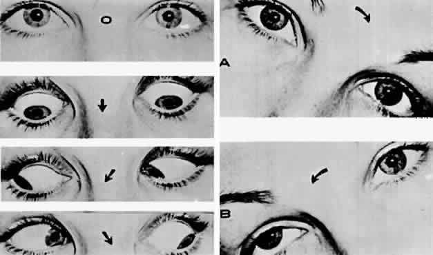
1. Warwick R: Representation of the extra-ocular muscles in the oculomotor complex. J Comp Neurol 98:449, 1953

2. Buttner-Ennever JA, Grob P, Akert K: A transsynaptic autoradiographic study of the pathways controlling the extraocular muscles using [125l]B-IIb tetanus toxin fragment. Ann NY Acad Sci 374:157, 1981

3. Porter JD, Guthrie BL, Sparks DL: Innervation of monkey extraocular muscles: localization of the sensory and motor neurons by retrograde transport of horseradish peroxidase. J Comp Neurol 218:208, 1983

4. Burde RM: The visceral nuclei of the oculomotor complex. Trans Am Ophthalmol Soc 81:533, 1983

5. Highstein SM, Baker R: Excitatory termination of abducens internuclear neurons on medial rectus motoneurons: relationship to syndrome of internuclear ophthalmoplegia. J Neurophysiol 41:1647, 1978

6. Destombes J, Horcholle-Bossavit G, Rouviere A: Données récentes concernant le noyau oculomoteur externe: centre du mouvement oculaire horizontal. J Fr Ophthalmol 6:605, 1983

7. Henn V, Lang W, Hepp K, Reisine H: Experimental gaze palsies in monkeys and their relation to human pathology. Brain 107:619, 1984

8. Crane TB, Yee RD, Baloh RW, Hepler RS: Analysis of characteristic eye movement abnormalities in internuclear ophthalmoplegia. Arch Ophthalmol 101:206, 1983

9. Gonyea EF: Bilateral internuclear ophthalmoplegia: association with occlusive cerebrovascular disease. Arch Neurol 31:168, 1974

10. Klintworth GK: The neuro-ophthalmic manifestations of transtentorial herniation. In Smith JL (ed): Neuro-Ophthalmology: Symposium of the University of Miami and the Bascom Palmer Eye Institute, Vol 4, pp 113–131. St. Louis, CV Mosby, 1972

11. Ishikawa H, Inagaki M, Kitano S: The oculomotor nerve in the cavernous sinus and orbit. Orbit 5:91, 1986

12. Kerr FW: The pupil: functional anatomy and clinical correlation. In Smith JL (ed): Neuro-Ophthalmology: Symposium of the University of Miami and the Bascom Palmer Eye Institute, Vol 4, pp 49–80. St. Louis, CV Mosby, 1968

13. Parkinson D, Johnston J, Chaudhuri A: Sympathetic connections of the fifth and sixth cranial nerves. Anat Record 191:221, 1978

14. Milisavljevi M, Marinkovi S, Loli-Dragani V, Kovacevi M: Oculomotor, trochlear, and abducens nerves penetrated by cerebral vessels: microanatomy and possible clinical significance. Arch Neurol 43:58, 1986

15. Lapresle J, Lasjaunias P: Cranial nerve ischaemic arterial syndromes: a review. Brain 109:207, 1986

16. Horton JC, Fishman RA: Neurovisual findings in the syndrome of spontaneous intracranial hypotension from dural cerebrospinal fluid leak. Ophthalmology 101:244, 1994

17. Perlman EM, Barry D: Bilateral sixth-nerve palsy after water-soluble contrast myelography. Arch Ophthalmol 102:1968, 1984

18. Nabors MW, McCrory ME, Fischer BA et al: Delayed abducens nerve palsies associated with cervical spine fractures. Neurology 37:1565, 1987

19. Barr HWK, Blackwood W, Meadows SP: Intracavernous carotid aneurysms: a clinical-pathological report. Brain 94:607, 1971

20. O'Connor PS, Glaser JS: Intracavernous aneurysms and isolated sixth nerve palsy. In Smith JL (ed): Neuro-Ophthalmology Focus, pp 155–159. New York, Masson, 1982

21. Newton TH, Hoyt WF: Dural arteriovenous shunts in the region of the cavernous sinus. Neuroradiology 1:71, 1970

22. Gutman I, Levartovski S, Goldhammer Y et al: Sixth nerve palsy and unilateral Horner's syndrome. Ophthalmology 93:913, 1986

23. Keane JR: Bilateral sixth nerve palsy. Arch Neurol 33: 681, 1976

24. Keane JR: Eye movement abnormalities in systemic lupus erythematosus. Arch Neurol 52:1145, 1995

25. Savino PJ, Hiliker JK, Casell GH, Schatz NJ: Chronic sixth nerve palsies: are they really harbingers of serious disease? Arch Ophthalmol 100:1442, 1982

26. Currie J, Lubin JH, Lessell S: Chronic isolated abducens paresis from tumors at the base of the brain. Arch Neurol 40:226, 1983

27. Volpe NJ, Lessell S: Remitting sixth nerve palsy in skull base tumors. Arch Ophthalmol 3:1391, 1993

28. Reisner SH, Perlman M, Ben-Tovim N et al: Transient lateral rectus muscle paresis in the newborn infant. J Pediatr 78:461, 1971

29. Knox DL, Clark DB, Schuster FF: Benign VI nerve palsies in children. Pediatrics 40:560, 1967

30. Nemet P, Ehrlich D, Lazar M: Benign abducens palsy in varicella. Am J Ophthalmol 78:859, 1984

31. Werner DB, Savino PJ, Schatz NJ: Benign recurrent sixth nerve palsies in childhood: secondary to immunization or viral illness. Arch Ophthalmol 101:607, 1983

32. Straussberg R, Cohen AH, Amir J, Versano I: Benign abducens palsy associated with EBV infection. J Pediatr Ophthalmol Strabismus 30:60, 1993

33. Bixenman WW, von Noorden GK: Benign recurrent VI nerve palsy in childhood. J Pediatr Ophthalmol Strabismus 18:29, 1981

34. Reinecke RD, Thompson WE: Childhood recurrent idiopathic paralysis of the lateral rectus. Ann Ophthalmol 13:1037, 1981

35. Boger WP III, Puliofito CA, Magoon EH et al: Recurrent isolated sixth nerve palsy in children. Ann Ophthalmol 16:237, 1984

36. Sullivan SC: Benign recurrent isolated VI nerve palsy of childhood. Clin Pediatr 24:160, 1985

37. Peatfield RC: Recurrent VI palsy in cluster headache. Headache 25:225, 1985

38. Robertson DM, Hines JD, Rucker CW: Acquired sixth-nerve paresis in children. Arch Ophthalmol 83:574, 1970

39. Harley RD: Paralytic strabismus in children: etiologic incidence and management of the third, fourth and sixth nerve palsies. Ophthalmology 87:24, 1980

40. Moster ML, Savino PJ, Sergott RC et al: Isolated sixth-nerve palsies in younger adults. Arch Ophthalmol 102: 1328, 1984

41. Shrader EC, Schlezinger NS: Neuro-ophthalmologic evaluation of abducens nerve paralysis. Arch Ophthalmol 63: 84, 1960

42. Rucker CW: The causes of paralysis of the third, fourth and sixth cranial nerves. Am J Ophthalmol 61:1294, 1966

43. Richards BW, Jones FR, Younge BR: Causes and prognosis in 4,278 cases of paralysis of the oculomotor, trochlear, and abducens cranial nerves. Am J Ophthalmol 113:489, 1992

44. Rose JW, Digre KB, Lynch SG, Marnsberger RH: Acute VIth cranial nerve dysfunction in multiple sclerosis. J Clin Neuro Ophthalmol 12:17, 1992

45. Kirkham TH, Bird AC, Sanders MD: Divergence paralysis with raised intracranial pressure: an electro-oculographic study. Br J Ophthalmol 56:776, 1972

46. Mays LE: Neural control of vergence eye movements: convergence and divergence neurons in the midbrain. J Neurophysiol 51:1091, 1984

47. Krohel GB, Tobin DR, Hartnett ME, Barrows NA: Divergence paralysis. Am J Ophthalmol 94:506, 1982

48. Stern RM, Tomsak RL: Magnetic resonance images in a case of “divergence paralysis.” Surv Ophthalmol 30:397, 1986

49. Lewis AR, Kline LB, Sharpe JA: Acquired esotropia due to Arnold-Chiari I malformation. J Neuro Ophthalmol 16:49, 1996

50. Moster ML, Bosley TM, Slavin ML, Rubin SE: Thyroid ophthalmopathy presenting as superior oblique paresis. J Clin Neuro Ophthalmol 12:94, 1992

51. Jacobson DM: Superior oblique palsy manifested during pregnancy. Ophthalmology 98:1874, 1991

52. Astle WF, Rosenblum AL: Familial congenital fourth cranial nerve palsy. Arch Ophthalmol 103:552, 1985

53. Aleksic S, Fudzilovich G, Choy A et al: Congenital ophthalmoplegia in oculoauriculovertebral dysplasia-hemifacial microsomia (Goldenhar-Gorlin syndrome): a clinicopathologic study and review of the literature. Neurology 26:638, 1976

54. Cowan WM, Fawcett JW, O'Leary DDM et al: Regressive events in neurogenesis. Science 225:1258, 1984

55. Pinchoff BS, Sandall G: Congenital absence of the superior oblique tendon in craniofacial dysostosis. Ophthalmic Surg 16:375, 1985

56. Helveston EM, Krach D, Plager DA, Ellis FD: A new classification of superior oblique palsy based on congenital variations in the tendon. Ophthalmology 99:1609, 1992

57. Berlit P: Isolated and combined pareses of cranial nerves III, IV and VI: a retrospective study of 412 patients. J Neurol Sci 103:10, 1991

58. Keane JR: Fourth nerve palsy: historical review and study of 215 patients. Neurology 43:2439, 1993

59. Grimson BS, Glaser JS: Isolated trochlear nerve palsies in herpes zoster ophthalmicus. Arch Ophthalmol 96:1233, 1978

60. Agostinis C, Caverni L, Moschini L et al: Paralysis of fourth cranial nerve due to superior cerebellar artery aneurysm. Neurology 42:457, 1992

61. Botelho PJ, Giangiacomo JG: Autosomal-dominant inheritance of congenital superior oblique palsy. Ophthalmology 103:1508, 1996

62. Lindenberg R: Significance of the tentorium in head injuries from blunt forces. Clin Neurosurg 12:129, 1966

63. Khawam E, Scott AB, Jampolsky A: Acquired superior oblique palsy. Arch Ophthalmol 77:761, 1967

64. Sydnor CF, Seaber JH, Buckley EG: Traumatic superior oblique palsies. Ophthalmology 89:134, 1982

65. Lee J, Flynn JT: Bilateral superior oblique palsies. Br J Ophthalmol 69:508, 1985

66. Trobe JD: Cyclodeviation in acquired vertical strabismus. Arch Ophthalmol 102:717, 1984

67. Keane JR: Vertical diplopia. Semin Neurol 6:147, 1986

68. Wallace DK, von Noorden GK: Clinical characteristics and surgical management of congenital absence of the superior oblique tendon. Am J Ophthalmol 118:63, 1994

69. Kushner BJ: ‘V’ esotropia and excyclotropia after surgery for bilateral fourth nerve palsy. Arch Ophthalmol 110: 1419, 1992

70. Gregersen E, Rindziunski E: Brown's syndrome: a longitudinal long-term study of spontaneous course. Acta Ophthalmol 71:371, 1993

71. Mombaerts I, Koornneef L et al: Superior oblique luxation and trochlear luxation as new concepts in superior oblique muscle weakening surgery. Am J Ophthalmol 120:83, 1995

72. Clarke WN, Noel LP, Agapitos PJ: Traumatic Brown's syndrome. Am Orthoptic J 37:100, 1987

73. Booth-Mason S, Kyle GM, Rossor M, Bradbury P: Acquired Brown's syndrome: an unusual cause. Br J Ophthalmol 69:791, 1985

74. Hickling P, Beck M: Brown's syndrome: an unusual ocular complication of rheumatoid arthritis. Ann Rheum Dis 50:66, 1991

75. Alonso-Valdivielso JL, Alvarez Lario B et al: Acquired Brown's syndrome in a patient with systemic lupus erythematosus. Ann Rheum Dis 52:63, 1993

76. Christiansen SP, Thomas AH: Postpartum Brown's syndrome. Arch Ophthalmol 112:23, 1994

77. Brazis PW, Miller NR, Henderer JD, Lee AG: The natural history and results of treatment of superior oblique myokymia. Arch Ophthalmol 112:1063, 1994

78. Martin TJ, Corbett JJ, Babikian PV et al: Bilateral ptosis due to mesencephalic lesions with relative preservation of ocular motility. J Neuro Ophthalmol 16:258, 1996

79. Keane JR, Zaias B, Itabashi HH: Levator-sparing oculomotor nerve palsy caused by a solitary midbrain metastases. Arch Neurol 14:210, 1984

80. Bryan JS, Hamed LM: Levator-sparing nuclear oculomotor palsy: clinical and magnetic resonance imaging findings. J Clin Neuro Ophthalmol 12:26, 1992

81. Van Dalen JTW, Van Mourek-Noordenbos AM: Isolated inferior rectus paresis: a report of six cases. Neuro Ophthalmology 4:89, 1984

82. Masucci EF: Bilateral ophthalmoplegia in basilar-vertebral artery disease. Brain 88:97, 1965

83. Acers TE: Oculomotor-corpus callosum dysplasia. Trans Am Ophthalmol Soc 80:172, 1982

84. Ksiazek SM, Slamovits TL, Rosen CE et al: Fascicular arrangement in partial oculomotor paresis. J Ophthalmol 118:97, 1994

85. Gauntt CD, Kashii S, Nagata I: Monocular elevation paresis caused by an oculomotor fascicular impairment. J Neuro Ophthalmol 15:11, 1995

86. Castro O, Johnson LN, Mamourian AC: Isolated inferior oblique paresis from brain-stem infarction: perspective on oculomotor fascicular organization in the ventral midbrain tegmentum. Arch Neurol 47:235, 1990

87. Trobe JD, Glaser JS, Quencer RC: Isolated oculomotor paralysis. Arch Ophthalmol 96:1236, 1978

88. Hyland HH, Barnett HJM: The pathogenesis of cranial nerve palsies associated with intracranial aneurysms. Proc R Soc Med 47:141, 1956

89. Ross JS, Masaryk TJ, Modic MT et al: Intracranial aneurysms: evaluation by MR angiography. AJNR 11:449, 1990

90. Capó H, Warren F, Kupersmith MJ: Evolution of oculomotor nerve palsies. J Clin Neuro Ophthalmol 12:21, 1992

91. Guy J, Savino PJ, Schatz NJ et al: Superior division paresis of the oculomotor nerve. Ophthalmology 92:777, 1985

92. Heinze J: Cranial nerve avulsion and other neural injuries in road accidents. Med J Aust 2:1246, 1969

93. Eyster EF, Hoyt WF, Wilson CB: Oculomotor palsy from minor head trauma: an initial sign of basal intracranial tumor. JAMA 220:1083, 1972

94. Neetens A: Extraocular muscle palsy from minor head trauma: initial sign of intracranial tumor. Neuro Ophthalmology 3:43, 1983

95. Walter KA, Newman NJ, Lessell S: Oculomotor palsy from minor head trauma: initial sign of intracranial aneurysm. Neurology 44:148, 1994

96. Ing EB, Sullivan TJ, Clarke MP, Buncic JR: Oculomotor nerve palsies in children. J Pediatr Ophthalmol Strabismus 29:331, 1992

97. Keane JR: Bilateral ocular motor signs after tentorial herniation in 25 patients. Arch Neurol 43:806, 1986

98. Acierno MD, Trobe JD, Cornblath WT, Gebarski SS: Painful oculomotor palsy caused by posterior-draining dural carotid cavernous fistulas. Arch Ophthalmol 113:1045, 1995

99. Schultheiss R, Kristol R, Schramm J: Complete removal of an oculomotor nerve neurinoma without permanent functional deficits. Ger J Ophthalmol 2:228, 1993

100. Kaye-Wilson LG, Gibson R, Bell JE et al: Oculomotor nerve neurinoma. Neuro-ophthalmology 14:37, 1994

101. Jacobson DM, McCanna TD, Layde PM: Risk factors for ischemic ocular motor palsies. Arch Ophthalmol 112:961, 1994

102. Sergott RC, Glaser JS, Buerger LJ: Simultaneous, bilateral diabetic ophthalmoplegia: report of two cases and discussion of differential diagnosis. Ophthalmology 91:18, 1984

103. Eshbaugh CG, Siatkowski RM, Smith JL, Kline LB: Simultaneous multiple cranial neuropathies in diabetes mellitus. J Neuro Ophthalmol 15:219, 1995

104. Weber RB, Daroff RB, Mackey EA: Pathology of oculomotor nerve palsy in diabetics. Neurology 20:835, 1970

105. Asbury AK, Aldredge H, Hershberg R et al: Oculomotor palsy in diabetes mellitus: a clinicopathological study. Brain 93:555, 1970

106. Breen LA, Hopf HC, Farris BK et al: Pupil-sparing oculomotor nerve palsy due to midbrain infarction. Arch Neurol 48:105, 1991

107. Pratt DV, Orengo-Nania S, Horowitz BL, Oram O: Magnetic resonance imaging findings in a patient with nuclear oculomotor palsy. Arch Ophthalmol 113:141, 1995

108. Position Statement: Tests of glycemia in diabetes. American Diabetes Association: Clinical practice recommendations. Diabetes Care, ADA, 1:S18, 1997

109. Sibony PA, Lessell S, Gittenger JW Jr: Acquired oculomotor synkinesis. Surv Ophthalmol 28:382, 1984

110. Sibony PA, Evinger C, Lessell S: Retrograde horseradish peroxidase transport after oculomotor nerve injury. Invest Ophthalmol Vis Sci 27:975, 1986

111. Lepore FE, Glaser JS: Misdirected revisited: a critical appraisal of acquired oculomotor nerve synkinesis. Arch Ophthalmol 98:2206, 1980

112. Wartenberg R: Associated movements in the oculomotor and facial muscles. Arch Neurol Psychiatry 55:439, 1946

113. Sibony PA, Lessell S: Transient oculomotor synkinesis in temporal arteritis. Arch Neurol 41:87, 1984

114. Schatz NJ, Savino PJ, Corbett JC: Primary aberrant oculomotor regeneration: A sign of intracavernous meningioma. Arch Neurol 34:29, 1977

115. Trobe JD, Glaser JS, Post JD: Meningiomas and aneurysms of the cavernous sinus: neuro-ophthalmologic features. Arch Ophthalmol 96:457, 1978

116. Cox TA, Wurster JB, Godfrey WA: Primary aberrant oculomotor regeneration due to intracranial aneurysm. Arch Neurol 36:570, 1979

117. Boghen D, Chartrand JP, LaFlamme P et al: Primary aberrant third nerve regeneration. Ann Neurol 6:415, 1979

118. Hepler RS, Cantu RC: Aneurysms and third nerve palsies: ocular status of survivors. Arch Ophthalmol 77:604, 1967

119. Czarnecki JSC, Thompson HS: The iris sphincter in aberrant regeneration of the third nerve. Arch Ophthalmol 96:1606, 1978

120. Miller NR: Solitary oculomotor nerve palsy in childhood. Am J Ophthalmol 83:106, 1977

121. Victor DI: The diagnosis of congenital unilateral third nerve palsy. Brain 99:711, 1976

122. Balkan R, Hoyt CS: Associated neurologic abnormalities in congenital third nerve palsies. Am J Ophthalmol 97:315, 1984

123. Varma R, Miller NR: Primary oculomotor nerve synkinesis caused by an extracavernous intradural aneurysm. Am J Ophthalmol 118:83, 1994

124. O'Day J, Billson F, King J: Ophthalmoplegic migraine and aberrant regeneration of the oculomotor nerve. Br J Ophthalmol 64:534, 1980

125. Packer AJ, Bienfang DC: Aberrant regeneration involving the oculomotor and abducens nerves. Ophthalmologica 189:80, 1984

126. Krzizok T, Gräf M: Acquired trigemino-abducens synkinesia. Proposed mechanisms and consequences for eye muscle surgery. Klin Monatsbl Augenheilkd 205:33, 1994

127. Pallini R, Fernandez E et al: Experimental repair of the oculomotor nerve: the anatomical paradigms of functional regeneration. J Neurosurg 77:768, 1992

128. Marsh RJ, Dulley B, Kelly V: External ocular motor palsies in ophthalmic zoster: a review. Br J Ophthalmol 61:677, 1977

129. Wexler I: Sequence of demyelination-remyelination in Guillain-Barré disease. J Neurol Neurosurg Psychiatry 46:168, 1983

130. Fisher CM: An unusual variant of acute idiopathic polyneuritis (syndrome of ophthalmoplegia, ataxia and areflexia). N Engl J Med 255:57, 1956

131. Asbury AK, Arnason BG, Adams R: The inflammatory lesion in idiopathic polyneuritis. Medicine 48:173, 1969

132. Grunnet ML, Lubow M: Ascending polyneuritis and ophthalmoplegia. Am J Ophthalmol 74:1155, 1972

133. Berlit P, Rakicky J: The Miller Fisher syndrome. J Clin Neuro Ophthalmol 12:59, 1992

134. Derakhshan I, Lotfi J, Kaufman B: Ophthalmoplegia, ataxia and hyporeflexia (Fisher's syndrome) with a midbrain lesion demonstrated by CT scanning. Eur Neurol 18:361, 1979

135. Ropper AH: The Guillain-Barré syndrome. N Engl J Med 326:1130, 1992

136. Chalmers AC, Miller RG: Chronic inflammatory polyradiculoneuropathy with ophthalmoplegia. J Clin Neuro Ophthalmol 6:166, 1986

137. Yuki N: Acute paresis of extraocular muscles associated with IgG anti-GQ1b antibody. Ann Neurol 39:668, 1995

138. Friedman AP, Harter DH, Merritt HH: Ophthalmoplegic migraine. Arch Neurol 7:320, 1962

139. Vijayan N: Ophthalmoplegic migraine: ischemic or compressive neuropathy? Headache 20:300, 1980

140. Stigmal EW, Ward TN, Harris RD: MRI findings in a case of ophthalmoplegic migraine. Headache 33:234, 1993

141. Straub A, Bondman O, Butter U, Schmidt H: A contrast enhances lesion of the III nerve on MR of a patient with ophthalmoplegic migraine as evidence for a Tolosa-Hunt syndrome. Headache 33:446, 1993

142. Østergaard JR, Moller HU, Christensen T: Recurrent ophthalmoplegia in childhood: diagnostic and etiologic considerations. Cephalgia 16:276, 1996

143. Imes RK, Monteiro MLR, Hoyt WF: Ophthalmoplegic migraine with proximal posterior cerebral artery vascular anomaly. J Clin Neuro Ophthalmol 4:221, 1984

144. Kandt RS, Goldstein GW: Steroid-responsive ophthalmoplegia in a child: diagnostic considerations. Arch Neurol 42:589, 1985

145. Osuntokun O, Osuntokun BO: Ophthalmoplegic migraine and hemoglobinopathy in Nigerians. Am J Ophthalmol 74:451, 1972

146. Sharf B, Hyams S: Oculomotor palsy following varicella. J Pediatr Ophthalmol 9:245, 1972

147. Chan CC, Sogg RL, Steinman L: Isolated oculomotor palsy after measles immunization. Am J Ophthalmol 89:446, 1980

148. Loewenfeld IE, Thompson HS: Oculomotor paresis with cyclic spasms: a critical review of the literature and a new case. Surv Ophthalmol 20:81, 1975

149. Bourgon P, LaFlamme P: Cited by Thompson HS: 13th Pupil Colloquium. Am J Ophthalmol 96:100, 1983

150. Bateman DE, Saunders M: Cyclic oculomotor palsy: description of a case and hypothesis of the mechanism. J Neurol Neurosurg Psychiatry 46:451, 1983

151. Dudim A, Othman A: Acute orbital swelling: evaluation and protocol. Pediatr Emerg Care 12:16, 1996

152. Bergin DJ, Wright JE: Orbital cellulitis. Br J Ophthalmol 70:174, 1986

153. Del Valle ZA, Suarez RA, Encinas PM et al: Mucormycosis of the sphenoid sinus in an otherwise health patient: case report and literature review. J Laryngol Otol 110:471, 1996

154. Yohai RA, Bullock JD, Aziz AA, Mirkert RJ: Survival factors in rhino-orbital cerebral mucormycosis. Surv Ophthalmol 39:3, 1994

155. Kennerdell JS, Dresner SC: The nonspecific orbital inflammatory syndromes. Surv Ophthalmol 29:93, 1984

156. Grimson BS, Simmons KB: Orbital inflammation, myositis and systemic lupus erythematosus. Arch Ophthalmol 101:36, 1983

157. Schonder AA, Clift RC, Brophy JW, Dane LW: Bilateral recurrent orbital inflammation associated with retroperitoneal fibrosclerosis. Br J Ophthalmol 69:783, 1985

158. Rootman J, Nugent R: The classification and management of acute orbital pseudotumors. Ophthalmology 89:1040, 1982

159. Siatkowski RM, Capó H, Byrne SF et al: Clinical and echographic findings in idiopathic orbital myositis. Am J Ophthalmol 118:343, 1994

160. Koorey DJ: Cranial arteritis: a twenty-year review of cases. Aust NZ J Med 14:143, 1984

161. Baricks ME, Traviesa DB, Glaser JS, Levy IS: Ophthalmoplegia in cranial arteritis. Brain 100:209, 1977

162. Cline RA, Rootman J: Enophthalmos: a clinical review. Ophthalmology 91:229, 1984

163. Keane JR: Cavernous sinus syndrome: analysis of 151 cases. Arch Neurol 53:967, 1996

164. Tolosa EJ: Periarteritic lesions of the carotid siphon with clinical features of carotid infraclinoid aneurysms. J Neurol Neurosurg Psychiatry 17:300, 1954

165. Hunt WE, Meagher JN, Lefever HE, Zeman W: Painful ophthalmoplegia: its relation to indolent inflammation of the cavernous sinus. Neurology 11:56, 1961

166. Thomas JE, Yoss RE: The parasellar syndrome: problems in determining etiology. Mayo Clin Proc 45:617, 1970

167. Spector RH, Fiandaca MS: The “sinister” Tolosa-Hunt syndrome. Neurology 36:198, 1986

168. Kline LB: The Tolosa-Hunt syndrome. Surv Ophthalmol 27:79, 1982

169. Campbell RJ, Okazaki H: Painful ophthalmoplegia (Tolosa-Hunt variant): autopsy findings in a patient with necrotizing intracavernous carotid vasculitis and inflammatory disease of the orbit. Mayo Clin Proc 62:520, 1987

170. Aktan S, Aykut C, Erzen C: Computed tomography and magnetic resonance imaging in three patients with Tolosa-Hunt syndrome. Eur Neurol 33:393, 1993

171. Takahashi Y, Abe T, Kojima K et al: Tolosa-Hunt syndrome with atypical intrasellar and juxtasellar lesions. Two case reports. Kurume Med J 43:165, 1996

172. Drevelengas A, Kalaitzoglou I, Tsolaki M: Tolosa-Hunt syndrome with sellar erosion: case report. Neuroradiology 35:451, 1993

173. Hama S, Arita K, Kurisu K et al: Parasellar chronic inflammatory disease presenting as Tolosa-Hunt syndrome, hypopituitarism and diabetes insipidus: a case report. Endocr J 43:503, 1996

174. Brazis PW, Capobianco DJ et al: Low flow dural arteriovenous shunt: another cause of “sinister” Tolosa-Hunt syndrome. Headache 34:523, 1994

175. Sanchez Pina C, Pascual-Castroviejo I et al: Burkitt's lymphoma presenting as Tolosa-Hunt syndrome. Pediatr Neurol 9:157, 1993

176. Montecucco C, Caporali R, Pacchetti C, Turla M: Is Tolosa-Hunt syndrome a limited form of Wegener's granulomatosis?: report of two cases with anti-neutrophil cytoplasmic antibodies. Br J Rheumatol 32:640, 1993

177. Masson C, Henin D, Hauw JJ et al: Cranial pachymeningitis of unknown origin: a study of seven cases. Neurology 17:1329, 1993

178. Vailati A, Marena C et al: Hashimoto's thyroiditis in association with Tolosa-Hunt syndrome: a case report. Thyroid 3:125, 1993

179. Bills DC, Meyer FB et al: A retrospective analysis of pituitary apoplexy. Neurosurgery 33:602, 1993

180. Vidal E, Cevallos R et al: Twelve cases of pituitary apoplexy. Arch Intern Med 152:1893, 1992

181. Weinberger LM, Adler FH, Grant FC: Primary pituitary adenoma and the syndrome of the cavernous sinus. Arch Ophthalmol 24:1197, 1940

182. Godtfredsen E, Lederman M: Diagnostic and prognostic roles of ophthalmoneurologic signs and symptoms in malignant nasopharyngeal tumors. Am J Ophthalmol 59:1063, 1965

183. Meadows SP: Intracavernous aneurysms of the carotid artery. Arch Ophthalmol 62:566, 1959

184. Lee AG, Mawad ME, Baskin DS: Fatal subarachnoid hemorrhage from the rupture of a totally intracavernous carotid artery aneurysm: case report. Neurosurgery 38:596, 1996

185. Keane JR, Talalla A: Post traumatic intracavernous aneurysm: epistaxis with monocular blindness preceded by chromatopsia. Arch Ophthalmol 87:701, 1972

186. Trobe JD, Glaser JS, Post JD: Meningiomas and aneurysms of the cavernous sinus: neuro-ophthalmologic features. Arch Ophthalmol 96:457, 1978

187. Rapport R, Murtagh FR: Ophthalmoplegia due to spontaneous thrombosis in a patient with bilateral cavernous carotid aneurysms. J Clin Neuro Ophthalmol 1:225, 1981

188. Kupersmith MJ, Hurst R, Berenstein A et al: The benign course of cavernous carotid artery aneurysms. J Neurosurg 77:690, 1992

189. Cushing H, Eisenhardt L: Meningiomas: Their Classification, Regional Behavior, Life History and Surgical End Results. New York, Hofner, 1962

190. Ojemann RG, Thornton AF, Harsh GR: Management of anterior cranial base and cavernous sinus neoplasms with conservative surgery alone or in combination with fractionated photon or stereotactic proton radiotherapy. Clin Neurosurg 42:71, 1995

191. Nugent GR, Sprinkle P, Bloor BM: Sphenoid sinus mucoceles. J Neurosurg 32:443, 1970

192. Valvassori GE, Putterman AM: Ophthalmological and radiological findings in sphenoidal mucoceles. Trans Am Acad Ophthalmol Otolaryngol 77:703, 1973

193. Casteels I, DeLoof E, Brock P et al: Sudden blindness in a child: presenting symptoms of sphenoid sinus mucocele. Br J Ophthalmol 76:502, 1992

194. El-Fiki ME, Abdel-Fattah HM, el-Deeb AK: Sphenoid sinus mucopyocele with marked intracranial extension. Surg Neurol 39:115, 1993

195. Post MJD, Mendez DR, Kline LB et al: Metastatic disease to the cavernous sinus: clinical syndrome and CT diagnosis. J Comput Assist Tomogr 9:115, 1985

196. Kline LB, Acker JD, Post JDM: Computed tomographic evaluation of the cavernous sinus. Ophthalmology 89:374, 1982

197. Moore CE, Hoyt WF, North JB: Painful ophthalmoplegia following treated squamous carcinoma of the forehead: orbital apex involvement from centripetal spread via the supraorbital nerve. Med J Aust 1:657, 1976

198. Catalano PJ, Sen C, Biller HF: Cranial neuropathy secondary to perineural spread of cutaneous malignancies. Am J Otol 16:772, 1995

199. ten Hove MW, Glaser JS, Schatz NJ: Occult perineural tumor infiltration of the trigeminal nerve. Diagnostic considerations. J Neuro Ophthalmol 17:170, 1997

200. Hayat G, Ehsan T, Selhorst JB, Manepali A: Magnetic resonance evidence of perineural metastasis. J Neuroimaging 5:122, 1995

201. Olson ME, Cornice NL, Posner JB: Infiltration of the leptomeninges by systemic cancer: a clinical and pathologic study. Arch Neurol 30:122, 1974

202. Little JR, Dale AJD, Okazaki H: Meningeal carcinomatosis: clinical manifestations. Arch Neurol 30:138, 1974

203. Wasserstrom WR, Glass JP, Posner JB: Diagnosis and treatment of leptomeningeal metastases from solid tumors: experience with 90 patients. Cancer 49:759, 1982

204. Watanabe M, Tanaka R, Takeda N: Correlation of MRI and clinical features in meningeal carcinomatosis. Neuroradiology 35:512, 1993

205. Stern BJ, Krumholz A, Johns C et al: Sarcoidosis and its neurological manifestations. Arch Neurol 49:909, 1985

206. Oksamen V: Neurosarcoidosis: clinical presentations and course in 50 patients. Acta Neurol Scand 73:283, 1986

207. Post MJD, Chan JC, Hensley GT et al: Toxoplasma encephalitis in Haitian adults with acquired immunodeficiency syndrome: a clinical-pathologic-CT correlation. Am J Neuroradiol 4:155, 1983

208. Berger JR, Flaster M, Schatz N et al: Cranial neuropathy heralding otherwise occult AIDS-related large cell lymphoma. J Clin Neuro Ophthalmol 13:113, 1993

209. Hamed LM, Schatz, NJ, Galetta SL: Brainstem ocular motility defects and AIDS. Am J Ophthalmol 106:437, 1988

210. Wadia NH, Wadia PN, Katrak SM, Misra VP: A study of the neurological disorder associated with acute hemorrhagic conjunctivitis due to enterovirus 70. J Neurol Neurosurg Psychiatry 46:559, 1983

211. Katiyar BC, Misra S, Singh RB et al: Adult polio-like syndrome following enterovirus 70 conjunctivitis (natural history of the disease). Acta Neurol Scand 67:263, 1983

212. Lesser RL: Ocular manifestations of Lyme disease. Am J Med 98:60, 1995

213. Rodriguez M, Gomez MR, Howard FM, Taylor WF: Myasthenia in children: long-term follow-up. Ann Neurol 13:504, 1983

214. Vincent A, Newsom-Davis J: Acetylcholine receptor antibody characteristics in myasthenia gravis: I. Patients with generalized myasthenia or disease restricted to ocular muscles. Clin Exp Immunol 49:257, 1982

215. Zimmerman CW, Eblen F: Repertoires of autoantibodies against homologous eye muscle in ocular and generalized myasthenia gravis. Clin Invest 71:445, 1993

216. Drachman DB, Adams RN, Josifek LF, Self SG: Functional activities of autoantibodies to acetylcholine receptors and the clinical severity of myasthenia gravis. N Engl J Med 307:769, 1982

217. Weinberg DA, Lesser RL, Vollmer TL: Ocular myasthenia: a protean disorder. Surv Ophthalmol 39:169, 1994

218. Grob D, Arsura EL, Brunner NG, Namba T: The course of myasthenia gravis, and therapies affecting outcome. Ann NY Acad Sci 505:472, 1987

219. Bever CT, Aquino AV, Penn AS et al: Prognosis of ocular myasthenia. Ann Neurol 14:526, 1983

220. Cogan DG: Myasthenia gravis: A review of the disease and a description of lid twitch as a characteristic sign. Arch Ophthalmol 74:217, 1965

221. Dutton GN, Garson JA, Richardson RB: Pupillary fatigue in myasthenia gravis. Trans Ophthalmol Soc UK 102:510, 1982

222. Glaser JS: Myasthenic pseudo-internuclear ophthalmoplegia. Arch Ophthalmol 75:363, 1966

223. Keane JR, Hoyt WF: Myasthenic (vertical) nystagmus: verification by edrophonium tonography. JAMA 212:1209, 1970

224. Barton JJS, Jama A, Sharpe JA: Saccadic duration and intrasaccadic fatigue in myasthenic and nonmyasthenic ocular palsies. Neurology 45:2065, 1995

225. Osher RH, Griggs RC: Orbicularis fatigue: the “peek” sign of myasthenia gravis. Arch Ophthalmol 97:677, 1979

226. Osher RH, Glaser JS: Myasthenic sustained gaze fatigue. Am J Ophthalmol 89:443, 1980

227. Retzlaff JA, Kearns TP, Howard FM Jr et al: Lancaster red-green test in evaluation of edrophonium effect in myasthenia gravis. Am J Ophthalmol 67:13, 1969

228. Seybold ME, Daroff RB: The office Tensilon test for ocular myasthenia. Arch Neurol 43:842, 1986

229. Coll GE, Demer JL: The edrophonium-Hess screen test in the diagnosis of ocular myasthenia gravis. Am J Ophthalmol 114:489, 1992

230. Siatkowski RM, Shah L, Feuer WJ: The effect of edrophonium chloride on muscle balance in normal subjects and those with nonmyasthenic strabismus. J Neuro Ophthalmol 17:7, 1997

231. Miller NR, Morris JE, Maguire M: Combined use of neostigmine and ocular motility measurements in the diagnosis of myasthenia gravis. Arch Ophthalmol 100:761, 1982

232. Oey PL, Wieneke GM, Hoogenraad TU, van Hofgelen AC: Ocular myasthenia gravis: the diagnostic yield of repetitive nerve stimulating and stimulated single fiber EMG of orbicularis oculi muscle and infrared reflection oculography. Muscle Nerve 16:142, 1993

233. Odel J, Winterkorn J, Behrens M: The sleep test for myasthenia gravis: a safe alternative. J Clin Ophthalmol 11:288, 1991

234. Janssen RS, Kaye AD, Lisak RP et al: Radiologic evaluation of the mediastinum in myasthenia gravis. Neurology 33:534, 1983

235. Sanders DB, Howard JF: Disorders of neuromuscular transmission. In Bradley WG, Daroff RB, Fenichel GM, Marsden CD (eds): Neurology in Clinical Practice, Chap 83, p 1983. Boston, Butterworth-Heinemann, 1996

236. Kupersmith MJ, Moster M, Bhuiyan S et al: Beneficial effects of corticosteroids on ocular myasthenia gravis. Arch Neurol 53:802, 1996

237. McEvoy KM: Diagnosis and treatment of Lambert-Eaton myasthenic syndrome. Neurol Clin 12:387, 1994

238. Molenaar PC, Newson-Davis J, Polak RL, Vincent A: Eaton-Lambert syndrome: acetylcholine and choline acetyltransferase in skeletal muscle. Neurology 32:1062, 1982

239. Tsuchiya N, Sato M et al: Lambert-Eaton myasthenic syndrome associated with Sjögren's syndrome and discoid lupus erythematosus. Scand J Rheumatol 22:302, 1993

240. Cruciger MP, Brown B, Denys EH et al: Clinical and subclinical oculomotor findings in the Eaton-Lambert syndrome. J Clin Neuro Ophthalmol 3:19, 1983

241. Dell'Osso LF, Ayyar DR, Daroff RB, Abel LA: Edrophonium test in Eaton-Lambert syndrome: quantitative oculography. Neurology 33:1157, 1983

242. Breen LA, Gutmann L, Birck JF, Riggs JR: Paradoxical lid elevation with sustained upgaze: a sign of Lambert-Eaton syndrome. Muscle Nerve 14:863, 1991

243. Grisold W, Drlicek M, Liszka-Setinek U, Wondrusch E: Anti-tumour therapy in paraneoplastic neurological disease. Clin Neurol Neurosurg 97:106, 1995

244. Katz LJ, Lesser RL, Merikangas JR, Silverman JP: Ocular myasthenia gravis after D-penicillamine administration. Br J Ophthalmol 73:1015, 1989

245. McQuillen MP, Cantor HE, O'Rourke JR: Myasthenic syndromes associated with antibiotics. Arch Neurol 18:402, 1968

246. Albert DM, Wong VG, Henderson ES: Ocular complications of vincristine therapy. Arch Ophthalmol 78:709, 1967

247. Blanton CL, Sawyer RA: Myasthenia gravis by another name: an elusive impostor [clinical conference]. Surv Ophthalmol 38:219, 1993

248. Hedges TR III, Jones A, Stark L, Hoyt WF: Botulin ophthalmoplegia: clinical and oculographic observations. Arch Ophthalmol 101:211, 1983

249. Schubart P, Kasperski S, Schroder P: Ocular involvement in botulism. Klin Monatsbl Augenheilkd 187:142, 1985

250. Hudson HL, Levin L, Feldon SE: Graves exophthalmos unrelated to extraocular muscle enlargement. Ophthalmology 98:1495, 1991

251. Hermann JS: Paretic thyroid myopathy. Ophthalmology 89:473, 1982

252. Gamblin FT, Galentine PG, Eil C: Intraocular pressure and thyroid disease. In Gorman CA, Waller RR, Dyer JA (eds): The Eye and Orbit in Thyroid Disease, pp 155–166. New York, Raven Press, 1984

253. Byrne SF, Glaser JS: Orbital tissue differentiation with standardized echography. Ophthalmology 90:1071, 1983

254. Kung AW, Yau CC, Cheng A: The incidence of ophthalmopathy after radioiodine therapy for Graves' disease: prognostic factors and the role of methimazole. J Clin Endocrinol Metabol 79:542, 1994

255. Gorman CA, Waller RR, Dyer JA: The Eye and Orbit in Thyroid Eye Disease. New York, Raven Press, 1984

256. Raikow RB, Tyutyunikov A et al: Correlation of serum immunoglobulin E elevations with clinical states of dysthyroid orbitopathy. Ophthalmology 99:361, 1992

257. Khuteta A, Mishra YC et al: Mesenchymal chondrosarcoma of orbit with intracranial extension (a rare case). Ind J Ophthalmol 40:92, 1992

258. Prummel MF, Mourits MP et al: Randomized double-blind trial of prednisone versus radiotherapy in Graves' ophthalmopathy. Lancet 342:949, 1993

259. Wilson WB, Prochoda M: Radiotherapy for thyroid orbitopathy. Arch Ophthalmol 113:1420, 1995

260. Shine CSK, Kendler DL et al: Radiotherapy in the management of thyroid orbitopathy. Arch Ophthalmol 111:819, 1993

261. Lueder GT, Scott WE, Kutschke PJ, Keech RV: Long-term results of adjustable suture surgery for strabismus secondary to thyroid ophthalmopathy. Ophthalmology 99:993, 1992

262. Kushner BJ: A surgical procedure to minimize lower-eyelid retraction with inferior rectus recession. Arch Ophthalmol 110:1011, 1992

263. Hudson HL, Feldon SE: Late overcorrection of hypotropia in Graves ophthalmopathy. Ophthalmology 99:356, 1992

264. Johns DR: Mitochondrial DNA and disease. N Engl J Med 333:638, 1995

265. Mitsumoto H, Aprille JR, Wray SH et al: Chronic progressive external ophthalmoplegia (CPEO): clinical, morphologic and biochemical studies. Neurology 33:452, 1983

266. Hyman BN, Patten BM, Dodson RF: Mitochondrial abnormalities in progressive external ophthalmoplegia. Am J Ophthalmol 83:362, 1987

267. Seibel P, Lauber J et al: Chronic progressive external ophthalmoplegia is associated with a novel mutation in the mitochondrial tRNA (Asn) gene. Biochem Biophys Res Commun 204:482, 1994

268. Hattori Y, Goto Y et al: Point mutations in mitochondrial tRNA genes: sequence analysis of chronic progressive external ophthalmoplegia (CPEO). J Neurol Sci 125:50, 1994

269. Laforet P, Lombes A, Eymard B et al: Chronic progressive external ophthalmoplegia with ragged-red fibers: clinical, morphological and genetic investigations in 43 patients. Neuromuscul Disord 5:399, 1995

270. Mullie MA, Harding AE, Petty RKH et al: Retinal manifestations of mitochondrial myopathy: a study of 22 cases. Arch Ophthalmol 103:1825, 1985

271. Rowland LP: Molecular genetics, pseudogenetics, and clinical neurology. Neurology 33:1179, 1983

272. Ogasahara S, Yorifuji S, Nishikawa Y et al: Improvement of abnormal pyruvate metabolism and cardiac conduction defect with coenzyme Q10 in Kearns-Sayre syndrome. Neurology 35:372, 1985

273. Coquet M, Vallat JM, Vital C et al: Nuclear inclusions in oculopharyngeal dystrophy: an ultrastructural study of six cases. J Neurol Sci 60:151, 1983

274. Satoyoshi E, Kinoshita M: Oculopharyngodistal myopathy: report of four families. Arch Neurol 34:89, 1977

275. Ionescu V, Thompson SH, Ionescu R et al: Inherited ophthalmoplegia with intestinal pseudo-obstruction. J Neurol Sci 59:215, 1983

276. Hsu CC, Chuang YH, Tsai JL et al: CPEO and carnitine deficiency overlapping in MELAS syndrome. Acta Neurol Scand 92:252, 1995

277. Flynn JT, Bachynski BN, Rodrigues MM et al: Hyperglycemic acidotic coma and death in Kearns-Sayre syndrome. Trans Am Ophthalmol Soc 83:131, 1985

278. Coulter DL, Allen RJ: Abrupt neurological deterioration in children with Kearns-Sayre syndrome. Arch Neurol 38:247, 1981

279. Litvan I, Agid Y, Caine D et al: Clinical research criteria for the diagnosis of progressive supranuclear palsy (Steele-Richardson-Olszewski syndrome). Neurology 47:1, 1996

280. Jackson JA, Jankovic J, Ford J: Progressive supranuclear palsy: clinical features and response to treatment in 16 patients. Ann Neurol 13:273, 1983

281. Dehaene J: Apraxia of lid opening in progressive supranuclear palsy. Ann Neurol 15:115, 1984

282. Cole DG, Growdon JH: Therapy for progressive supranuclear palsy: past and future. J Neural Transm 42(suppl): 283, 1994

283. DiMonte DA, Harati Y et al: Muscle mitochondrial ATP production in progressive supranuclear palsy. J Neurochem 62:1631, 1994

284. Savoiardo M, Girotti F, Strada L, Ciceri E: Magnetic resonance imaging in progressive supranuclear palsy and other parkinsonian disorders. J Neural Transm 42(suppl):93, 1994

285. Calabrese VP, Hadfield MG: Parkinsonism and extraocular motor abnormalities with unusual neuropathological findings. Mov Disord 6:257, 1991

286. Knox DL, Green WR, Troncoso JC et al: Cerebral ocular Whipple's disease: a 62-year odyssey from death to diagnosis. Neurology 45:617, 1995

287. Rabiah PK, Bateman JB, Demer JL, Perlman S: Ophthalmologic findings in patients with ataxia. Am J Ophthalmol 123:108, 1997

288. Shelbourne P, Johnson K: Myotonic dystrophy: another case of too many repeats? Hum Mutat 1:183, 1992

289. Lessell S, Coppeto J, Samet S: Ophthalmoplegia in myotonic dystrophy. Am J Ophthalmol 71:1231, 1971

290. Burian HM, Burns CA: Ocular changes in myotonic dystrophy. Am J Ophthalmol 63:22, 1967

291. Thompson HS, Van Allen MW, von Noorden GK: The pupil in myotonic dystrophy. Invest Ophthalmol 3:325, 1964

292. Hayasaka S, Kiyosawa M, Katsumata S et al: Ciliary and retinal changes in myotonic dystrophy. Arch Ophthalmol 102:88, 1984

293. Pryse-Phillips W, Johnson GJ, Larsen B: Incomplete manifestations of myotonic dystrophy in a large kinship in Labrador. Ann Neurol 11:582, 1982

294. Ter Bruggen JP, Bastiaensen LA, Tyssen CC, Gielen G: Disorders of eye movement in myotonic dystrophy. Brain 113(pt 2):463, 1990

295. Ono S, Kanda F, Takahashi K et al: Neuronal loss in the medullary reticular formation in myotonic dystrophy: a clinicopathological study. Neurology 46:228, 1996

296. Schults WT, Hoyt WF, Behrens M et al: Ocular neuromyotonia: a clinical description of six patients. Arch Ophthalmol 104:1028, 1986

297. Lessell S, Lessell IM, Rizzo JF: Ocular neuromyotonia after radiation therapy. Am J Ophthalmol 102:766, 1986

298. Newman SA: Gaze-induced strabismus (clinical conference). Surv Ophthalmol 38:303, 1993

299. Barroso L, Hoyt WF: Episodic exotropia from lateral rectus neuromyotonia: appearance and remission after radiation therapy for a thalamic glioma. J Pediatr Ophthalmol Strabismus 30:56, 1993

300. Frohman EM, Zee DS: Ocular neuromyotonia clinical features, physiological mechanisms, and response to therapy. Ann Neurol 37:620, 1995

301. Ezra E, Spalton D, Sanders MD et al: Ocular neuromyotonia. Br J Ophthalmol 80:350, 1996

302. Seybold ME, Yoss RE, Hollenhorst RW, Moyer NJ: Pupillary abnormalities associated with tumors of the pineal region. Neurology 21:232, 1971

303. Gressel MG, Hanson MR, Tomsak RL: Reversible Sylvian aqueduct syndrome following jejunoileal bypass. In Smith JL (ed): Neuro-ophthalmology Focus, pp 231–233. New York, Masson, 1981

304. Daras M, Koppel BS, Samkoff L, Marc J: Brain stem toxoplasmosis in patients with acquired immunodeficiency syndrome. J Neuroimaging 4:85, 1994

305. Paulus W, Straube A, Bauer W, Harding AE: Central nervous system involvement in Leber's optic neuropathy. J Neurol 240:251, 1993

306. Neville BGR, Lake BD, Stephens R, Sanders MD: A neurovisceral storage disease with vertical supranuclear ophthalmoplegia, and its relationship to Niemann-Pick disease: a report of nine patients. Brain 96:97, 1973

307. Yan-Go FL, Yanagihara T, Pierre RV, Goldstein NP: A progressive neurologic disorder with supranuclear vertical gaze paresis and distinctive bone marrow cells. Mayo Clin Proc 59:404, 1984

308. Shulman LM, David NJ, Weiner WJ: Psychosis as the initial manifestation of adult-onset Niemann-Pick disease type C. Neurology 45:1739, 1995

309. Palmer M, Green WR, Maumenee IH et al: Niemann-Pick disease—type C. Arch Ophthalmol 103:817, 1985

310. Kirkham TH, Kamin DF: Slow saccadic eye movements in Wilson's disease. J Neurol Neurosurg Psychiatry 37:191, 1974

311. Hoyt CS, Bilson FA, Alpins N: The supranuclear disturbances of gaze in kernicterus. Ann Ophthalmol 10:1487, 1978

312. Edis RH, Mastaglia FL: Vertical gaze palsy in barbiturate intoxication. Br Med J 1:144, 1977

313. Hamed LM, Mancuso A: Inferior rectus muscle contracture syndrome after retrobulbar anesthesia. Ophthalmology 98:1506, 1991

314. Hamilton SM, Elsas FJ, Dawson TL: A cluster of patients with inferior rectus restriction following local anesthesia for cataract surgery. J Pediatr Ophthalmol Strabismus 30:288, 1993

315. Hunter DG, Lam GC, Guyton DL: Inferior oblique muscle injury from local anesthesia for cataract surgery. Ophthalmology 102:501, 1995

316. Muñoz M: Letter to the Editor: Inferior rectus muscle overaction after cataract extraction. Am J Ophthalmol 118:664, 1994

317. Capó H, Roth E, Johnson T et al: Vertical strabismus after cataract surgery. Ophthalmology 103:918, 1996

318. Muñoz M, Parrish RK II: Strabismus following implantation of Baerveldt drainage devices. Arch Ophthalmol 111: 1096, 1993

319. Smith SL, Starita RJ, Fellman RL, Lynn JR: Early clinical experience with the Baerveldt 350-mm2 glaucoma implant and associated extraocular muscle imbalance. Ophthalmology 100:914, 1993

320. Cornblath WT, Elner V, Rolfe M: Extraocular muscle involvement in sarcoidosis. Ophthalmology 100:501, 1993

321. Patel AS, Kelman SE, Duncan GW, Rismondo V: Painless diplopia caused by extraocular muscle sarcoid. Arch Ophthalmol 112:879, 1994

322. Hakin KN, McNab AA, Sullivan TJ: Spontaneous hemorrhage within the rectus muscle. Ophthalmology 101:1631, 1994

323. Harris GJ, Murphy ML et al: Orbital myositis as a paraneoplastic syndrome. Arch Ophthalmol 112:380, 1994

324. Leib ML, Odel JG, Cooney MJ: Orbital polymyositis and giant cell myocarditis. Ophthalmology 101:950, 1994

325. Atabay C, Tyutyunikov A et al: Serum antibodies reactive with eye muscle membrane antigens are detected in patients with nonspecific orbital inflammation. Ophthalmology 102: 145, 1995
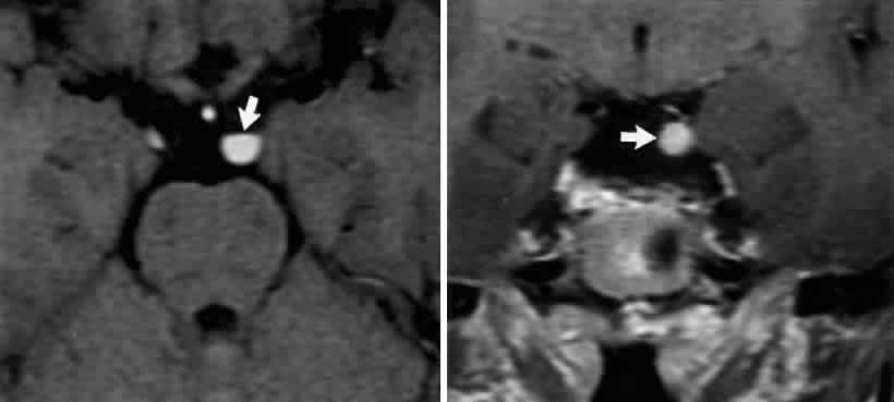
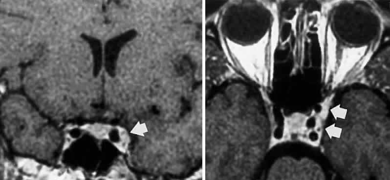
Back to Top