|
|
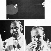
| Fig. 14. Use of colored objects to detect and plot central scotomas. A. The limits of the defect are most easily defined when the target subjectively increases in color intensity as it is moved out of scotoma. B. Two identical, colored targets are used for simultaneous comparison, one centrally (on nose), the other at approximately 10°. Normally, the target fixated centrally appears brighter. C. Use of brightly colored bottle tops (mydriatic red) for color comparison. |