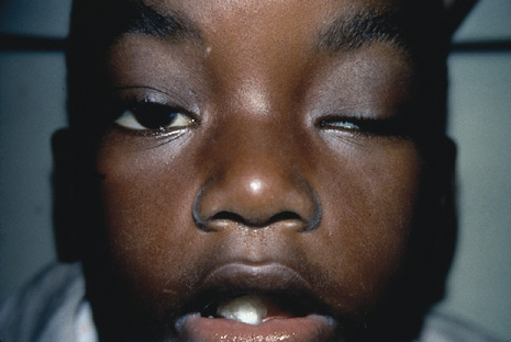
Fig. 23.
Automated perimetry demonstrating funcitonal visual field loss.
A.
Normal right eye field.
B.
Left eye field demonstrating dense interotemporal defect.
C.
Binocular perimetry shows persistence of field defect and absence of blind spot.