Chapter 71A
Embryology and Anatomy of Human Lenses
J.R. KUSZAK and M.J. COSTELLO
Main Menu   Table Of Contents

Search

LENS EMBRYOLOGY
LENS SUTURES
LENS ANATOMY
LENS ULTRASTRUCTURE
SUMMARY
ACKNOWLEDGMENT
REFERENCES

The cornea and the lens are the principal refractive elements of the eye. They are responsible for, respectively, stationary and variable refraction. However, although both must be transparent to function properly, the basis of their transparency is different. The cornea relies on the continuous pumping of interstitial fluid across its semipermeable surface membranes and a supramolecular organization of collagen fibrils for its clarity. Lens transparency is the result of a unique arrangement of its fiber-like cells, or fibers, and a gradient of refractive index produced by a variable crystallin protein concentration within these fibers. In fact, alterations in lens morphology affected during specific periods of development and growth, lead to quantifiable improvements in lens optical quality. Furthermore, lens structural anomalies affected during the same periods of development and growth, lead to quantifiable degradation in lens optical quality. Thus, the human lens is a prime example of form following function and malformation leading to malfunction.

In this chapter we describe lens anatomy throughout development, growth, and aging. In addition, we discuss how variations in lens structure influence its optics, physiology, and pathology.

Back to Top
LENS EMBRYOLOGY

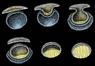
PRIMARY FIBER FORMATION

Lens formation begins in the latter part of the first trimester as surface ectodermal cells, immediately overlying the developing optic vesicle, are induced to thicken and form the lens placode (Fig. 1). As the optic vesicle begins to fold inward and form the optic cup, the lens placode invaginates toward the developing optic cup until it eventually pinches off as an inverted (inside-out) lens vesicle. The apical surfaces of the lens vesicle cells are directed toward the lumen, whereas their basal surfaces are directed toward the outer surface. Throughout life, the basal surfaces of lens cells produce a progressively thicker basement membrane, the lens capsule, that envelops the lens. Lens development proceeds as cells of the lens vesicle approximating its retinal half are induced to terminally differentiate. A striking consequence of lens terminal differentiation is that the originally cuboidal lens vesicle cells are transformed into long fiber-like cells, or fibers. Fiber elongation proceeds until they fill in the lumen of the lens vesicle. Because these fibers are the first lens cells to be transformed into fibers, they are referred to as the primary fibers.

Fig. 1. Schematic diagrams of key structural events in lens development. Upper left. The thickening of surface ectodermal cells to form the lens placode (yellow). Upper middle and right. The invagination of the lens placode toward the developing optic cup (blue). Lower left. The inverted, or “inside out,” lens vesicle. Lower middle. The elongation of posterior lens vesicle cells as they terminally differentiate to form primary lens fibers. Lower right. The obliteration of the lumen of the lens vesicle by fully elongated primary fibers. Note the anterior lens vesicle cells (white) remain as an undifferentiated monolayer epithelium covering the lens fiber mass. (Adapted from Kuszak JR, Brown HG: Embryology and anatomy of the lens. In Principles and Practice of Ophthalmology: Basic Sciences. Philadelphia, WB Saunders, 1993, p. 83.)

SECONDARY FIBER FORMATION

The cells of the lens vesicle that were not induced to form primary fibers remain as a monolayer, the lens epithelium, that covers the anterior surface of the primary fiber mass. Lens development and growth continues throughout life, in a manner similar to other stratified epithelia with the lens epithelium constituting the basal layer. However, whereas typically stratified epithelia have their progenitor cells distributed throughout the basal layer, the lens is unique in that its progenitor cells are sequestered as a distinct subpopulation within the lens epithelium known as the germinative zone (GZ). The remainder of the lens epithelial cells are sequestered in three additional regions known as the central zone (CZ), pregerminative zone (PGZ), and transitional zone (TZ). CZ epithelial cells comprise a broad polar cap of the lens epithelium covering most (approximately 80%) of the anterior surface of the lens. These cells are arrested in the G0 stage of the cell cycle and are not recruited to terminally differentiate into fibers.1 PGZ cells comprise a narrow, latitudinal band (approximately 5% of the lens epithelium) peripheral to the CZ. Although a small number of these cells undergo mitotic division, only rarely are any of their daughter cells induced to terminally differentiate and become additional fibers. Rather, these daughter cells add to the lens epithelial population as the anterior surface of the lens increases in size because of continued growth and aging.2,3 GZ cells comprise a narrow, latitudinal band (approximately 10% of the lens epithelium) peripheral to the PGZ. These cells undergo mitotic division, and some daughter cells are selected to terminally differentiate into additional fibers. Because these are the second fibers to develop, they are referred to as secondary fibers. Finally, TZ cells comprise a narrow, latitudinal band (approximately 5% of the lens epithelium) peripheral to the GZ. These cells are the nascent fibers having already begun the process of elongation.

FIBER ELONGATION

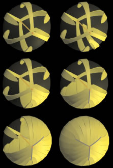
The secondary fibers comprise the layers, or strata, of the lens. An understanding of how fibers are added onto the existing lens throughout life is necessary to comprehend the complex structure of the lens that is paramount in the establishment and maintenance of its transparency (Fig. 2).

Fig. 2. Schematic diagram showing secondary fiber development or lens growth at selected stages. Lens epithelial cells in the peripheral GZ undergo mitosis and some daughter cells are selected to terminally differentiate into secondary fibers. As these cells migrate posteriorly, their shape changes from low cuboidal to high columnar and finally to elongate, crescent fiber. However, it is most important to note that the in addition to a crescent curvature, the ends of most secondary fibers have additional curvature in opposite directions away from the polar axis but within growth shells.

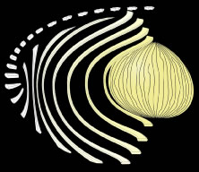
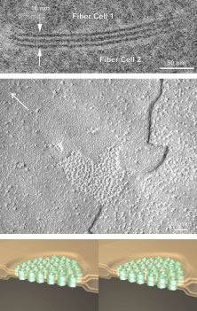
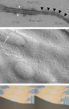
As the cells within the TZ migrate posteriorly, they rotate 90 degrees about their polar axis while elongating bidirectionally. As fiber elongation continues, the anterior ends are insinuated between the lens epithelium and the primary fiber mass while the posterior ends are insinuated between the primary fiber mass and the posterior lens capsule. Elongation is complete when the anterior and posterior ends of newly formed secondary fibers break contact with the apical surfaces of CZ epithelial cells anteriorly and the capsule posteriorly, respectively.The fact that all fibers detach from the basement membrane, the lens capsule, is direct evidence that the lens is a stratified, rather than a simple epithelium because in simple epithelia, all cells permanently retain contact with the basement membrane.

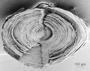
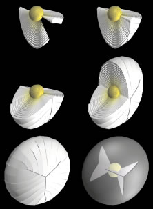
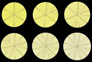
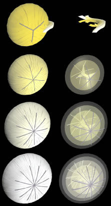
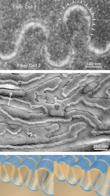
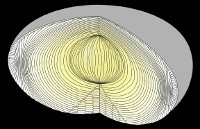
The anterior ends of fully elongated fibers abut and overlap with one another, as do the posterior ends, to form a growth shell. Because secondary fiber formation, or stratification, occurs throughout life, the net result is the establishment of successive growth shells, surrounding the original primary fiber mass. Thus, a view through a lens split along its anteroposterior or visual axis reveals concentric growth shells and/or radial cell columns (Fig. 3). The inside-out development scheme of the lens dictates that growth shells become progressively more internalized and, therefore, fibers cannot be sloughed off. In fact, all primary and secondary fibers formed are retained and must be supported for a lifetime. Failure to preserve the viability of any fiber is presumed to lead to pathology.

Fig. 3. When a forming fetal human lens is separated along the visual axis, it is readily apparent that lens growth (secondary fiber development) results in fetal fibers becoming arranged in age-defined growth shells and radial cell columns. However, although the opposite-end curvature of fetal fibers is less apparent in this view, the arrangement of their fiber ends to form offset continuous suture planes between concentric growth shells is facilitated.

After fiber elongation is complete, terminal differentiation continues as the newly formed fibers routinely eliminate their nuclei, Golgi bodies, rough endoplasmic reticulum, and most smooth endoplasmic reticulum and mitochondria.4–11 The removal of these organelles is often described as necessary because their retention would cause a significant diffraction of light and thereby compromise lens function. However, it should be noted that other stratified epithelia (e.g., skin) also routinely eliminate these same organelles from cells in upper or older layers as a function of terminal differentiation. Fiber maturation continues with the production of specialized cytoplasmic proteins, the crystallins, as well as specialized cytoskeletal and plasma membrane components. As fibers age, the structural and biochemical nature of the previously described fiber characteristics are altered.12

Back to Top
LENS SUTURES
A common misconception is that all fully elongated fibers are meridians, crescent-shaped, fusiform cells that are widest at their midportion and tapered to a point at their ends. A logical extension of this misconception is that the ends of all secondary fibers in any growth shell extend to confluence at the anterior and posterior lens poles. Although such a description of gross fiber shape, length, and end-to-end organization is fairly accurate for some vertebrate lenses, most notably avian, it grossly oversimplifies the intricate variations in fiber shape and length that result in the highly ordered arrangement necessary for a human lens to function properly.

Human fibers are not meridians. They neither extend from pole to pole nor have tapered ends. In fact, human fibers are more than three times as wide, or flared at their ends, than at their midportions. Furthermore, the entire length of most fibers does not lay within a plane coincident with the visual axis as defined by its equatorial location. Instead, most fibers have two types of curvature: a convex-concave curvature that conveys the fiber's crescent appearance and end curvature, the curvature of anterior and posterior end segments of fibers, within any growth shell in opposite directions away from the polar axis. Together, these two curvatures convey on fibers a three-dimensional (3D) S shape (see Fig. 2).

Y SUTURE OF THE FETAL NUCLEUS

The ends of fibers abut and overlap within and between growth shells to form distinct latitudinal arc segments. Individually these arc segments are referred to as suture branches, and collectively they make up defined “suture patterns” within each growth shell. Fiber shape, end taper or flare, and the extent of end curvature combine to determine the number of suture branches and resulting suture patterns (Fig. 4).

Fig. 4. Scanning electron microscopy (SEM) micrograph of a human fetal nucleus dissected out of a 36-year-old adult lens and split along the visual axis to reveal radial cell columns, growth shells, and continuous Y suture planes (asterisks).

Growth shells are composed of two types of secondary fibers: straight and S-shaped fibers. A straight fiber is crescent-shaped, with its entire length lying within a plane passing through the visual axis as defined by its equatorial location (Fig. 5, upper left). However, only one end of a straight fiber extends to a pole. Throughout embryonic development, each growth shell normally contains six straight fibers. In the anatomic position, these straight fibers are normally positioned equidistantly around the lens equator beginning with the most superior equatorial location. Thus, each growth shell is effectively subdivided into sextets.

Fig. 5. 3D-computer assisted drawings (CADs) showing key structural elements in the formation of Y sutures during fetal development. Upper left. After the primary fiber mass (dark gold embryonic nucleus) has been formed, six straight fibers normally positioned equidistantly around the equator, separate growth shells into equal sextants composed of S-shaped fibers (upper right and middle left). The ends of the S-shaped fibers abut and overlap to form suture branches (blue lines) that extend to confluence at the poles (middle right and lower right and left). Note that the length and location of suture branches are defined by the ends of straight fibers. The end result is an upright Y anterior suture (lower right) and an inverted posterior Y suture (partially shown in lower left).

In addition to their crescent shape, S-shaped fibers have anterior and posterior end segments that exhibit precise curvature away from the poles inopposite directions (see Fig. 5, upper right). Neither of the ends of an S fiber extends to a pole nor does its entire length lie within a plane passed through the visual axis as defined by its equatorial location. S fibers are arranged as distinct groups, positioned between straight fibers. Because of opposite-end curvature, within any embryonic growth shell, the anterior and posterior ends of S fibers become aligned as offset anterior and posterior latitudinal arc lengths (see Fig. 5, middle left). The anterior ends of proximal groups of S fibers abut and overlap within and between successive growth shells to produce anterior suture branches (see Fig. 5, middle right). However, although the anterior ends of fibers in proximal groups form anterior suture branches, opposite-end curvature dictates that their posterior ends form precisely offset posterior branches with different groups (see Fig. 5, lower left). The origin of anterior suture branches is defined by the ends of straight fibers that extend to confluence at the posterior pole but not to the anterior pole. Similarly, the origin of posterior suture branches is defined by the ends of straight fibers that extend to confluence at the anterior pole but not to the posterior pole. All suture branches extend to confluence at the poles and combine to form discrete anterior and posterior suture patterns. Throughout development the three anterior suture branches are normally oriented at 120 longitudinal degrees to one another to form a Y suture pattern (see Fig. 5, lower right). It follows then that the three posterior suture branches are also normally oriented at 120 degrees to one another but because of opposite-end curvature are offset 60 degrees to the anterior suture branches to form an inverted Y suture pattern.

As additional growth shells are added throughout fetal development, each growth shell normally consists of radially longer and identically shaped secondary fibers (Fig. 6, upper left). Thus, as the Y suture branches of each growth shell become overlain in successive shells, the anterior and posterior ends of all fibers become arranged as right triangle-shaped suture planes extending from the primary fiber mass to the lens periphery (see Fig. 6, upper right, middle, and lower rows). The three anterior suture planes are normally oriented at 120 degrees to one another as are the three posterior suture planes. However, as a result of opposite-end curvature, all six of the Y suture planes are normally offset by 60 degrees to one another.

Fig. 6. 3D-computer assisted drawings (CADs) showing key structural elements in the production of continuous Y suture planes. As additional secondary fibers (white to yellow fibers) are added throughout fetal development, radial cell columns and concentric growth shells (upper and middle rows) are formed consisting of identically shaped but radially longer fibers. As a result, the ends of all fetal secondary fibers become aligned at suture branches that are overlain in successive growth shells to form continuous triangular suture planes extending from the embryonic nucleus to the lens periphery (lower row).

STAR SUTURES OF THE JUVENILE AND ADULT NUCLEI AND CORTEX

After birth and for the rest of life, primate lenses normally form progressively more complex “star suture” patterns during infancy, adolescence, and adulthood. The evolution of the “simple star suture,” produced during infancy, is depicted in Figure 7. As additional growth shells are added after birth, specific S-shaped fibers are overlain by a pair of new straight fibers. Because the ends of straight fibers delimit suture branches, new groups of S-shaped fibers with less end curvature are defined on either side of the new straight fibers. The ends of these fibers abut and overlap to form two new evolving suture branches that extend to confluence at one of the original three Y suture branches (see Fig. 7, upper middle). At this point S-shaped fibers are arranged in eight unequal groups positioned between eight straight fibers.

Fig. 7. Scale 3D-computer assisted drawings (CADs) showing key structural elements in the production of a primate lens six branch star suture formed from birth through infancy. Upper left. At birth, six straight fibers (gold), normally positioned equidistantly around the equator, separate growth shells into equal groups of S-shaped fibers with ends that abut and overlap to form three Y suture branches (blue) that extend to confluence at the poles. The length and location of suture branches are defined by the ends of straight fibers. Upper middle and right and lower left and middle. In successive growth shells, additional straight fibers positioned nonequidistantly around the equator are added that separate the newly added growth shells into unequal groups of S-shaped fibers with ends that abut and overlap to form additional suture branches that extend to confluence at the original suture branches. Lower right. By the end of the infantile period, 12 straight fibers, positioned equidistantly around the equator, separate growth shells into equal groups of S-shaped fibers with ends that abut and overlap to form a simple star suture. (Adapted from Kuszak JR: The development of lens sutures. Progress in Retinal and Eye Research 1412:580, 1994.)

As more growth shells are added (see Fig. 7, upper right), the new pair of straight fibers are overlain by longer straight fibers, and a second new pair of straight fibers are also added. The S-shaped fiber groups on either side of the original new pair of straight fibers are longer, forming two longer evolving suture branches that extend to confluence farther along an original Y suture branch. At the same time the ends of the second new pair of straight fibers delimit additional new evolving suture branches that extend to confluence at a second of the original three Y suture branches. At this point, the S-shaped fibers are arranged into 10 unequal groups positioned between 10 straight fibers.

As more growth shells are added (see Fig. 7, lower left), the first new pair of straight fibers are overlain by even longer straight fibers, the second new pair of straight fibers are overlain by longer straight fibers, and a third new pair of straight fibers are added. The ends of the S-shaped fibers positioned on either side of the first pair of straight fibers now form two complete new suture branches that extend to confluence at the poles. At the same time, the ends of S-shaped fiber positioned on either side of the second pair of new straight fibers continue forming evolving suture branches that extend to confluence farther along the second of the original three Y suture branches. The ends of the third new pair of straight fibers delimit additional new evolving suture branches that extend to confluence at the third of the original three Y suture branches. At this point, S-shaped fibers are arranged into 12 unequal groups positioned between 12 straight fibers.

As more growth shells are added (see Fig. 7, lower middle), the second new pair of straight fibers are overlain by longer straight fibers, and the third new pair of straight fibers are overlain by longer straight fibers. The groups of S-shaped fibers positioned on either side of the second new pair of straight fibers, now form two additional new suture branches that extend to confluence at the poles. At the same time, the ends of the third pair of new straight fibers form longer evolving suture branches that extend to confluence farther along the third of the original three Y suture branches. As the final infantile growth shells are added (see Fig. 7, lower right), all three new pairs of straight fibers continue to be overlain by straight fibers. All the groups of S-shaped fibers positioned on either side of all the new pairs of straight fibers form a symmetrical, six-branch simple star anterior and posterior suture that are offset by 30 degrees.

As a result of forming a simple star suture over the period of infantile development, radial cell columns now contain fibers that have neither identical shape nor lengths that are a simple radial function (Fig. 8, first row). The ends of fibers, and by extrapolation suture branches, are out of register in successive growth shells. Thus, discontinuous suture planes are formed from the fetal nucleus to the lens periphery (see Fig. 8, second row).

Fig. 8. Scale 3D-computer assisted drawings (CADs) showing key structural elements in the production of discontinuous suture planes in human lenses. Upper row. From birth through infancy secondary fibers in radial cell columns (the juvenile nucleus) are neither of uniform shape nor simply longer as a function of radial location. Upper middle row. Consequently, the 12 suture branches constituting the simple star suture of the juvenile nucleus, form “discontinuous” suture planes throughout the juvenile nucleus. Lower middle row. Similarly, the 18 suture branches constituting the star suture of the adult nucleus continue to form discontinuous suture planes throughout the adult nucleus to the lens periphery, and, finally, (bottom row) the 24 suture branches constituting the complex star suture of the cortex continue to form discontinuous suture planes extending from the adult nucleus to the lens periphery.

Throughout adolescence and adulthood, star (see Fig. 8, third row and Fig. 9) and complex star sutures (see Fig. 8, fourth row), respectively, the third and fourth generations of sutures in primate lenses, are formed in a similar manner. The fully formed anterior and posterior star sutures, offset by 20 degrees because of opposite-end curvature, are normally characterized by 18 groups of S-shaped fibers, with even less opposite-end curvature, positioned on either side of 18 straight fibers. Finally, the fully formed anterior and posterior complex star sutures, only offset by 15 degrees because of the least amount of opposite-end curvature, are normally characterized by 24 groups of S-shaped fibers positioned on either side of 24 straight fibers.

Fig. 9. Low magnification scanning electron microscopy montage of a young adult human lens star suture (see Fig. 8).

ZONES OF DISCONTINUITY

The zones of discontinuity, seen in human lenses with transillumination slit-lamp biomicroscopy, contain the four progressively more complex generations of sutures. Thus, abnormal slit-lamp profiles of cataractous lenses (e.g., diabetic and cortical) contain a record of abnormal suture development.13 Slit-lamp biomicroscopy also reveals a thin equatorial band, or region of minimal light scatter, commonly referred to as the central sulcus. The thickness of the central sulcus is the thickness of the embryonic nucleus. Thus, it only includes embryonic nuclear fibers and portions of the midsegments of fetal, infantile, juvenile, and adult nuclear fibers, all aligned parallel to the anteroposterior axis. As aresult, the central sulcus is characterized by a complete lack of sutures within its boundaries.

SUTURES AND LENS OPTICAL QUALITY

The functional significance of increasingly complex suture patterns formed throughout life is as follows: As light rays pass through uniformly shaped fibers arranged in radial cell columns, they repeatedly encounter fiber membrane, cytoplasm filled with crystallin proteins, fiber membrane, extracellular space, and so forth in relatively constant increments. Cumulatively, the lens is transformed into a series of coaxial refractive surfaces or into a structure that minimizes light diffraction.14 This theory of lens function has been proven to be accurate by correlating lens sharpness of focus, ascertained by low power helium neon laser scan analysis, with lens structure, as assessed by light (LM) and electron microscopic (EM) analysis.15–18 However, suture branches are formed by the overlap of irregularly shaped fiber ends. In the Y suture, formed before birth, branches are overlain in successive growth shells to form continuous, irregular suture planes extending from the embryonic nucleus to the lens periphery. Thus, light rays passing through these planes would repeatedly encounter fiber membrane, cytoplasm with crystallins, fiber membrane, extracellular space, and so forth in increments that are not constant, causing a reduction in lens sharpness of focus. However, as lenses grow and age, branches are not overlain in successive growth shells, and, thus, they form discontinuous suture planes extending from the fetal nucleus to the lens periphery. Then as light rays pass through the lens, they encounter fewer instances of constant increments of fiber membrane, cytoplasm with crystallins, fiber membrane, extracellular space, and so forth characteristic of nonprimate lens suture planes. This explains in part why primate lenses are optically superior to nonprimate lenses.

SUTURES AND LENS PHYSIOLOGY

Lenses lack a blood supply. Nourishment derived from the aqueous and vitreous humors enters the lens mass through simple diffusion. However, the effective depth for nutrient diffusion is restricted to the outer 10% of the lens radius.19 Thus, only the lens epithelium and the peripheral superficial cortical fibers, which have full complements of organelles, have effective access to critical nutrients. In contrast, the cortical and nuclear fibers, which lack organelles, have limited access to nutrients, and this nutrient delivery problem is amplified as the lens mass grows throughout life. Lens sutures may play an important role in supporting an internal circulatory system in the lens.

It has been proposed that current circulates around and through the lens, inward at the poles and outward at the equator.20 The inward movement of fluid is along the polar intercellular clefts of fibers at the polar caps, or by definition at the sutures, convecting glucose to the innermost fibers where it used for anaerobic metabolism. The outward movement of fluid is described as intracellular, convecting waste products of metabolism out of the lens at the equator presumably through gap junctions. If fiber end segments, or sutures, are characterized by pumps, transporters, and channels as is typical of the apicolateral and basolateral membrane of other epithelia, then diffusional transport throughout a lens may be enhanced at suture branches. This leads to speculation that the level of sutural complexity is a factor in determining how efficiently fluids can move into lenses. Primate lenses with more extensive suture patterns (e.g., star sutures) would be more efficient at fluid transport than would nonprimate lenses with less extensive suture patterns (e.g., umbilical, line, and Y sutures). This may also explain, in part, why primates develop progressively more complex suture patterns as a function of age.

Numerous gap junctions connecting fiber midportions, or those segments of fibers not involved in sutures, are proposed to be responsible for outward movement of fluid (fiber ultrastructure is shown in greater detail in a subsequent section). However, the density of fiber gap junctions varies considerably between species.21–23 The midportions of chick, rat, frog, and human fibers have 65%, 33%, 12% and 5%, respectively, of their membrane specialized as gap junction. This implies that fiber gap junction density will also be a factor in determining how efficiently fluids can move out of lenses in different species and/or as a function of age. In relation to other vertebrate lenses, primate lenses have the most complex suture patterns and a low density of fiber gap junctions. In contrast, avian lenses have the simplest suture patterns and a high density of gap junctions. It remains to be determined how and/or if these two structural parameters are balanced to insure optimal efficiency of an internal fluid circulation network in different lens types.

Back to Top
LENS ANATOMY

LENS AND FIBER SHAPE

The anterior surface of a normal adult human lens is the equivalent of one half of a 15-degree spheroid, whereas its posterior surface is the equivalent of one half of a 30-degree spheroid (Fig. 10). Lens thickness is the measure of a lens from anterior to posterior poles along the visual axis, whereas lens width is the span of its equator perpendicular to the visual axis. Most fibers are hexagonal in cross section with two broad and four narrow faces (see Fig. 2). Fibers are arranged in growth shells, and/or radial cell columns, with their broad faces oriented parallel to the lens surface (see Fig. 3). The measure between broad faces constitutes fiber thickness, whereas the perpendicular distance defined by the furthest extent of the paired narrow faces constitutes fiber width. At the equator fiber thickness is approximately 1.0 to 1.5 μm, whereas fiber width measures on average 10.0 to 12.0 μm.

Fig. 10. A scale schematic diagram showing the relative sizes of the cortex and nucleus in a newborn (upper) and middle-aged (lower) human lens. Note the following: The thickness of the peripheral cortex is essentially constant throughout life. However, because of continuous lens growth, the size of the lens nucleus increases as a function of development and age.

LENS CORTEX AND NUCLEUS

A cortex can be defined as the outer part or external layers of an internal organ, and a nucleus is defined as anything serving as the center of development or growth. Given the inside-out development and growth scheme of the lens, it is entirely appropriate to consider any vertebrate lens to consist of a cortex and a nucleus, each comprised of cortical and nuclear fibers, respectively (see Fig. 10). In any lens, by definition the primary fibers are the most internal fibers and are, thus, referred to as embryonic nuclear fibers. Similarly, the initial growth shells of secondary fibers comprise the initial external layers of the lens and are, thus, the original cortical fibers. However, because the lens grows throughout life, most secondary fibers become nuclear fibers on completion of the fiber maturation process. The types of nuclear fibers that secondary fibers become are determined by their time of origin (period of development, growth, or age).

The embryonic nucleus is comprised solely of primary fibers. Primary fibers do not form sutures. Their long axis is oriented essentially parallel to the anteroposterior axis. Their average cross-sectional area is large with a high standard deviation. Thus, within any one equatorial cross section, embryonic nuclear fibers with large cross-sectional areas are seen adjacent to others with very small cross-sectional area.24,25 As a result, the embryonic nucleus is the only lens region where fibers are not organized into defined growth shells or radial cell columns.

The fetal nucleus is comprised of all the secondary fibers formed until birth. Fetal nuclear fibers have more uniform cross-sectional measures and shape and have on average smaller cross sections than primary fibers.24 The organization of fetal nuclear fibers into defined radial cell columns evolvesover the period of fetal development. The initial shells of fetal nuclear fibers are characterized by irregularly shaped polygonal fibers (e.g., rhombic, pentagonal, hexagonal, etc.). As additional layers are formed, new fetal nuclear fibers are more uniform in shape (hexagonal), and, thus, they become arranged in radial cell columns. Because fetal nuclear fibers are secondary fibers, they are either straight or S-shaped, and their ends abut and overlap to form sutures. Therefore, the beginning of suture formation signals the beginning of the fetal nucleus.

The fibers formed after birth and through sexual maturation are juvenile nuclear fibers.24 Because the length of time to complete the sexual maturation process is not constant, juvenile nuclear dimensions have broader boundaries compared with that of the fetal and embryonic nuclei. Juvenile nuclear fibers are consistently hexagonal with minimal cross-sectional size variation, as compared with fetal nuclear fibers. Juvenile nuclear fibers terminate in suture patterns that transition from three to six main branches (see Fig. 7) through infancy and from six to nine branches through adolescence (see Figs. 8 and 9).

The adult lens nucleus is comprised of all the secondary fibers formed after sexual maturation minus the fibers of the cortex. All elongating fibers are superficial cortical fibers and all fully elongated fibers in the process of eliminating their organelles are intermediate cortical fibers. After eliminating their organelles, intermediate cortical fibers are transformed into deep cortical fibers as they alter their cytoplasm and plasma membrane.

The boundary between the human cortex and nucleus can be visualized by both slit-lamp and darkfield biomicroscopy. When using white transmitted light, it is apparent that the human adult lens nucleus has a uniform yellow coloration clearly distinct from the colorless cortex24 (Fig. 11). Based on the diameter of the chromatic region, the average thickness of the cortex in adult human lenses (age range 49 to 73 years) is 1.13 mm plus or minus 0.15 mm (n = 10; unpublished data). The thickness of the lens cortex does not appear to change with age.

Fig. 11. A normal adult human lens as seen through a surgical microscope. Note that although the entire lens is transparent, the peripheral cortex (approximately 1 mm thick) is readily distinguishable from the central nucleus by its lack of yellow coloration.

LENS CELL HOMEOSTASIS

The size of any tissue cell population is determined by both the rate at which new cells are added (the cell birth rate, KB) and the rate at which cells are lost (the cell loss rate, KL).26 Although KB can be readily assessed in most tissues by such methods as metaphase blocking,27 KL is more difficult to measure because of exfoliation, cell death, and migration. In simple terms, if KB is greater than KL, then the tissue is growing; if KB is less than KL, then the tissue is regressing; and finally, if KB = KL, then the tissue is in kinetic equilibrium.

Throughout life the cumulative fiber total increases without a concomitant loss of the previously formed fibers. Thus, as regards the fiber mass, the lens grows throughout life because KB is greater than KL. From fetal development, and continuing throughout life, the lens produces more cortical fibers that eventually become nuclear fibers. In addition, because the rate of adult lens growth is very slow, the nucleus of any lens comprises a much larger proportion of the lens mass than the cortex (see Fig. 10). By relating lens and fiber dimensions, reasonable estimates of the number of fibers can be calculated as a function of age. The data shown in Table 1 clearly show that lens growth continues throughout life.


Table 1. Approximate Number of Cells in Different Regions of a 60-Year-Old Human Lens and the Average Regional Growth Rate Per Year*


RegionCell Dimensions (μm)Number of CellsGrowth Rate† (cells/year)
Cortex2.24 × 14‡665,000133,000
Adult nucleus0.75 × 7.5‡4,460,000101,000
Juvenile nucleus1.7 × 9.0‡640,00053,000
Fetal nucleus5.0§700,0001,400,000
Embryonic nucleus10.0§8003,500

*Measurements of fiber cross-sectional size were taken from thick sections through the equatorial plane.
†The number of years required to form each region corresponds to accepted periods of development (primary and secondary fiber formation during fetal development), growth (infancy and childhood), and aging (beyond puberty).
‡Thickness × width of an average fiber.
§Diameter of an average equivalent circular cell perimeter.
(Adapted from Taylor VI, Al-Ghoul KJ, Lane CW et al: Morphology of the normal human lens. Invest Ophthalmol Vis Sci 37:1396, 1996.)


LENS EPITHELIAL CELL HOMEOSTASIS

Anterior surface area increases as a result of lifelong lens growth in primate lenses. The size of the GZ and CZ, as well as epithelial cell size as a function of age, are shown in Table 2. At birth, GZ and CZ cells are essentially identical in size. As the lens grows and ages, epithelial cell size varies as a function of zonal location.3,28,29 From birth to adulthood, the CZ area increases on average by 10%, whereas from adulthood to old age it increases by 30%. In contrast, GZ area increases by 80% from birth to adult and by 50 % from adult to old age. Consequently, GZ KB is greater than GZ KL, and, therefore, the GZ is a growing cell population throughout life. GZ growth is due to the addition of daughter cells at the periphery that did not enter into terminal differentiation to become fibers.2


Table 2. Average Central Zone (CZ) and Germinative Zone (GZ) Lens Epithelial Cell Size and Number As a Function of Development, Growth, and Age in an Aged (30 year old) Primate Lens


 At BirthAdultAged
Total anterior surface area31.65 × 106 μm257.15 × 106 μm281.76 × 106 μm2
CZ surface area25.32 × 106 μm245.72 × 106 μm265.41 × 106 μm2
GZ surface area3.165 × 106 μm25.715 × 106 μm28.176 × 106 μm2
Average CZ epithelial cell size77.43 μm292.21 μm2193.97 μm2
Average GZ epithelial cell size77.43 μm283.76 μm99.58 μm2
CZ epithelial cell number287,701435,390290,696
GZ epithelial cell number121,718203,316254,744
Total epithelial cell number408,799638,069545,440


The average age-related increase in CZ cell size is more pronounced than GZ cell size. By adulthood, the average CZ cell has increased in surface area by 20%. However, by old age, the average percent size increase is 150%. Consequently, although CZ KB is greater than CZ KL from birth through adulthood, it is less from adulthood through old age. Therefore, although the CZ is a growing cell population throughout adulthood, it is a regressing cell population for the rest of life. Failure to consider the zonal variation in lens epithelial cell density leads to a gross overestimation of total cell number.30

LENS EPITHELIAL CELL APOPTOSIS

The lens epithelium eliminates some cells by apoptosis31 throughout life. Gorthy and Anderson32 have shown that as CZ cells increase in size, there is a concomitant increase in the number of their intracellular lysosomal bodies as a function of age. These increasingly large and prominent lysosomal bodies likely represent breakdown of apoptotic cells and fragments. Indeed, macrophages could not play such a role in the lens because it is completely enclosed by the capsule.

Back to Top
LENS ULTRASTRUCTURE

LENS EPITHELIAL CELLS

The lens epithelium is composed of low cuboidal cells (Fig. 12). These cells have large indented nuclei with two nucleoli and numerous pores. As the lens ages, these cells become more flattened although they cannot be accurately described as squamous. There are nominal numbers of ribosomes and polysomes, smooth and rough endoplasmic reticulum, and Golgi bodies. The mitochondria of these cells are small with irregular cristae. In addition, lysosomes, dense bodies, and glycogen particles have been noted.

Fig. 12. Low-magnification transmission electron micrograph of central zone adult human lens epithelial cells. The complex lateral membranes of these cells is apparent.

Cytoskeletal elements found in lens epithelial cells include actin, intermediate filaments (vimentin); microtubules; and the proteins spectrin, actinin, and myosin. As in all eukaryotic cells, it is presumed that these elements are interconnected to produce a well-defined cytoskeleton or microtrabecular latticework that compartmentalizes the components of the cell's interior. Lens epithelial cells are remarkable in that they have a prominent well-characterized cytoskeletal network consisting of actin filaments and myosin in the form of polygonal arrays or “geodomes” located subjacent and attached to their apical membrane. All the cytoskeletal components become more dense as the cells age.

The polarized lens epithelial cells have apical, lateral, and basal membranes. The smooth basal membranes underlie and produce the lens capsule, a replicated basal lamina. The lateral membranes of lens epithelial cells are markedly enfolded. Na,K-adenosine triphosphatase (ATPase) and acid phosphatase are localized on the apicolateral membrane. Acid phosphatase, involved in cellular breakdown and removal, increases with age. The lateral membranes also feature a small number of gap junctions composed of connexin 43. Desmosomes also exist between lateral membranes of lens epithelial cells. An uncommon feature of lens epithelial cell apicolateral membrane is that it lacks effective tight or occluding junctions. Simple interlaced linear arrays of intramembrane particles are present between some lens epithelial cells at the apicolateral border. However, physiologically they present no significant barrier to extracellular flow and are, thus, considered to be very leaky tight junctions.33,34

The apical membrane of lens epithelial cells is planar and interfaces with the apical membranes of elongating fiber cells as they migrate to their sutural destinations. This epithelial-fiber interface (EFI) is characterized by transcytotic events. Numerous micropinocytotic and clathrin-coated vesicles can be found at and immediately subjacent to the EFI. Thus, nutrients, ions, essential metabolites, and presumably other receptor-mediated substances can be exchanged across the EFI via transcytosis. Both gap junctions and square array membrane, areas of membrane characterized by groups of orthogonally arranged aquaporins, are extremely rare at the EFI.

THE EFI AND LENS PHYSIOLOGY

Despite the results of numerous structural35–37 and physiologic (electrotonic and dye coupling38–42) studies confirming that there is limited communication across the EFI, it can still be reasonably argued that cell-cell coupling across this interface is important to lens physiology.43 Epithelial-epithelial and fiber-fiber coupling have been shown by correlative structural and functional studies to be essentially 1:1. Therefore, every epithelial cell does not need to be conjoined directly to an underlying elongating fiber. Structural analysis shows that the elongating or superficial cortical fiber that breaks contact with either the CZ epithelium at the anterior pole or the posterior capsule at the posterior pole is approximately 200 μm deep at the equator.4,5,44,45 The equatorial diameter of a human lens at birth is approximately 6 mm, and this dimension increases to more than 9 mm by adulthood. Thus, the percent of fibers in direct contact with the epithelium at the EFI will vary as a function of size and/or age. Estimates of the percent of fibers in direct contact with the epithelium at the EFI can be derived from standard mensuration formulas for spheroidal geometry.46 When calculating such estimates, it is important to consider that lenses are spheroids and not spheres. Failure to accurately factor in lens shape correctly results in a gross overestimation of lens volume.

The shape and relevant axial parameters of newborn and adult human lenses used to estimate the percent of fibers in direct contact with the epithelium at the EFI are shown in Figures 10 and 11. Considering that the lens is an asymmetrical oblate spheroid, an estimate of its total volume can be calculated as the sum of the anterior lens volume, defined as (4/3 π a12 b1)/2, and the posterior lens volume, defined as (4/3 π a1 b22)/2, where a1 is the major equatorial radius, b1 is the minor anterior radius, and b2 is the minor posterior radius. The same formulas can be used to estimate the percent of fibers in direct contact with the epithelium at the EFI. However, in this case, a1 is replaced by a2, the major equatorial radius minus the 200 μm contribution of the elongating fibers, the only fibers in direct contact with the epithelium anteriorly and with the capsule posteriorly. Because the size of the elongating fiber zone is consistent throughout life, then at birth slightly more fibers (13%) are in direct contact with epithelial cells at the EFI then in adulthood (9%).

FIBERS

Because every fiber is maintained for a lifetime, the lens presents an ideal model for studies of cellular senescence. In normal transparent lenses, the cytoplasm of fibers in all the developmental regions is smooth and homogeneous. This fact can be confirmed by Fourier transform analysis.47 The principal components of fiber cytoplasm are the specialized lens crystallin proteins and the lens cytoskeleton. The crystallins provide a medium of high refractive index. With increased age, a variable concentration of the different types of crystallins is related to the higher water content in the lens cortex compared with the nucleus. The breakdown of crystallins, and their subsequent aggregation or cross-linking, is believed to be responsible for some opacities. The fiber cytoskeleton contains all the elements of the lens epithelial cells, as well as beaded filaments. It is presumed that these components are collectively arranged into a supportive network that pervades the cell to organize the lens crystallins and maintain fiber shape as a function of age and accommodation. With increased age, most formed cytoskeletal elements are modified and are not found in the lens nucleus.48

Differentiating fibers are separated by a well-defined extracellular space except at intercellular junctions. As a result of terminal differentiation, the extracellular space between mature fibers is minimized as fiber plasma membranes are transformed into a complex topology involving different intercellular junctional contacts. To facilitate an understanding of the structural differences between fiber intercellular junctions, we present representative thin-section and freeze-etch replica electron microscopic views followed by interpretative 3D-computer assisted drawings (CADs).

A representative fiber gap junction as seen by thin-section electron microscopy is shown in Figure 13, upper. This lens fiber gap junction is typically 16 nm thick and is formed by transmembrane proteins from the connexon family, connexins (Cx46 and Cx50). These proteins, arranged in apposed regions from neighboring fibers, are conjoined across a narrowed extracellular space, or gap, measuring 1 to 2 nm. Freeze-etch analysis reveals fiber gap junctions to be composed of complementary aggregates of loosely packed connexons (see Fig. 13, middle). As described previously, fiber gap junctions occur primarily between the midsegments of fibers, and their density varies considerably between species.21–23 A 3D-CAD drawing of a fiber gap junction is shown in Figure 13 lower. In this view, it is apparent that each connexon is a protein channel connected to a matched connexon across conjoined cells. Fiber gap junction pore size allows passage of 1000 to 1500 dalton (Da) molecules. Thus, charge- and size-restricted substances that can pass freely from fiber to fiber without an energy requirement. As described earlier, this is a major consideration for lens physiology because most fibers lack organelles and are far removed from their source of nutrition, the aqueous and vitreous humors. Fiber gap junctions differ from typical gap junctions (e.g., gap junctions between hepatocytes or even between lens epithelial cells49) in that their amino acid sequence, their ultrastructure, and their gating and permeation properties are not identical. Tissue specific differences between gap junctions reflect the terminal differentiation process rather than a major physiologic distinction.

Fig. 13. Complementary views of a fiber gap junction as seen by (upper) thin-section electron microscopy, (middle) freeze-etch electron microscopy (arrow in the upper left indicates direction of shadow), and (lower) a 3D-computer assisted drawings (CAD) stereopair based on the previous images and other analytical techniques. By thin-section analysis it is apparent that two apposite segments of plasma membrane from neighboring fibers are conjoined across a narrowed extracellular space or gap. By freeze-etch analysis it is further apparent that two apposing regions of plasma membrane from neighboring fibers are composed of complementary aggregates of transmembrane proteins, or connexons, conjoined across a narrowed extracellular gap. Finally, 3D-CADs show that each hexameric connexon is a channel protein, and, thus, conjoined connexons from neighboring fibers permit size and charge related transport of substances between fibers without an energy requirement.

A representative low-amplitude thin asymmetric junction (TAJ50) as seen by thin-section electron microscopy is shown in Figure 14, upper. Although this junction has a pentilaminar profile, similar in appearance to fiber gap junctions, it is typically only 11 nm thick and is formed by transmembrane proteins from the aquaporin (AQP0) family, closely apposed to lipid rich region from a neighboring fiber without any intervening extracellular space. Freeze-etch analysis reveals fiber TAJs to be composed of aggregates of orthogonally packed AQP0s (see Fig. 14 middle). These proteins were originally and are still often referred to as major intrinsic membrane protein 26K (MIP26). In these thin-section and freeze-etch micrographs, the TAJs occur on low-amplitude undulating membrane. 3D-CADs of fiber TAJs are shown in Figure 14 lower. Fiber TAJs are likely to serve one of two functions as a consequence of their arrangement between fibers. The AQP0s can serve as single membrane water transport channels that remove water from the extracellular space.50,51 In addition, the close association of apposed undulating membrane in the TAJs subserves an adhesive function.50,52,54

Fig. 14. Complementary views of low-amplitude thin asymmetric junctions (TAJs) as seen by (upper) thin-section electron microscopy, (middle) freeze-etch electron microscopy (arrow in the upper left indicates direction of shadow), and (lower) 3D-computer assisted design (CAD) stereopairs based on the previous images and other analytical techniques. By thin-section analysis it is apparent that two apposite segments of TAJ membranes from neighboring fibers are also conjoined across an even narrower extracellular space than fiber gap junctions. By freeze-etch analysis it is further apparent that two apposing TAJ membranes (arrowheads) from neighboring fibers are composed of “noncomplementary” aggregates of transmembrane proteins, or AQP0, offset across an essentially eliminated extracellular space (arrows delimit a fiber gap junction). Finally, this 3D-CAD stereopair shows that each AQP0, is a tetrameric water transport particle, and, thus, water can flow in and out of the extracellular space in response to local electromotive and osmotic conditions.

Representative high-amplitude TAJs as seen by thin-section microscopy is shown in Figure 15 upper. These TAJs are also typically 11 nm thick and have a pentilaminar profile. However, the lipid rich membrane from one of the apposed fibers is often not preserved, leaving a trilaminar profile of AQP0s (see Fig. 15, upper, arrows). These fiber junctions were originally termed square array junctions.50 Freeze-etch analysis reveals these junctions are also composed of aggregates of orthogonally packed AQP0s, but in this case they exist on high-amplitude undulating membranes (see Fig. 15, middle). 3D-CADs of high-amplitude TAJs are shown in Figure 15, lower. In this view, it is apparent that the aggregates of AQP0s only exist in one of the paired membranes. This arrangement of AQP0s further suggests that these junctions subserve an adhesive role.

Fig. 15. Complementary views of high-amplitude thin asymmetric junctions (TAJs) as seen by (upper) thin-section electron microscopy, (middle) freeze-etch electron microscopy (arrow in the upper left indicates direction of shadow), and (lower) 3D-computer assisted design (CAD) stereopairs based on the previous images and other analytical techniques. By thin-section analysis it is apparent that apposing segments of human nuclear fibers have been transformed into furrowed membrane domains (see also Fig. 16). By freeze-etch analysis it is further apparent that furrows from neighboring nuclear fibers are characterized by noncomplementary and offset aggregates of transmembrane proteins, or AQP0. Finally, this 3D-CAD stereopair shows that although each AQP0 is a water transport particle in nuclear fibers, the staggered aggregates of AQP0s may also serve an adhesive function. Although this schematic represents AQP0s as tetramers forming a central channel, in fact each of the tetrameric subunits contains a patent water channel.63 (Middle adapted from Kuszak JR, Brown HG: Embryology and anatomy of the lens. In Albert DM, Jacobiec FA (eds): Principles and practice of ophthalmology: Basic sciences. Philadelphia, WB Saunders, 1994, 94.)

Although scanning electron microscopy does not provide sufficient resolution to resolve the previously described junctional complexes, it does provide a superior overview of the density of the different gross fiber membrane configurations (flat, low- and high-amplitude fiber membranes) characterized by fiber gap junctions and TAJs, respectively. In all lenses, after the elongation and terminal differentiation processes are completed, the smooth lateral membranes of fibers are radically altered. Scanning electron microscopy (SEM) reveals that mature and aged fiber membranes become characterized by numerous polygonal domains of furrowed membrane (Fig. 16). These domains are the high-amplitude undulating membranes as seen in thin-section and freeze-etch electron microscopy.53,54 The fact that the character of fiber membrane varies as a function of maturity and age has led to speculation that this is the result of the AQP0 channels being redistributed within the plane of the membrane. The functional significance of such a dramatic remodeling of fiber membrane is unknown.

Fig. 16. Scanning electron micrographs showing the surface morphology of (left) cortical and (right) nuclear fibers. Cortical fibers are characterized by complex lateral interdigitations arising at the angles formed by the six lateral membrane faces and arrayed along a fiber's length. In addition, small ball and socket-like interdigitations are randomly found on and within the lateral faces. In contrast, nuclear fiber lateral faces are transformed into numerous furrowed membrane domains by the reorganization of aquaporin 0s.

LENS FIBER CELL-TO-CELL FUSION

Closely apposed regions of lateral membrane from neighboring fibers are frequently fused for a variable distance along the fiber length.25,55–57 Fusion zones are frequently seen to occur between the anterior segments or posterior segments of fiber cells as they approach their sutural locations. The frequency of cell-to-cell fusion zones near sutures is interpreted as a means for fibers to change their direction or curvature to conform to the precise modeling of sutures required for proper lens function. Cell-to-cell fusion zones also provide large patent pathways for intercellular transport between fibers for substances too large to pass through fiber gap junctions.

AGING NUCLEAR FIBERS

As fibers age, cytoskeletal changes include the loss of selected cytoskeletal elements48,58 and increased condensation through the modification and crosslinking of crystallins and through the loss of water.59,60 These processes result in nuclear fiber compaction that is most dramatic in primate lenses causing a decrease in fiber cross-sectional area by a factor of three.24 Because the loss of water occurs without a concomitant elimination of membrane surface area, an increased complexity of fiber surface membrane signals the transformation of a deep cortical fiber into a nuclear fiber.24 A redistribution of the integral membrane proteins of nuclear fibers creates regions rich in AQP0 in the form of high-amplitude TAJs.50,53,54 As a consequence of all of the previously mentioned factors, the nucleus is a hardened region as compared with the cortex.

AGE-RELATED FIBER COMPACTION

Because the lens grows throughout life, it is necessary to compact the fiber mass so that the lens does not outgrow its place within the eye. Compaction, although not evident in young humans or animals, is particularly evident in aging humans.61

The age-related changes in fiber compaction within the different developmental regions are best appreciated by relating equatorial thin section (in which all fibers are cut in cross section at their midpoints) to SEM analysis of lenses split along their anteroposterior axis to reveal the end-to-end arrangement of nuclear fibers in growth shells and radial cell columns. In thick and thin sections, taken through the lens equatorial plane, the reduction in fiber thickness as a result of fiber compaction is most evident at the transition from cortex to adult nucleus.24 However, fiber compaction occurs in the juvenile and fetal nuclei, although to a lesser extent in the fetal nucleus.62 Comparable thick and thin sections through the embryonic nucleus also suggest that age-related compaction in the oldest region of lenses appears to be insignificant.

However, significant age-related compaction in the fetal and embryonic nucleus is readily apparent by SEM analysis.61 Young human lenses have a thicker embryonic nucleus and less acute fetal nuclear fiber angle (more spherical shape) than older lenses. In fact, fibers from older lenses have accordion-like folds that decrease the overall thickness of the embryonic nucleus along the optic axis. SEM also reveals the complex pattern of furrowed membranes that correspond exactly to the undulating membranes visible in transmission electron microscopy (TEM) thin sections or freeze-etch replicas (see Figs. 15 and 16). Finally, the knobs that appear on the fibers in the SEM images correspond to the collection of edge processes in TEM images that are exposed when adjacent fibers are split apart during the preparation of SEM specimens. An important conclusion is that a full appreciation of the complex topology of the fibers is only possible when both SEM and TEM techniques are employed.

Back to Top
SUMMARY
In summary, the lens is a prime example of how an organ modifies its structure through terminal differentiation to accomplish its function. Lenses produce fibers of defined shape and size and arrange these cells into radial cell columns and growth shells throughout a lifetime. It uses an abundance of specialized cytoplasmic crystallin proteins, cytoskeletal elements, intercellular contacts (gap junctions and square array membrane), and cell-to-cell fusion to produce a structure that will remain viable throughout decades. The crystallin proteins provide a continuous gradient of refraction between fibers. The extracellular space, a likely source of diffraction, is essentially eliminated by conjoining fibers via low- and high-amplitude TAJs and gap junctions. In addition, gap junctions and fusion zones provide intercellular pathways between the fibers that become positioned farther away from their source of nutrition with the addition of each growth shell. But ultimately, age-related changes in lens crystallins, cytoskeleton, and membrane render the lens incapable of preserving its necessary structure-function regimen indefinitely.
Back to Top
ACKNOWLEDGMENT
The authors gratefully acknowledge the contributions of H. Brown, M.D., Ph.D., K.J. Al-Ghoul, Ph.D., R.K. Zoltoski, Ph.D., L. Novak, K.L. Peterson, A.J. Kuszak, R. Nordgren, J. Chrisman, C.F. Freel, K.O. Gilliland, C.W. Lane, and H. Mekeel. JRK has been supported by NIH NEI grant EY06642, the Regenstein Foundation, the Louise C. Norton Trust and the Alcon Research Institute. MJC has been supported by NIH NEI grants EY08148 and EY05722. Human lenses were obtained from the NDRI and the North Carolina Eye Bank. Primate lenses were obtained from the Regional Primate Center in Seattle, Washington, and the Yerkes Primate Center in Atlanta, Georgia.
Back to Top
REFERENCES

1. Harding CV, Reddan JR, Unakar NJ et al: The control of cell division in the ocular lens. Int Rev Cytol 31:215, 1971

2. Rafferty NS, Rafferty KA Jr: Cell population kinetics of the mouse lens epithelium. J Cell Physiol 107:309, 1981

3. Kuszak JR: A re-examination of primate lens epithelial cell size, density and structure as a function of development, growth and age. Nova Acta Leopoldina NF 75:45, 1997

4. Bassnett S, Beebe DC: Coincident loss of mitochondria and nuclei during lens fiber cell differentiation. Dev Dyn 194:85, 1992

5. Bassnett S: Mitochondrial dynamics in differentiating fiber cells of the mammalian lens. Curr Eye Res 11:1227, 1992

6. Bassnett S: The fate of the Golgi apparatus and the endoplasmic reticulum during lens fiber cell differentiation. Invest Ophthalmol Vis Sci 36:1793, 1995

7. Bassnett S: Chromatin degradation in differentiating fiber cells of the eye lens. J Cell Biol 137:37, 1997

8. Modak SP, Morris G, Yamada T: DNA synthesis and mitotic activity during early development of chick lens. Develop Biol 17:544, 1968

9. Modak SP, Bollum FJ: Detection and measurement of single-strand breaks in nuclear DNA in fixed lens sections. Exp Cell Res 75:544, 1972

10. Papaconstantinou J: Molecular aspects of lens cell differentiation. Science 156:338, 1967

11. Kuwabara T, Imaizumi M: Denucleation process of the lens. Invest Ophthalmol 13:973, 1974

12. Garland DL, Duglas-Tabor Y, Jimenez-Asensio J et al: The nucleus of the human lens: Demonstration of a highly characteristic protein pattern by two-dimensional electrophoresis and introduction of a new method of lens dissection. Exp Eye Res 62:285, 1996

13. Kuszak JR, Al-Ghoul KJ, Costello MJ: Pathology of age-related human cataracts. In Tasman W, Jaeger EA (eds): Duane's Clinical Ophthalmology. Philadelphia, Lippincott Williams & Wilkins, 1998, pp 1–14

14. Trokel S: The physical basis for transparency of the crystalline lens. Invest Ophthalmol 1:493, 1962

15. Kuszak JR, Sivak JG, Weerheim JA: Lens optical quality is a direct function of lens sutural architecture. Invest Ophthalmol Vis Sci 32:2119, 1991

16. Sivak JG, Herbert KL, Peterson KL et al: The inter-relationship of lens anatomy and optical quality. I. Non-Primate lenses. Exp Eye Res 59:505, 1994

17. Priolo S, Sivak JG, Kuszak JR: Effect of age on the morphology and optical quality of the avian crystalline lens. Exp Eye Res 69:629, 1999

18. Priolo S, Sivak JG, Kuszak JR et al: Effect of experimentally induced ametropia on the morphology and optical quality of the avian crystalline lens. Invest Ophthalmol Vis Sci 41:3516, 2000

19. Fischbarg J, Diecke FP, Kuang K et al: Transport of fluid by lens epithelium. Am J Physiol 276:C548, 1999

20. Mathias RT, Rae JL, Baldo GJ: Physiological properties of the normal lens. Physiol Rev 77:21, 1997

21. Kuszak JR, Maisel H, Harding CV: Gap junctions of chick lens fiber cells. Exp Eye Res 27:495, 1978

22. Kuszak JR, Shek YH, Carney KC et al: A correlative freeze-etch and electrophysiological study of communicating junctions in crystalline lenses. Curr Eye Res 4:1145, 1985

23. Lo WK, Harding CV: Structure and distribution of gap junctions in lens epithelium and fiber cells. Cell Tiss Res 244:253, 1986

24. Taylor VI, Al-Ghoul KJ, Lane CW et al: Morphology of the normal human lens. Invest Ophthalmol Vis Sci 37:1396, 1996

25. Shestopalov VI, Bassnett S: Three dimensional organization of primary lens fiber cells. Invest Ophthalmol Vis Sci 41:1, 2000

26. Budtz PE: Epidermal homeostasis: A new model that includes apoptosis. In Tomei LD, Cope FO (eds): Apoptosis II: The Molecular Basis of Apoptosis in Disease. Plainview, Cold Springs Harbor Laboratory Press, 1994, pp 165–183

27. Wright NA, Appleton DR: The metaphase arrest technique. A critical review. Cell Tissue Kinet 13:643, 1980

28. Fagerholm PP, Philipson BT: Human lens epithelium in normal and cataractous lenses. Invest Ophthalmol Vis Sci 21:408, 1981

29. Karim AKA, Jacob TJC, Thompson GM: The human anterior lens capsule: Cell density, morphology and mitotic index in normal and cataractous lenses. Exp Eye Res 45:865, 1987

30. Kuszak JR, Brown HG: Embryology and anatomy of the crystalline lens. In Dowling JE, Raviola E (eds): Basic Sciences. Philadelphia, WB Saunders, 1994, pp 82–96

31. Ishizaki Y, Voyvodic JT, Burne JF et al: Control of lens epithelial survival. J Cell Biol 121:899, 1993

32. Gorthy WC, Anderson JW: Special characteristics of the polar regions of the rat lens: Morphology and phosphatase histochemistry. Invest Ophthalmol Vis Sci 19:1038, 1980

33. Rae JL, Stacey TR: Lanthanum and procion yellow as extracellular markers. Exp Eye Res 28:1, 1979

34. Zampighi GA, Eskandari S, Kreman M: Epithelial organization of the mammalian lens. Exp Eye Res 71:415, 2000

35. Brown HG, Ireland M, Pappas GD et al: Ultrastructural, biochemical and immunological evidence of receptor-mediated endocytosis in the crystalline lens. Invest Ophthalmol Vis Sci 31:2579, 1990

36. Bassnett S, Kuszak JR, Reinisch L et al: Intercellular communication between epithelial and fiber cells of the eye lens. J Cell Sci 107:799, 1994

37. Kuszak JR, Novak LA, Brown HG: An ultrastructural analysis of the epithelial-fiber interface (EFI) in primate lenses. Exp Eye Res 61:579, 1995

38. Schuetze SM, Goodenough DA: Dye transfer between cells of the embryonic chick lens becomes less sensitive to CO2 treatment with development. J Cell Biol 92:694, 1982

39. Rae JL, Kuszak JR: The electrical coupling of epithelium and fibers in the frog lens. Exp Eye Res 36:317, 1983

40. Miller T, Goodenough DA: Evidence for two physiologically distinct gap junctions expressed by the chick lens epithelial cell. J Cell Biol 102:194, 1986

41. Prescott AR, Duncan G, Rawlins D et al: Dye communication properties in three regions of the intact frog lens. In Vrensen GFJM, Clauwert J (eds): Eye Lens Membrane and Aging. The Netherlands, Eurage, 1991, pp 59–71

42. Prescott A, Duncan G, Van Marle J et al: A correlated study of metabolic cell communication and gap junction distribution in the adult frog lens. Exp Eye Res 58:737, 1994

43. Rae JL, Bartling C, Rae J et al: Dye transfer between cells of the lens. J Membr Biol 150:89, 1996

44. Kuszak JR, Rae JL: Scanning electron microscopy of the frog lens. Exp Eye Res 35:499, 1982

45. Kuszak JR, Bertram BA, Rae JL: The ordered structure of the crystalline lens. In Hilfer SR, Sheffield JB (eds): Cell and Developmental Biology of the Eye. Development of Order in the Visual System. New York, Springer-Verlag 1986, pp 35–60

46. Eves H: Geometry: Mensuration formulas. In Beyer WH (ed): CRC Handbook of Mathematical Sciences. Boca Raton, FL, CRC Press, 1978, pp 188–210

47. Taylor VL, Costello MJ: Fourier analysis of textural variations in human normal and cataractous lens nuclear fiber cell cytoplasm. Exp Eye Res 69:163, 1999

48. Alcala J, Maisel H: Biochemistry of lens plasma membrane and cytoskeleton. In Maisel H (ed): The Ocular Lens: Structure, Function and Pathology. New York, Marcel Dekker 1985, pp 169–222

49. Peracchia C, Girsch SJ, Bernardini G et al: Lens junctions are communicating junctions. Curr Eye Res 4:1155, 1985

50. Costello MJ, McIntosh TJ, Robertson JD: Distribution of gap junctions and square array junctions in the mammalian lens. Invest Ophthalmol Vis Sci 30:975, 1989

51. Chandy G, Zampighi GA, Kreman M et al: Comparison of the water transporting properties of MIP and AQP1. J Membr Biol 159:29, 1997

52. Fotiadis D, Hasler L, Muller DJ et al: Surface tongue-and-groove contours on lens MIP facilitate cell-to-cell adherence. J Mol Biol 300:779, 2000

53. Lo WK, Harding CV: Square arrays and their role in ridge formation in human lens fibers. J Ultrastruct Res 86:228, 1984

54. Zampighi GA, Hall JE, Ehring GR et al: The structural organization and protein composition of lens fiber junctions. J Cell Biol 108:2255, 1989

55. Kuszak JR, Macsai MS, Bloom KJ et al: Cell-to-cell fusion of lens fiber cells in situ: Correlative light, scanning electron microscopic, and freeze-fracture studies. J Ultrastruct Res 93:144, 1985

56. Shestopalov VI, Bassnett S: Expression of autofluorescent proteins reveals a novel protein permeable pathway between cells in the lens core. J Cell Sci 113:1913, 2000

57. Kuszak JR: The ultrastructure of lens epithelial and fiber cells in the crystalline lens. Int Rev Cytol Surv Cell Biol 163:305, 1995

58. Blankenship TN, Hess JF, Fitzgerald PG: Development and differentiation dependent reorganization of intermediate filaments in fiber cells. Invest Ophthalmol Vis Sci 42:735, 2001

59. Kenworthy AK, Magid AD, Oliver TN et al: Colloid osmotic pressure of steer alpha- and beta-crystallins: Possible functional roles for lens crystallin distribution and structural diversity. Exp Eye Res 59:11, 1994

60. Tardieu A, Veretout F, Krop B et al: Protein interactions in the calf eye lens: Interactions between beta-crystallins are repulsive whereas in gamma-crystallins they are attractive. Eur Biophys J 21:1, 1992

61. Al-Ghoul KJ, Nordgren RK, Kuszak AJ et al: Structural evidence of nuclear fiber compaction as a function of aging and cataractogenesis. Exp Eye Res 72:199, 2001

62. Al-Ghoul KJ, Costello MJ: Light microscopic variation of fiber cell size, shape and ordering in the equatorial plane of bovine and human lenses. Mol Vision 5: May 6, 6, 1999

63. Murata K, Mitsuoka K, Hirar T et al: Structural determinants of water permeation through aquaporin-1. Nature 407:599, 2000

Back to Top