|
|
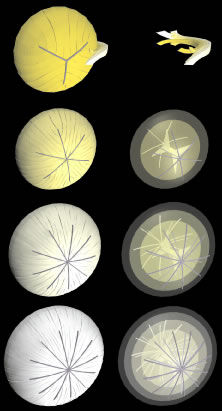
| Fig. 8. Scale 3D-computer assisted drawings (CADs) showing key structural elements in the production of discontinuous suture planes in human lenses. Upper row. From birth through infancy secondary fibers in radial cell columns (the juvenile nucleus) are neither of uniform shape nor simply longer as a function of radial location. Upper middle row. Consequently, the 12 suture branches constituting the simple star suture of the juvenile nucleus, form “discontinuous” suture planes throughout the juvenile nucleus. Lower middle row. Similarly, the 18 suture branches constituting the star suture of the adult nucleus continue to form discontinuous suture planes throughout the adult nucleus to the lens periphery, and, finally, (bottom row) the 24 suture branches constituting the complex star suture of the cortex continue to form discontinuous suture planes extending from the adult nucleus to the lens periphery. |