|
|
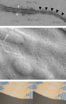
| Fig. 14. Complementary views of low-amplitude thin asymmetric junctions (TAJs) as seen by (upper) thin-section electron microscopy, (middle) freeze-etch electron microscopy (arrow in the upper left indicates direction of shadow), and (lower) 3D-computer assisted design (CAD) stereopairs based on the previous images and other analytical techniques. By thin-section analysis it is apparent that two apposite segments of TAJ membranes from neighboring fibers are also conjoined across an even narrower extracellular space than fiber gap junctions. By freeze-etch analysis it is further apparent that two apposing TAJ membranes (arrowheads) from neighboring fibers are composed of “noncomplementary” aggregates of transmembrane proteins, or AQP0, offset across an essentially eliminated extracellular space (arrows delimit a fiber gap junction). Finally, this 3D-CAD stereopair shows that each AQP0, is a tetrameric water transport particle, and, thus, water can flow in and out of the extracellular space in response to local electromotive and osmotic conditions. |