|
|
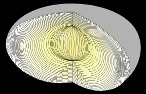
| Fig. 3. When a forming fetal human lens is separated along the visual axis, it is readily apparent that lens growth (secondary fiber development) results in fetal fibers becoming arranged in age-defined growth shells and radial cell columns. However, although the opposite-end curvature of fetal fibers is less apparent in this view, the arrangement of their fiber ends to form offset continuous suture planes between concentric growth shells is facilitated. |