|
|
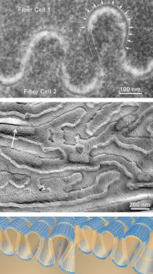
| Fig. 15. Complementary views of high-amplitude thin asymmetric junctions (TAJs) as seen by (upper) thin-section electron microscopy, (middle) freeze-etch electron microscopy (arrow in the upper left indicates direction of shadow), and (lower) 3D-computer assisted design (CAD) stereopairs based on the previous images and other analytical techniques. By thin-section analysis it is apparent that apposing segments of human nuclear fibers have been transformed into furrowed membrane domains (see also Fig. 16). By freeze-etch analysis it is further apparent that furrows from neighboring nuclear fibers are characterized by noncomplementary and offset aggregates of transmembrane proteins, or AQP0. Finally, this 3D-CAD stereopair shows that although each AQP0 is a water transport particle in nuclear fibers, the staggered aggregates of AQP0s may also serve an adhesive function. Although this schematic represents AQP0s as tetramers forming a central channel, in fact each of the tetrameric subunits contains a patent water channel.63 (Middle adapted from Kuszak JR, Brown HG: Embryology and anatomy of the lens. In Albert DM, Jacobiec FA (eds): Principles and practice of ophthalmology: Basic sciences. Philadelphia, WB Saunders, 1994, 94.) |