|
|
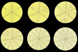
| Fig. 7. Scale 3D-computer assisted drawings (CADs) showing key structural elements in the production of a primate lens six branch star suture formed from birth through infancy. Upper left. At birth, six straight fibers (gold), normally positioned equidistantly around the equator, separate growth shells into equal groups of S-shaped fibers with ends that abut and overlap to form three Y suture branches (blue) that extend to confluence at the poles. The length and location of suture branches are defined by the ends of straight fibers. Upper middle and right and lower left and middle. In successive growth shells, additional straight fibers positioned nonequidistantly around the equator are added that separate the newly added growth shells into unequal groups of S-shaped fibers with ends that abut and overlap to form additional suture branches that extend to confluence at the original suture branches. Lower right. By the end of the infantile period, 12 straight fibers, positioned equidistantly around the equator, separate growth shells into equal groups of S-shaped fibers with ends that abut and overlap to form a simple star suture. (Adapted from Kuszak JR: The development of lens sutures. Progress in Retinal and Eye Research 1412:580, 1994.) |