|
|
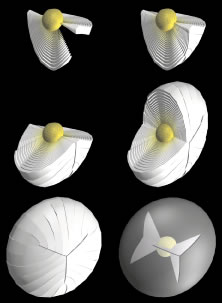
| Fig. 6. 3D-computer assisted drawings (CADs) showing key structural elements in the production of continuous Y suture planes. As additional secondary fibers (white to yellow fibers) are added throughout fetal development, radial cell columns and concentric growth shells (upper and middle rows) are formed consisting of identically shaped but radially longer fibers. As a result, the ends of all fetal secondary fibers become aligned at suture branches that are overlain in successive growth shells to form continuous triangular suture planes extending from the embryonic nucleus to the lens periphery (lower row). |