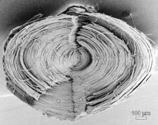
Fig. 4.
Scanning electron microscopy (SEM) micrograph of a human fetal nucleus dissected out of a 36-year-old adult lens and split along the visual axis to reveal radial cell columns, growth shells, and continuous Y suture planes (
asterisks
).