|
|
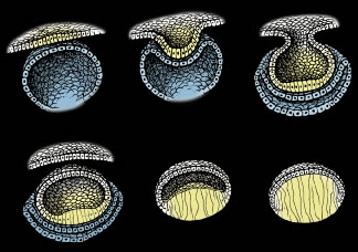
| Fig. 1. Schematic diagrams of key structural events in lens development. Upper left. The thickening of surface ectodermal cells to form the lens placode (yellow). Upper middle and right. The invagination of the lens placode toward the developing optic cup (blue). Lower left. The inverted, or “inside out,” lens vesicle. Lower middle. The elongation of posterior lens vesicle cells as they terminally differentiate to form primary lens fibers. Lower right. The obliteration of the lumen of the lens vesicle by fully elongated primary fibers. Note the anterior lens vesicle cells (white) remain as an undifferentiated monolayer epithelium covering the lens fiber mass. (Adapted from Kuszak JR, Brown HG: Embryology and anatomy of the lens. In Principles and Practice of Ophthalmology: Basic Sciences. Philadelphia, WB Saunders, 1993, p. 83.) |