|
|
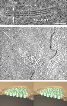
| Fig. 13. Complementary views of a fiber gap junction as seen by (upper) thin-section electron microscopy, (middle) freeze-etch electron microscopy (arrow in the upper left indicates direction of shadow), and (lower) a 3D-computer assisted drawings (CAD) stereopair based on the previous images and other analytical techniques. By thin-section analysis it is apparent that two apposite segments of plasma membrane from neighboring fibers are conjoined across a narrowed extracellular space or gap. By freeze-etch analysis it is further apparent that two apposing regions of plasma membrane from neighboring fibers are composed of complementary aggregates of transmembrane proteins, or connexons, conjoined across a narrowed extracellular gap. Finally, 3D-CADs show that each hexameric connexon is a channel protein, and, thus, conjoined connexons from neighboring fibers permit size and charge related transport of substances between fibers without an energy requirement. |