Chapter 86
Orbital Surgery
ROBERT C. KERSTEN and JEFFREY A. NERAD
Main Menu   Table Of Contents

Search

PREOPERATIVE EVALUATIONS
PREOPERATIVE CONSIDERATIONS
INTRAOPERATIVE CONSIDERATIONS
INSTRUMENTATION
COMPUTERIZED IMAGE-GUIDED INTRAOPERATIVE NAVIGATION
HEMOSTASIS
INTRAOPERATIVE FLEXIBILITY
CARE OF THE SURGICAL SPECIMEN
CHOICE OF ORBITOTOMY APPROACH
INCISIONAL VERSUS EXCISIONAL BIOPSY
THE SURGICAL SPACES
DEEP VERSUS ANTERIOR ORBITAL APPROACHES
DEEP ORBITAL APPROACHES
DEEP MEDIAL ORBITOTOMY
DEEP INFERIOR ORBITOTOMY
ANTERIOR ORBITAL APPROACHES
SUPERIOR ANTERIOR ORBITOTOMIES
MEDIAL ANTERIOR ORBITOTOMIES
LATERAL ANTERIOR ORBITOTOMY
ANTERIOR INFERIOR ORBITOTOMIES
COMBINED APPROACHES
REFERENCES

Neoplasms involving the orbit are uncommon. Their proper treatment requires a detailed knowledge of orbital anatomy and the clinical manifestations of orbital diseases, and appropriate diagnostic assessment of these manifestations. A thorough understanding of the natural history of the various orbital disorders is essential to determine the need for appropriate surgical intervention and the optimal surgical approach.
Back to Top
PREOPERATIVE EVALUATIONS

HISTORY AND PHYSICAL EXAMINATION

A wide range of signs and symptoms may alert the ophthalmologist to involvement of the orbit by a pathologic process. These include proptosis or globe displacement; compressive optic neuropathy with visual field or visual acuity loss; refractive changes such as progressive hyperopia, restricted eye movements, and diplopia; optic disc changes, including pallor or edema; and fundus changes such as choroidal folds. These manifestations may be the consequences of a primary intraorbital disease or may occur secondary to involvement of the orbit by a process extending from the overlying soft tissues, periorbital sinuses, or intracranial cavity.

All patients with suspected orbital pathology should have a thorough ophthalmic history and physical examination. Detailed questioning about the nature, onset, and progression of the chief complaint is necessary to formulate a differential diagnosis. Previous ocular and general medical history related to diseases, operations, medications, and family history should be obtained. A general review of systems will identify risk factors for related systemic diseases, such as hyperthyroidism, metastatic cancer, or lymphoma. A complete examination of the eye and periocular tissues is essential. Abnormal protrusion or displacement of the globe, extraocular motility disturbances, soft-tissue changes, and decreased corneal or periorbital cutaneous sensation should be specifically sought. Special attention always should be directed to evaluation of optic nerve function. Visual acuity testing alone is not sufficient because visual acuity often is preserved initially in patients with compressive optic neuropathy. All patients should be examined closely for detection of an afferent pupillary defect, abnormal color vision, or loss of contrast sensitivity. Formal perimetry should be performed to rule out peripheral field loss. If the presentation has been gradual, evaluation of old photographs may help establish the time course of the pathologic process. In selected cases, appropriate blood work to help identify orbital inflammatory or neoplastic processes also should be obtained.

ORBITAL IMAGING

Valuable information about the nature and extent of an orbital pathologic process is provided by imaging studies such as computed tomography (CT) scan, magnetic resonance imaging (MRI), and orbital echography. Better spatial resolution, ready accessibility, and lower cost make CT the preferred choice for orbital imaging in most cases. Orbital fat provides a natural contrast between most adjacent orbital structures on CT scanning, and orbital bones are visualized well. Computed tomography is essential for evaluation of the orbital bones because they cannot be imaged with MRI. Direct coronal or sagittal images are important to identify the relationship of a lesion to the optic nerve so that the surgical approach can be planned to avoid traversing the optic nerve (Fig. 1).

Fig. 1. A. Axial CT scan demonstrating a large, well-encapsulated lesion in the orbital apex. Coronal (B) and sagittal (C) scans demonstrate that the mass lies inferior and medial to the optic nerve within the intraconal space. This information is useful in planning the surgical approach to the mass, which should avoid traversing the optic nerve.

Newer multislice helical CT scanners allow continuous acquisition of data so that a single rapid pass allows formatting in axial, coronal, and sagittal planes. Multislice CT provides shorter examination times with reduced motion artifact and radiation exposure when compared with conventional CT.

Magnetic resonance imaging is useful in selected cases, especially when evaluating the orbital cranial junction. Significant bony artifact and a lack of orbital fat in the orbital apex make CT scan resolution of the orbital apex structures poor. Because cortical bone has low signal on MRI, there is no bone artifact when viewing the orbital apex on MRI. The lack of intervening fat in the apex to provide contrast is overcome on MRI because contrast is provided by the individual nuclear characteristics of each tissue so that the orbital apex structures are visualized well. Consequently, conditions that affect the optic nerve and chiasm, such as optic nerve meningioma and glioma, generally are evaluated with MRI rather than CT scan (Fig. 2).

Fig. 2. A. Axial orbital CT scan of right optic nerve meningioma. Note lack of detail in orbital apex. B. Axial orbital MR image, same patient. Note increased detail in orbital apex owing to lack of bone artifact. C. MRI with gadolinium contrast. Note extension into brain not easily appreciated with CT scanning or MRI without contrast.

Use of a contrast agent such as gadolinium can further enhance the T1 signal intensity of lesions and is particularly helpful in imaging areas where there has been a breakdown in the blood–brain barrier. Because orbital fat produces a bright signal on T1-weighted images, orbital MR usually is performed with “fat suppression” to prevent masking of lesions. However, incomplete fat suppression can produce artifacts that may be misinterpreted as abnormal enhancement.

A few other orbital conditions are better visualized with MRI than CT scanning.1–3 Often, organic foreign bodies, such as wood, are not imaged well with CT scan. These foreign bodies often are visible with MRI. However, care must be taken when imaging any metallic intraocular or intraorbital foreign body because the strong magnetic field of the MR scanner may cause the foreign body to shift position and damage surrounding structures. In some tumors and vascular anomalies, high blood flow is appreciated on MRI. This is caused by a lack of signal, known as a flow void, created by blood flowing rapidly through larger vessels. Tumors that originate in the brain and extend into the orbit secondarily, such as sphenoid wing meningioma, also are visualized well with MRI. However, bony detail and calcification within the meningioma are not imaged well. In unusual circumstances such as this, CT scan and MRI may prove to be complementary, and both techniques may be required to fully evaluate the orbital disease process.

Orbital echography can provide useful information in the evaluation of orbital disorders. Because each tissue has its own acoustic characteristics, echography can provide a high degree of tissue contrast. Spatial resolution also is quite good. In some situations, echography can provide information not available on CT scan or MRI. These instances include detection and measurement of blood flow4 and intraoperative localization of small foreign bodies.5 Orbital echography requires specialized equipment and a large amount of expertise to obtain the detailed information that is readily available with CT scan or MRI; consequently, its use has decreased. Recent advances in orbital imaging have been summarized by Lee and colleagues.6

Back to Top
PREOPERATIVE CONSIDERATIONS
As in any surgical procedure, appropriate preoperative communication with all members of the surgical team is necessary. This communication starts with an informed consent to the patient. An explanation of the procedure as well as the specific goals and risks of the orbitotomy are explained. Any questions are answered. In most cases, a formal consent form is signed. Appropriate general preoperative laboratory tests are undertaken, depending on the patient's age and type of anesthetic used. Communication with the operating room staff about additions to the routine instrumentation help to avoid delays once the operation has started. Preoperative discussion with the anesthesiologist may be required to review the patient's specific medical condition or operative concerns regarding intraoperative blood pressure or possible blood loss. In some cases, collaboration with other surgical specialists, such as a neurosurgeon or otolaryngologist, is necessary. The surgical plan should be discussed before the team enters the operating room.
Back to Top
INTRAOPERATIVE CONSIDERATIONS
Each surgical approach to the orbit offers its own specific challenges; however, there are several general considerations that make any orbitotomy more successful. These include adequate exposure, proper instrumentation, and good hemostasis. It is also important to have intraoperative flexibility to alter the surgical plan when necessary. Finally, surgical specimens must be handled carefully and submitted properly to obtain the maximum amount of information from the orbitotomy.

Incisions around the orbit are planned with two key considerations in mind. The first is to allow the most direct access to the lesion, and the second is to allow optimal scar camouflage. Incisions may be hidden by placing them within the hairline or brow, in the bulbar conjunctiva, or in the palpebral conjunctiva of the lower lid. A1ternatively, external incisions may be disguised by placing them in preexisting upper or lower eyelid creases, or in lateral canthal rhytides (Fig. 3).

Fig. 3. Incisions frequently used for orbital surgery. Deep approaches: A, Stallard-Wright lateral orbitotomy incision; B, lid crease with lateral extension; C, modified Berke lateral canthotomy incision; D, transcaruncular incision; E, frontoethmoidal “Lynch” incision. Anterior approaches: F, upper lid crease incision, G, vertical lid split incision; H, transconjunctival medial orbitotomy; I, lateral canthotomy incision; J, lower lid percutaneous incision; K, transconjunctival lower lid incision.

Adequate exposure begins with marking the appropriate conjunctival or skin incision. The majority of conjunctival incisions heal without visible scar tissue. However, every skin incision leaves a somewhat visible scar. Thus, there is a tendency to mark as small a skin incision as possible. In some cases, the incision is too small to adequately expose the orbital mass. Hesitation in making a long enough skin incision can be minimized by camouflaging skin incisions as described. Once the skin incision is marked, local anesthetic containing 1:200,000 epinephrine is injected into the adjacent skin. This is done before the surgeon scrubs so that there is sufficient time for vasoconstriction to occur before the skin incision is made.

While the surgeon scrubs, the operating room personnel prepare and drape the patient. Because many orbitotomies are performed with the assistance of ophthalmic nurses, there is a tendency to drape an inappropriately small operating field. The operating room staff should be instructed to prepare and drape a generous surgical field. It is usually helpful to prepare and drape both orbits within the field so that the contralateral side may be used as a reference to judge symmetry. Skin incisions usually are made with a No. 15 scalpel blade or a Colorado microdissector needle. Although an adequate-length incision should be made, it is often surprising how extensively the deeper structures can be exposed through a small skin incision when there is wide elevation and retraction of deeper soft tissues overlying the periosteum. If the choice of incision or length of incision is inadequate, the surgical plan should be modified to allow appropriate exposure. Often it is possible to start with a shorter incision, lengthening it if it restricts access.

Visualization of deeper orbital tissues involves appropriate retraction of overlying soft tissues. Individual 4-0 silk sutures placed in the subcutaneous tissues offer a large degree of flexibility in retracting the skin and subcutaneous tissues. A variety of orbital retractors should be available for each case. The value of an experienced assistant to provide adequate retraction and exposure cannot be overemphasized.

Back to Top
INSTRUMENTATION
Orbital surgery requires special instrumentation. Familiarity with and use of these instruments are essential for efficient orbital surgery. Proper instrumentation begins with illumination. Overhead operating room lights provide useful illumination for superficial parts of the orbitotomy. Except for somewhat superficial anterior orbitotomies, these overhead lights provide inadequate illumination to complete the orbitotomy. A fiberoptic headlight is extremely helpful for working more deeply within the orbit.

To detect the subtle differences in color and texture of the many orbital tissues, magnification is helpful. Surgical operating loupes are used routinely by most surgeons. Many varieties of surgical loupes are available. Custom-fit operating loupes with the surgeon's individual near and distant prescription provide the best fit and comfort. Magnification of 2.5× with a working distance of 14 to 16 inches is standard for most orbital procedures. Higher-powered operating loupes are available, but they are somewhat awkward to work with and limit the field of view. An operating microscope is recommended for higher levels of magnification. Maximum benefit is obtained with a microscope on a counter-weighted stand, similar to those used by most neurosurgeons. These stands allow upright positioning of the microscope on the X, Y, and Z axes. The use of a 300-mm objective lens attached to the microscope permits the longer instruments used in orbital surgery to be placed into the operative field without contamination by inadvertent contact with the microscope. One of the most significant benefits of an intraoperative microscope is that it allows both the surgeon and the assistant to simultaneously visualize the depths of the orbital incision. The operating microscope also can be attached to a video monitor so that the operating room staff can be more involved in the procedure, and the operation also may be videotaped.

A variety of specialized orbital instruments, including retractors, dissectors, and bone-cutting tools, are available. Standard orbital retractors include several sizes of Sewall and malleable ribbon retractors. Retractors are useful in dissecting through the orbital fat; in addition, they offer exposure once the mass is identified. Placement of neurosurgical cottonoids beneath the retractor helps to keep orbital fat from prolapsing into the wound. Several types of periosteal elevators are useful; the most common is a Freer periosteal elevator. Numerous air-driven bone saws and drills are available. Bone rongeurs of several sizes should be on the surgical instrument tray. One of the various orbital miniplating or microplating systems may be useful for reattaching bone flaps and reconstructing bony defects. A retinal or glaucoma cryoprobe is helpful for applying traction to encapsulated solid or cystic orbital masses to facilitate their dissection from surrounding orbital tissues. For combined sinus and orbital surgery, the use of a nasal endoscope sometimes is helpful. Techniques involving many of these instruments frequently are not emphasized in ophthalmic residency training. Familiarity in these instruments must be obtained before beginning orbital surgery.

Back to Top
COMPUTERIZED IMAGE-GUIDED INTRAOPERATIVE NAVIGATION
In recent years, computerized image guided systems have been devised to display the location of anatomic structures intraoperatively, often referred to as surgical navigation technology. The position of a probe in the surgical field is visualized in real time on a computer screen adjacent to the operating table that displays the patient's preoperative imaging. The most common applications are where the anatomy is complex with many vital structures are situated in close proximity. The technique is fascinating and worthy of explanation.

There are three steps in the image guidance process: (a) preoperative imaging, (b) intraoperative registration of anatomic landmarks with the scans, and (c) intraoperative navigation. Prior to preoperative imaging, markers (known as fiducials) are placed on the patient's skin, usually behind and above the ear. The position of the fiducials can be seen on the scans. In the operating room, the patient, the probe and the computer are linked in the registration process. A “camera” linked to the computer “sees” the probe as the surgeon touches a fiducials on the patient with a probe. The exact three-dimensional position of the probe is recorded in the computer. The registration process continues in a similar manner, linking each fiducial on the patient with its position on the computer image. Additional registration points are linked using anatomic landmarks such as the tragus. The more points that are registered the higher the accuracy of the subsequent navigation. The operation proceeds in normal fashion. When confirmation of the location of any anatomic structure is desired, the probe is placed into at that position, the camera “sees” the probe, and the computer shows the position of the probe on the monitor (Fig. 4). Different probe configurations allow simultaneous positioning of more than one probe. It is possible to use a drill or suction catheter as a probe so that the intraoperative position of these devices can be visualized while being used. The accuracy of intraoperative navigation can be in the submillimeter range if high-resolution scans and accurate registration are used.

Fig. 4. Computerized image guided intraoperative navigation system. Surgeon holds probe in wound. Infrared camera detects position of probe tip and sends information to the computer. The position of the probe is seen on the CT scan or MR image on the computer screen.

Functional endoscopic sinus surgery and skull base operations are examples of procedures in which complex anatomy exists, often with variation from patient to patient or even side to side. Disease processes such as tumors or polyps can distort the anatomy as well. In these cases, the time spent in setting up the equipment and registering the system can be worthwhile. Surgical navigation has been used in orbital surgery, especially intraorbital foreign body removal or those orbital cases involving combined sinus and intracranial approaches. No doubt new innovations in imaging and computer technology will expand the uses of image guidance systems.

Back to Top
HEMOSTASIS
Control of bleeding is one of the most important skills an orbital surgeon must learn. Obtaining hemostasis begins in the office with specific questioning of the patient about the use of aspirin, nonsteroidal anti-inflammatory drugs, and vitamins and dietary supplements. Frequently, the patient will not regard these over-the-counter medications as important. Discontinuation of anticoagulants and antiplatelet medications may have serious adverse outcomes and should be undertaken in conjunction with the patient's medical doctor. Although the need to routinely discontinue anticoagulant medications before performing all ophthalmic plastic procedures has been questioned,7 orbital hemorrhage carries a considerable risk of visual loss, and all attempts to optimize the patient's coagulation parameters should be made before elective orbital surgery. Warfarin generally is stopped 4 to 5 days before surgery, and the patient's international normalization ratio (INR) should be checked immediately preoperatively to ensure adequate hemostasis. At times it may be necessary to replace warfarin with heparin, which can be reversed immediately preoperatively if the patient is at high risk of thromboembolic complications. Patients being considered for elective orbital surgery should discontinue aspirin and other nonreversible platelet inhibitors for 1 week before surgery. Nonsteroidal medications are reversible platelet inhibitors whose effect diminishes as they are cleared from the circulation. The duration of the platelet often depends on the half-life of the drug, but it is usually recommended that the medication be discontinued 1 to 2 days preoperatively. Of course, systemic hypertension should be brought under optimal medical control in the preoperative period.

Once in the operating room, the patient should be placed in the reverse Trendelenburg position to decrease venous pressure. The patient's blood pressure should be strictly controlled in most cases, with the goal of maintaining the blood pressure at or slightly below the patient's normal range. In patients with particularly vascular tumors, consideration should be given to hypotensive anesthesia with placement of an intra-arterial blood pressure monitor.

The vasoconstriction provided by the dilute epinephrine (1:100,000 to 1:200,000) mixed with local anesthetic helps to prevent bleeding when 1 or 2 ml is injected 10 minutes before incision. Small vessels are coagulated with bipolar cautery. When the surgeon is working away from the eyelids or orbit, unipolar cautery may be used to cut or coagulate the bleeding vessels. In general, unipolar cautery should not be used within the confines of the orbit because it may preferentially conduct along myelinated nerve fibers. When large arteries are encountered, aneurysm clips may be applied. Long-handled bayonet microsurgical bipolar cautery instruments are available for intraorbital and intracranial coagulation. Visualization is improved with Q-tips or “peanut” type gauze dissectors to absorb small amounts of blood on the field. When bleeding is more vigorous, suction may be required to visualize bleeding vessels. Suction is facilitated by placing the suction tip against a cottonoid rather than on the orbital fat. Specialized suction tips are useful in the sinus. These suction devices may include irrigation or unipolar cautery. Bone wax, Gelfoam (absorbable gelatin sponge), Surgicel (oxidized regenerated cellulose), Avitene (microfibrillar collagen), and Thrombogen (thrombin) also are available to facilitate coagulation.

In most orbitotomy cases, blood products are not required. In some cases where tumor resection involves other parts of the cranium or face, blood replacement may be required. In cases where resection of particularly vascular tumors is necessary, preoperative angiography with arterial embolization may be helpful. If there is concern about postoperative bleeding, a flexible vacuum drain may be used. Atraumatic extubation of the patient at the completion of the procedure is important to prevent increased venous pressure, which increases the risk of orbital hemorrhage.

Back to Top
INTRAOPERATIVE FLEXIBILITY
The ability to modify the surgical plan intraoperatively is necessary in orbital surgery. Inadequate exposure may be improved by extending the surgical incision or by more aggressive soft-tissue and periosteal elevation. Unexpected difficulties in exposing or making a safe biopsy of an orbital mass may require the use of combined surgical approaches, such as the addition of lateral orbitotomy, to a planned medial orbitotomy. Larger orbital procedures involving tumors that extend outside the orbit require a team approach in cooperation with surgeons from other disciplines. In these cases, as well as all orbital procedures, preoperative contingency plans for intraoperative modifications will make the orbitotomy more successful. In all types of surgery, the ability to modify the initial plan becomes easier with experience.
Back to Top
CARE OF THE SURGICAL SPECIMEN
Orbital tumors are rare. Frequently, the amount of tissue that is available is small. Consequently, great care must be taken in obtaining and handling the orbital biopsy specimen to ensure the most accurate diagnosis. Preoperative consultation with the ocular pathologist is advisable when an unusual diagnosis is suspected. Intraoperative frozen-section analysis of the biopsy tissue may be helpful in confirming that appropriate tissue has been sampled. Frozen-section diagnosis should be confirmed with permanent section analysis before decisions are made about disfiguring procedures such as orbital exenteration. Fresh tissue for flow cytometry and immunopathologic analysis should be obtained for all lymphoid lesions and is often of benefit in the diagnosis of many unusual orbital tumors. Occasionally, electron microscopy may be helpful. When the orbitotomy is performed for diagnostic reasons, the tissue must be treated delicately to avoid crush and drying artifact. Attention to these details will increase the diagnostic accuracy of the procedure.
Back to Top
CHOICE OF ORBITOTOMY APPROACH
To select the best surgical approach to an orbital lesion, the surgeon must know the location of the lesion and the structures to be traversed in reaching it, and have a properly formulated working diagnosis. The diagnosis anticipates whether the lesion is expected to be encapsulated or diffuse, benign or malignant, and whether complete excision of the lesion is preferred or a biopsy followed by nonsurgical treatment is more appropriate. The orbital examination and review of orbital imaging studies usually suggests that the lesion is either benign or malignant; in many cases a specific diagnosis is suggested. In general, well-outlined masses with smooth contours are likely to be benign. Tumors with diffuse borders and evidence of invasion into the surrounding tissues, including orbital fat or bone, are likely to be malignant. Exceptions to these rules exist (e.g., the smooth, well-outlined mass frequently seen in malignant lymphoma, or the diffuse borders apparently infiltrating into orbital fat found in idiopathic inflammatory disease of the posterior orbit).
Back to Top
INCISIONAL VERSUS EXCISIONAL BIOPSY
A thorough knowledge of orbital disorders guides the decision to treat medically or perform an incisional or an excisional biopsy. In some cases, such as suspected Graves' ophthalmopathy or idiopathic inflammatory “pseudotumor,” medical treatment may be prescribed without the need for surgery. In other cases, orbitotomy may be required before treatment can be initiated. The differential diagnosis suggests whether the lesion is thought to be amenable to surgical excision or is a process in which surgery to establish a diagnosis is better followed by medical management or radiation therapy.

An incisional biopsy is performed when a representative piece of tissue is removed but some of the lesion is left behind. This type of biopsy is used to confirm a clinical diagnosis before proceeding with medical therapy or radiation therapy (e.g., lymphoma, orbital pseudotumor, rhabdomyosarcoma). It also may be used to obtain a tissue diagnosis before planning a definitive surgical treatment such as exenteration or extensive orbitocranial resection (e.g., malignant lacrimal gland tumors). In some cases, incisional biopsy provides a tissue diagnosis and also is used to debulk a tumor. Diffuse infiltrative orbital lesions usually require incisional biopsy to confirm the diagnosis before other therapy can begin.

An excisional biopsy is performed when an orbital mass is removed in its entirety. Excisional biopsy is performed on lesions that are believed to be benign, well localized, and easy to remove without damage to surrounding orbital tissues. As a general rule, well-circumscribed and encapsulated lesions are treated with excisional biopsy. This is especially important in cases of benign mixed tumors of the lacrimal gland or orbital dermoid cysts where incision through an intact capsule dramatically increases the chance of recurrence and orbital morbidity. In general, malignant lesions of the orbit are not amenable to excisional biopsies, because tumor-free microscopic margins cannot be obtained in orbital fat.

Back to Top
THE SURGICAL SPACES

The other important consideration in planning orbitotomy is the position of the tumor within the orbit. Lesions generally may be categorized as either anterior (superficial) or posterior (deep). They can be further categorized by their relationship to the optic nerve (medial, lateral, superior, or inferior to the optic nerve). It may be helpful to further classify the location of lesions relative to adjacent anatomic structures in the orbit. Many surgeons prefer to conceptualize orbital pathologic processes as being located within one or more of seven “surgical spaces” within or around the orbit (Fig. 5). These spaces include:

Fig. 5. Axial drawing of anatomic spaces of the orbit. (1, intraconal space; 2, extraocular muscles; 3, peripheral surgical space; 4, subperiosteal space; 5, preaponeurotic space; 6, Tenon's space; 7, periorbital tissues.)

  1. Central surgical space (intraconal space)
  2. Extraocular muscles
  3. Peripheral surgical space (extraconal space)
  4. Subperiosteal space
  5. Preaponeurotic space
  6. Tenon's space
  7. Periorbital tissues

The central surgical space is bounded by the intermuscular septum, which connects the four rectus muscles in the coronal plane. This space also is described as the retrobulbar or intraconal space, and it contains the optic nerve, intraconal fat, and various vascular and neural structures. Primary orbital tumors such as cavernous hemangiomas or optic nerve meningiomas originate in the central surgical space.

Many orbital processes involve the extraocular muscles, primarily or secondarily, making it useful to classify the extraocular muscles as residing in their own separate surgical “space.” Conditions affecting the extraocular muscles include thyroid orbitopathy, myositis, and a variety of neoplastic conditions.

The peripheral surgical space, also known as the extraconal space, lies outside the intermuscular septum but within the periorbita. This space contains a scant amount of orbital fat, the superior oblique muscle and trochlea, inferior oblique muscle, and lacrimal gland. Other important nerves and vessels extend into the space, such as the superior ophthalmic vein. A variety of pathologic processes may encroach on the peripheral surgical space.

The subperiosteal space is a potential space lying between the orbital bones and periorbita. The periorbita may provide a barrier to extension of neoplastic and infectious processes originating in the adjacent sinuses or intracranial cavity. Frequently, the subperiosteal space may fill with blood after orbital fracture or infection when associated with a paranasal sinusitis.

The preaponeurotic space is actually an anterior extension of the extraconal space. Because this space is superficial and readily accessible, sometimes it is referred to as its own orbital compartment. Frequently, neoplasms affecting the lacrimal gland, such as lymphoma, extend forward into the preaponeurotic space.

Tenon's space is a potential space between the sclera and Tenon's capsule. Tumors originating in the eye, such as choroidal melanoma, may extend into Tenon's space.

Secondary orbital tumors may arise from periorbital tissues and secondarily invade the orbit. Cutaneous malignancies arising in the eyelid or facial skin may extend posteriorly through the orbital septum, whereas nasal and paranasal sinus tumors may grow through the orbital bones into or through the subperiosteal space. Tumors arising within the cranial cavity, such as sphenoid wing meningiomas, also may affect the orbit secondarily.

Back to Top
DEEP VERSUS ANTERIOR ORBITAL APPROACHES
In general, approaches to the orbit can be divided into two categories: those in which a lesion is sufficiently superficial that access may be achieved without removing the orbital walls, and those for deeper lesions in which orbital bones must be removed to gain access. The location of the lesion relative to the optic nerve further dictates the optimal approach. It is always desirable to avoid traversing the nerve while approaching a lesion so as to minimize traction on or trauma to the nerve, which may result in visual dysfunction.

Historically, an exploratory orbitotomy, often with removal of the lateral orbital wall, frequently was required to establish a diagnosis in patients with proptosis and a presumed orbital mass. Fortunately, modern advances in orbital imaging have allowed much more precise and sophisticated refinement of surgical approaches to the orbit.

A large number of innovative anterior surgical approaches have been developed over the years, and whenever possible, anterior approaches to the orbit are preferred to avoid the extra time and potential morbidity of bone removal and refixation. Although deeper orbital lesions may require access by either lateral orbitotomy with removal of the lateral wall, transcranial orbitotomy with removal of the orbital roof, medial orbitotomy with removal of the ethmoid sinus (medial orbital wall), or inferior orbitotomy with removal of the orbital floor, increasingly lateral orbitotomy with elevation and incision of the lateral periorbita allow deep orbital access without the need to routinely remove the lateral bony wall.

There may be considerable overlap when characterizing lesions as either deep or superficial, and it is not always possible to determine if a given external incision approach will or will not require removal of underlying bone to achieve the goals of the procedure. Although one cannot overemphasize the need for intraoperative flexibility when performing orbital surgery, for purposes of discussion here we segregate descriptions of deep and anterior orbital approaches.

Back to Top
DEEP ORBITAL APPROACHES

LATERAL ORBITOTOMY

Lateral orbitotomy originally was popularized by Kronlein8 in 1888. The incision described by Kronlein was a reverse C-shaped incision placed over the lateral rim and extending superiorly toward the hairline and inferiorly toward the ear. This resulted in an unsightly scar and a high likelihood of damage to the seventh cranial nerve. Subsequently, a number of superior skin incisions have been devised to allow exposure of the bony lateral orbital wall and access to the lateral retrobulbar space.9–11 Currently, the lateral wall is most often approached through either a canthotomy incision (modified Berke),12 or an upper eyelid crease incision extending into a lateral “laugh line.”13 Rarely, a coronal incision in the hairline with subgaleal dissection of a scalp flap carried down to the lateral rim is useful.14,15

Although percutaneous dissection and orbital bone removal can be performed satisfactorily under local anesthesia, deeper orbital manipulation requires retrobulbar injection, which would alter postoperative pupillary evaluation and visual acuity checks. Because visual loss is a well-recognized complication of orbital surgery,16 most lateral orbitotomies are performed under general anesthesia to allow 
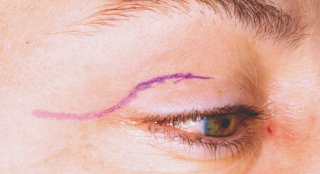
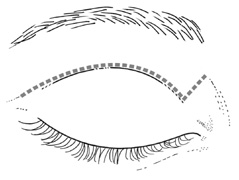
close postoperative monitoring of vision.

EYELID CREASE LATERAL ORBITOTOMY

This approach affords excellent exposure of the lateral orbital rim through an incision placed within the upper eyelid crease, which may extended 
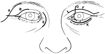
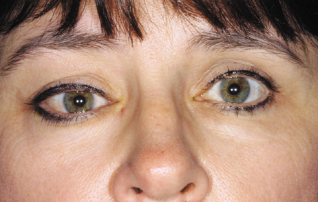
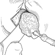
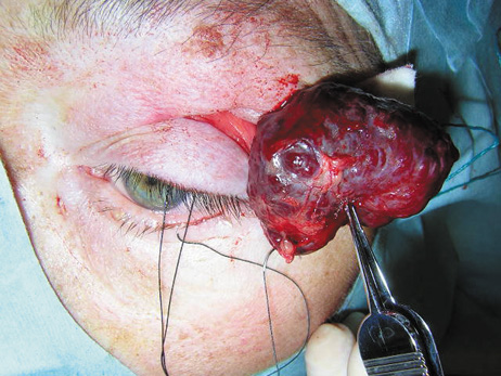
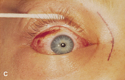
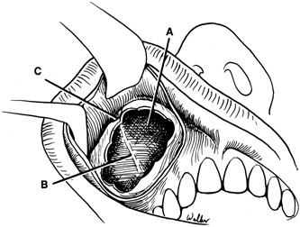
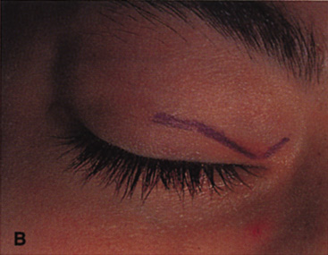
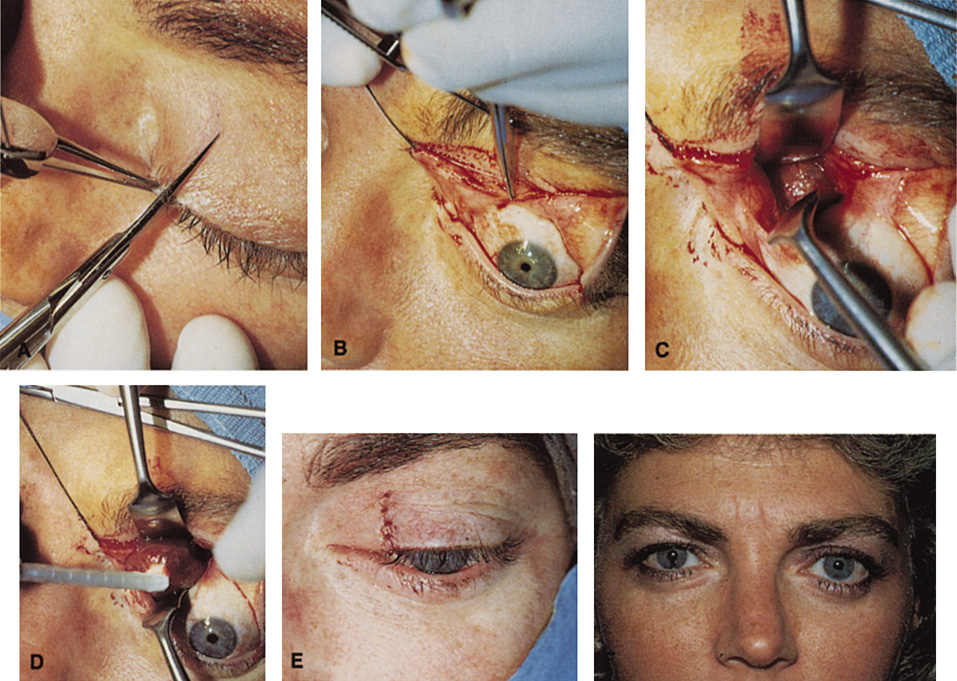
laterally into a temporal “laugh line” (Fig. 6). Infiltration with local anesthetic with epinephrine (1:200,000) at least 10 minutes before performing the skin incision minimizes bleeding.

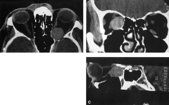
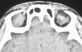
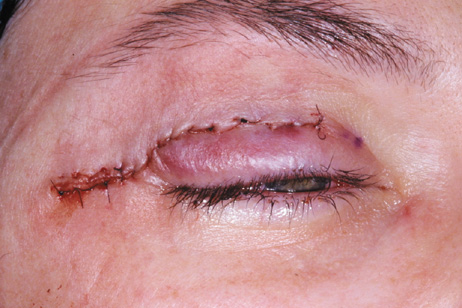
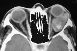
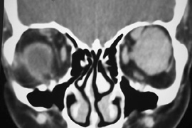
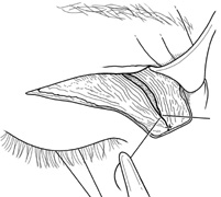
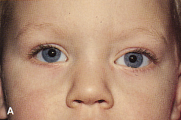
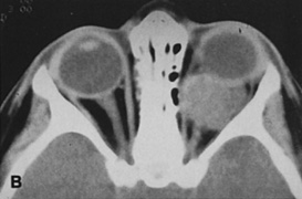
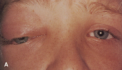
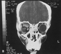
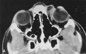
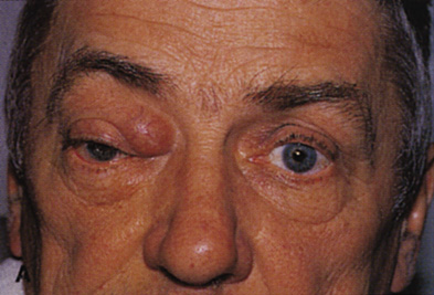
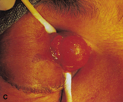
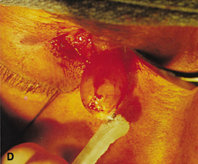
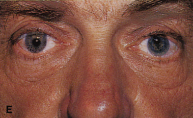
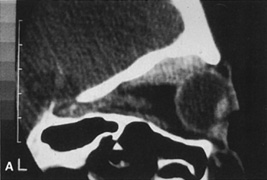
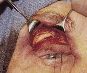
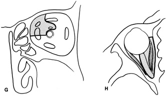
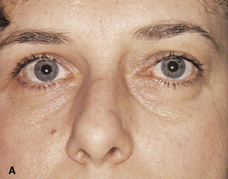
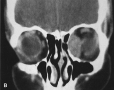
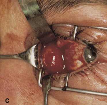
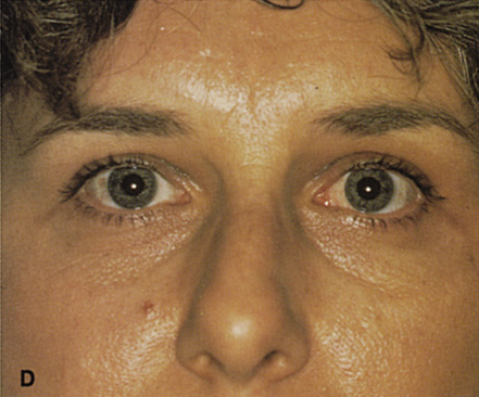
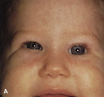
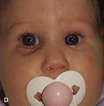
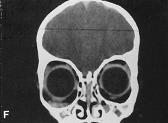
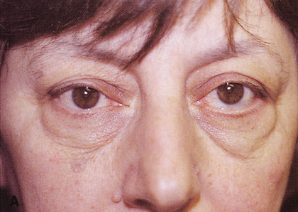
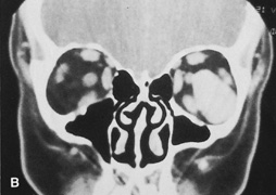
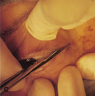
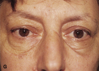
Fig. 6. Lateral orbitotomy through upper eyelid skin crease A. Photo demonstrating right globe ptosis present for more than 2 years. B. Axial CT scan showing a well outlined oval lesion in the lacrimal gland 
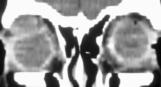
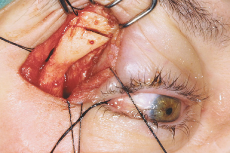
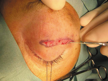
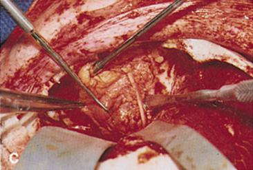
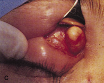
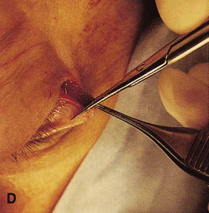
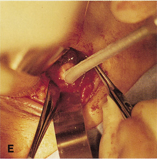
fossa. C. Coronal CT showing lesion pushing globe inferiorly. D. Skin crease excision marked for lateral orbitotomy. E. Lateral orbital rim exposed. Bone cuts made above frontozygomatic suture 
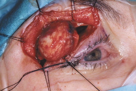
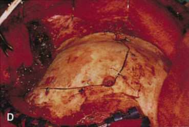
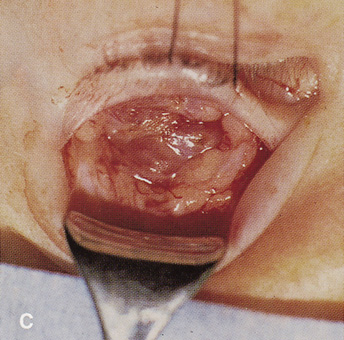
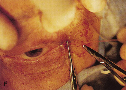
and at zygomatic arch. F. Lateral wall removed. Subperiosteal space exposed. Hard tumor could be 
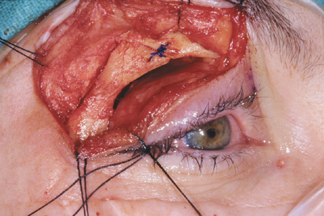
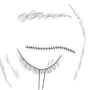
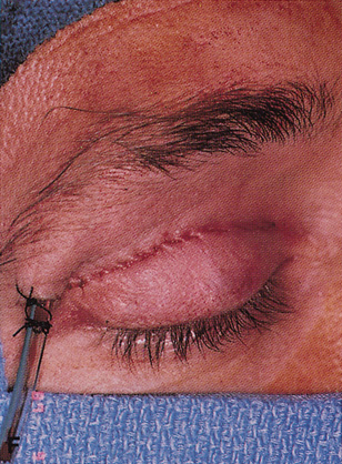
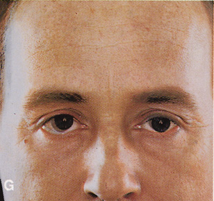
palpated in area of lacrimal gland. G. Benign mixed tumor of lacrimal gland removed. H. Bone sutured into place. I. Skin crease closed.

The incision is made in the skin to the depth of the orbicularis muscle, and skin and muscle dissected superficial to the orbital septum. Elevation of a skin–muscle flap superiorly and laterally allows exposure of the underlying superior and lateral orbital rims. Dissection in this plane, deep to orbicularis muscle, can be carried inferiorly to the level of the inferior orbital rim. In this fashion the superior and lateral orbital rim can be exposed from a point just lateral to the supraorbital nerve, superiorly, down to the junction of the lateral wall and orbital floor, inferiorly. The periosteum then is incised with the unipolar cautery just posterior to the arcus marginalis along the orbital rim. A Cottle periosteal elevator can be used to elevate periosteum. If the lateral bony rim is to be removed (see Fig. 6), then periosteum usually is elevated first over the external surface of the lateral rim to expose the underlying bone. The lateral rim periosteum fuses with the superficial temporal fascia at the posterior border of the lateral orbital rim. As periosteal elevation progresses posteriorly, the superficial temporal fascia is encountered and cut with unipolar cautery and the anterior temporalis muscle is elevated to expose the temporal fossa. The temporalis muscle may be bluntly swept out of the large temporal fossa by use of gauze wrapped around the surgeon's index finger or a large periosteal elevator. Elevation of periosteum and temporalis muscle is carried superiorly to a point 1 cm above the zygomaticofrontal suture and inferiorly to the level of the zygomatic arch, which is even with the orbital floor (see Fig. 6E).

Once the outer surface of the lateral rim has been exposed, the periorbita along the mesial surface of the lateral wall is similarly elevated posteriorly within the orbit so that the bony lateral orbital rim can be completely bared along its external and internal surfaces. The periorbita is tightly adherent at the inner orbital rim (arcus marginalis), especially over the lateral orbital tubercle. Periorbital elevation in this area must be performed carefully to avoid buttonholing and prolapse of orbital fat. Once the elevation has proceeded along the inner surface posterior to the lateral orbital tubercle, the periorbita elevates quite freely from the bone. The zygomaticotemporal and zygomaticofacial neurovascular bundles are encountered about 1 cm posterior to the rim (Fig. 7). Transecting the neurovascular bundles results in a small area of postoperative hypesthesia over the lateral rim. These bundles usually are cut with a unipolar cautery to allow periorbital elevation to continue back to the inferior orbital fissure. The periorbita enters the inferior orbital fissure at the junction of the lateral wall and floor and is a landmark to establish the depth of dissection along the inner 
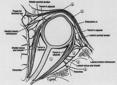
surface of the lateral rim.

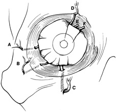
Fig. 7. Coronal schematic view demonstrating major vessels penetrating periorbita and traversing the extraperiosteal space that may be encountered during periorbital elevation. (A, zygomaticotemporal artery; B, zygomaticofacial artery; C, communicating branch of infraorbital artery: D, supraorbital artery: E, posterior ethmoidal artery: F, anterior ethmoidal artery.)

After the periorbita has been elevated internally, and the periosteum reflected laterally and superiorly to completely expose the lateral rim, a large malleable retractor is placed between the orbital contents and the mesial surface of the lateral orbital rim. This protects the orbital contents while an air-driven saw is used to make two osteotomies. One is placed superiorly about 0.5 cm above the zygomaticofrontal suture. Aligning the saw blade toward the upper molars on the opposite side (an angle of 45°) produces a wide opening while avoiding inadvertent entry into the overlying frontal cranial fossa by the saw blade. Finger palpation of the orbital roof beneath the brow reminds the surgeon of the location of the frontal lobe and helps ensure that the saw is not angled in a fashion to allow intracranial penetration. A second osteotomy is made inferiorly through the lateral rim just adjacent to the floor of the orbit above the zygomatic arch. The osteotomies may be shifted superiorly or inferiorly depending on the exact location of the orbital lesion, but an opening in the lateral wall of 2.5 to 3 cm is desirable. After the saw cuts have been made, a wire-passing drill bit is used to preplace drill holes above and below each of the two osteotomies so that the bone can be refixated at the completion of the procedure. Care is taken again to angle the superior drill hole to avoid intracranial penetration and to protect orbital contents with a wide malleable retractor. The lateral rim is out-fractured with large rongeur by gently “rocking” the rim outward, and remaining strands of temporalis muscle at the posterior edge of the bone fragment may be cut with the unipolar cautery. The rim is removed and placed off the field in moist gauze. Great care is taken to avoid inadvertent contamination of the lateral rim as it will be replaced at the end of the procedure. After out-fracture of the lateral rim, the thinner bone behind the rim can be removed with a rongeur to provide better access to the orbital apex. Bone removal usually is carried posteriorly to the pterion, the thicker wedge of cancellous bone that constitutes the body of the greater wing of the sphenoid. Hemorrhage from the cancellous bone may be brisk and may require tamponade with bone wax for control. The temporalis fascia is retracted posteriorly with broad malleable retractors so that the apical periorbita can be exposed. Relaxing incisions can be made with the unipolar cautery through the superior temporalis muscle if it is difficult to adequately expose the 
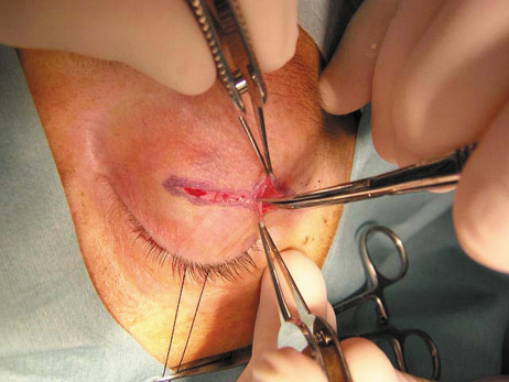
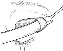
apex.

The lateral rectus muscle and lacrimal gland lie just beneath the periorbita along the lateral orbit (see Fig. 6F), and damage to these structures is avoided by opening the periorbita with blunt-tipped scissors. A traction suture placed transconjunctivally beneath the insertion of the muscle can be manipulated to help identify the belly of 
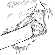
the lateral rectus posteriorly in the orbit. A longitudinal periorbital incision beginning anteriorly and extending back to the orbital apex will allow extensive retraction of the periorbita. Additional vertical relaxing incisions through the periorbita may be made anteriorly. The periorbita is retracted, and the lateral rectus muscle and lacrimal gland are identified. If the lesion lies in the intraconal space, sharp dissection to release the intermuscular septum, either above or below the lateral rectus muscle, is usually required to gain access to the retrobulbar space. Sewall orbital retractors provide retraction of the lacrimal gland and lateral rectus muscle. Depending on the location of the lesion superiorly or inferiorly within the orbit, dissection is carried out above the superior border or below the inferior border of the retracted lateral rectus muscle. Deep orbital dissection requires fiberoptic illumination and loupe magnification at a minimum, and often it is best performed with the operating microscope to aid in identification of the vital vascular and neural structures within the orbit.

After removal or biopsy of the proposed lesion, the orbital rim is reinserted and fixated with sutures passed through the predrilled holes, or with rigid screw and microplate fixation (see Fig. 6H). Usually it is not necessary or desirable to close the periorbita because this allows for decompression of any postoperative retro-orbital hemorrhage into the temporal fossa. After the bone has been resutured in place, the anterior cut edges of periorbita and periosteum along the lateral rim may be loosely reapposed over it to prevent the overlying orbicularis-skin flap from adhering to the bare bony rim. The eyelid crease incision can be closed with a running suture (see Fig. 6I) as one would after upper blepharoplasty. A drain may be placed in the temporal fossa and brought through the lateral aspect of the incision or through a separate stab wound and connected to external suction for the first 24 hours postoperatively if there is any question about 
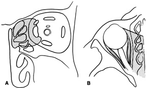
complete hemostasis.

Indications

Lateral orbitotomy provides excellent access to deep lesions in the subperiosteal, peripheral, or 
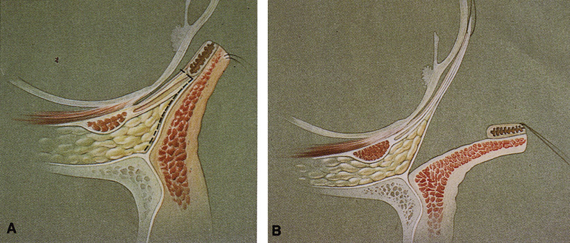
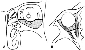
intraconal space lateral to the optic nerve (Fig. 8A, B).

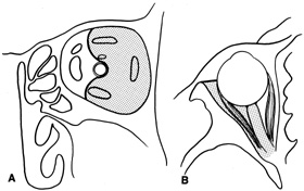
Fig. 8. Coronal (A) and axial (B) views in an illustration of areas (shaded) amenable to lateral orbitotomy.

Although intraconal lesions medial to the nerve sometimes can be approached laterally, great care to identify and protect the optic nerve is required during deep orbital dissection. Because the eyelid crease incision allows such wide exposure of the superolateral orbit, it is often possible to remove fairly large orbital lesions without removing the lateral orbital wall (Fig. 9). Surgery in this case proceeds as described to exposure of the superior and lateral bony orbital rims. It is not necessary to reflect periosteum over the external surface of the rim. Instead, once periosteum at the rim is exposed, it is cut with cautery and then only the mesial periorbita need be elevated internally to expose orbital contents with subsequent intra-orbital dissection carried out with the lateral rim in place. Often it is preferable to initially attempt to remove intraconal or lacrimal fossa lesions in this fashion. If exposure proves inadequate, the periosteum over the external surface of the lateral orbital rim can be elevated and osteotomies and removal of the lateral
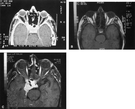
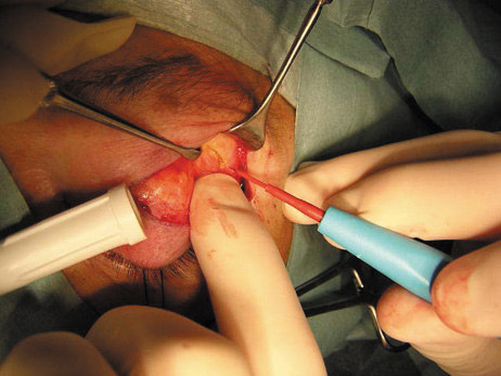
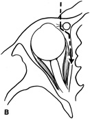
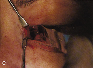
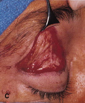
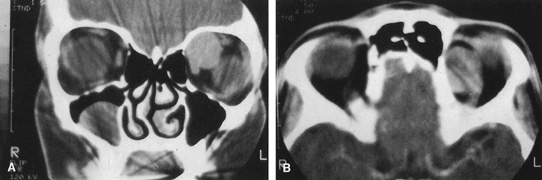
 wall still can be carried out.

   
Fig. 9. A,B. Coronal and axial CT images of a large intraconal neoplasm. C. Because it was felt to represent a well-encapsulated cavernous hemangioma, this lesion was a candidate for removal via an eyelid crease orbitotomy 
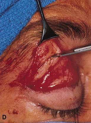
without bone removal. The eyelid crease incision marked. D. Incision made with scalpel. E. Orbicularis muscle is tented up and incised to expose the underlying septum. F. Dissection of a skin-muscle flap deep to orbicularis exposes the orbital septum and superior orbital bony rim. G. Cutting cautery is used to incise periosteum along the superior and lateral 
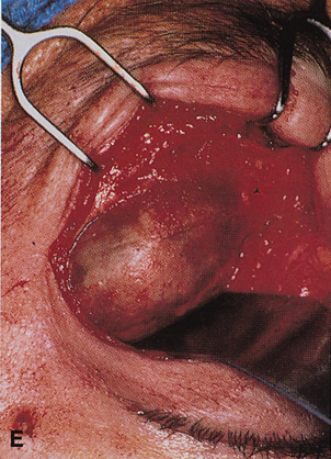
rims; finger palpation of the bone helps to direct this incision. H. Periorbita is elevated along the mesial surface of the lateral orbital rim in order to expose the deep orbital tissues. I. The cavernous hemangioma is visualized in the wound. Retraction is provided by one or more malleable retractors. J. Cryoprobe is affixed to the hemangioma to facilitate manipulation of the lesion. K. Large cavernous hemangioma after removal through the eyelid crease incision which was accomplished without bone removal. L. Periorbita is reattached over the lateral rim. M. The eyelid crease incision is closed with a running suture.

CANTHOTOMY APPROACH

Many deep orbital lesions requiring removal of the lateral orbital rim can be approached through a smaller lateral canthotomy incision. Although Berke17 initially described a fairly extensive lateral canthal incision extending back over the zygomatic arch for 5 to 7 cm, this longer incision often leaves an unsightly scar and may risk damage to the seventh cranial nerve. Because of the extensibility of the periocular tissues, exposure of the lateral orbital rim usually can be accomplished through a small lateral canthotomy incision measuring 1 to 1.5 cm in length (Figs. 10, 11, and 12). With wide undermining in the suborbicularis plane and retraction of the skin and orbicularis, superior and inferior osteotomies can be made quite readily despite the small external incision. The incision 
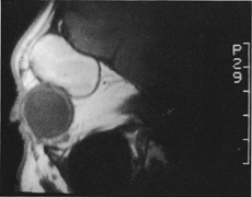
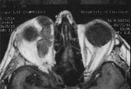
always can be extended farther posteriorly if exposure is inadequate.

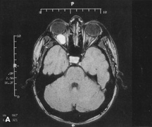
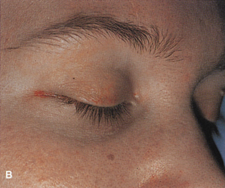
Figure 10. A. Large, well-encapsulated intraconal mass on MR scan. B. Small lateral canthotomy incision will be used to perform lateral orbitotomy and remove the intraconal mass.

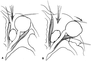
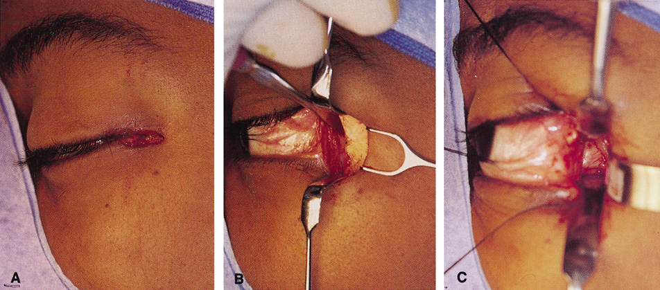
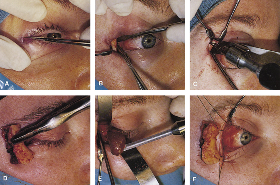
Fig. 11. A. Lateral canthotomy incision is made with straight iris scissors. B. Periosteum is elevated off of the lateral orbital rim. C. Wide undermining allows retraction of the skin incision to permit superior and inferior osteotomies to be made with the air-driven saw. D. The bony rim has been outfractured. Because of the distensibility of the skin, it is possible to remove a large bone flap through the small canthotomy incision. E. The intraconal mass is extracted with the aid of the cryoprobe. F. The bone fragment is positioned for resuturing.

Fig. 12. A. The lateral canthotomy incision is reapproximated with simple closure of the superior and inferior crura of the lateral canthal tendon. A drain from the temporal fossa has been brought out through a separate stab incision posteriorly. 12B. Excellent postoperative scar camouflage is obtained by this approach.

Procedure

The lateral canthotomy is made with straight iris scissors or a No. 15 blade, and the unipolar cautery then can be used to extend the incision deeply through orbicularis to expose periosteum along the length of the lateral rim (see Fig. 11A). The periosteum is opened vertically along the length of the rim, and large periosteal elevators are used to elevate the periosteum superiorly and inferiorly to completely expose the bony lateral rim. The inferior and superior crura of the lateral canthal tendon generally are left attached to the periosteum during dissection. If further exposure is needed, they can be sharply disinserted. Once the bony lateral rim has been fully exposed, the subsequent steps are otherwise identical to the eyelid crease approach as described.

At the completion of the procedure, the lateral canthus is reapproximated with a single double-armed absorbable suture passed through the superior and inferior crura of the lateral canthal tendon (see Fig. 12). It is not necessary to refixate the canthus to the periosteum except in older patients with pre-existing significant lower lid laxity. The skin is then closed with one or two interrupted absorbable sutures. If a drain is required in the temporal fossa, it can be brought out through a separate stab incision posterior to the canthotomy or at the posterior edge of the canthotomy incision.

Indications

The majority of lateral orbital lesions can be approached with this limited skin incision. However, very large tumors, apical orbital lesions, and lacrimal fossa lesions requiring periosteal removal and a wide field (e.g., benign mixed tumors of the lacrimal gland; see Fig. 8) are better approached with a more extensive eyelid crease incision

Transcoronal Lateral Orbitotomy

Access to the lateral orbital wall also can be achieved through an extended incision placed at or behind the hairline in the scalp.14,15 The incision is carried full-thickness through skin and subcutaneous tissue down to pericranium, and dissection then is carried forward deep to the galea aponeurotica and frontalis muscle to expose the lateral orbital rim and overlying temporalis muscle. Raney clips may be used to tamponade bleeding vessels along the extended scalp incision. Inferior extension of the coronal incision to the preauricular area usually is required to sufficiently relax the coronal flap to allow exposure of the lateral rim. The temporalis muscle must be disinserted along its anterior and superior origin and retracted extensively so that the usual osteotomies can be made through the lateral rim. Once the rim has been exposed, osteotomies and bone removal are performed.

Indications

This approach provides access for removal of the lateral wall and access to lateral orbital lesions. It has several disadvantages, however. It requires a lengthy incision and extensive undermining with increased potential for blood loss. Although it provides excellent scar camouflage in patients with an intact hairline, the potential for male-pattern hair recession makes it less satisfactory in male patients. There is also the possibility of damage to the seventh cranial nerve if the dissection plane strays too superficially over the temporalis muscle. Extensive elevation and retraction of the temporalis muscle usually are required, and this may be associated with cosmetically objectionable temporalis atrophy in the postoperative period. Although the coronal approach is ideal for lesions requiring removal of the orbital roof or extensive bony reconstruction of the superior orbital rim, it is rarely indicated for routine lateral orbitotomy, where upper eyelid crease or lateral canthotomy incisions leave a virtually imperceptible scar while avoiding these complications

TRANSCRANIAL ORBITOTOMY

The superior orbit can be approached through a number of orbitotomy incisions; however, the orbital apex, intracanalicular optic canal, and chiasmal region may be adequately exposed only with use of a transcranial orbitotomy. An anterior orbitotomy through the upper lid may be used for the anterior one-half of the superior orbit. A lateral orbitotomy with bone removal gives exposure of the lateral superior orbit, but the nerves and vessels in the superior orbital fissure limit the extent of the posterior exposure. A frontoethmoidal or transcaruncular incision with removal of the ethmoid sinus gives a limited view of the orbital apex. Only a transcranial orbitotomy provides extensive exposure 
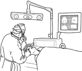
of the orbital apex, especially superior and medial to the optic nerve.

Procedure

The transcranial orbitotomy uses a frontal craniotomy with removal of a portion of the orbital roof to expose the orbital apex or superior orbit. This is best performed by a neurosurgeon familiar with skull-base surgical approaches. In most cases, the supraorbital rim over the involved side is removed en bloc with the frontal bone flap (Fig. 13). The anterior one-half or two-thirds of the orbital roof breaks free with removal of the rim and frontal bone flap, and the remaining posterior portion of the roof can be removed with rongeurs. Historically, it was suggested that all orbital tumors be removed via craniotomy, because before the imaging era it was difficult to anticipate the intraorbital location of a mass.18 The transfrontal approach was first described by Jones10 in 1970. Jane and colleagues19 proposed the current technique in 1982. Refinements have been discussed 
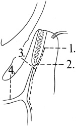
by Maroon and Kennerdell9 and Housepian.20 This operation has been termed the panoramic orbitotomy by Rootman21 because of the wide area of exposure offered by this procedure.

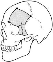
Fig. 13. Schematic diagram for transcranial orbitotomy in which the supraorbital rim is removed en bloc with the frontal bone flap. This provides extensive exposure to the superior and lateral orbit.

Selected steps in the procedure are demonstrated in Figure 14. A coronal skin incision is made 2 to 3 cm behind the normal hairline. A frontal scalp flap is raised, elevating pericranium off the frontal bone. The supraorbital nerve is identified and, if exiting the skull through a foramen rather than a notch, is chiseled out, mobilizing the nerve. The subperiosteal plane is followed over the superior orbital rim, elevating periorbita from the orbital roof and lateral orbital wall. The temporalis muscle is dissected anteriorly out of the temporalis fossa to expose the temporal bone. Once these soft tissues have been separated from the bone, a high-speed drill is used to cut the bone flap.

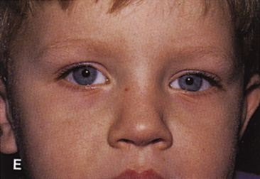
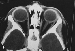
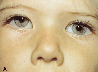
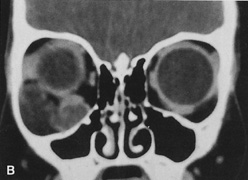
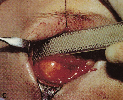
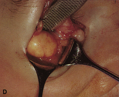
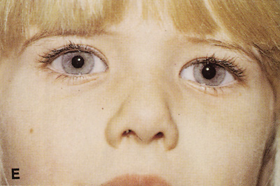
Fig. 14. A,B. Large intraorbital lymphangioma causing proptosis and optic nerve compression in a 2-year-old child. C. View of the left orbit from above after removal of the frontal bone flap, including the supraorbital rim and orbital roof. An extensive exposure of the entire superior and lateral orbit is afforded. The levator and superior rectus complex is being retracted laterally with a muscle hook, whereas the Freer elevator retracts the superior oblique muscle medially. The frontal nerve can be seen running from posterior to anterior over the superior orbit. The orbital mass is exposed in this fashion. D. The fronto-orbital bone flap is wired back in place after completion of 
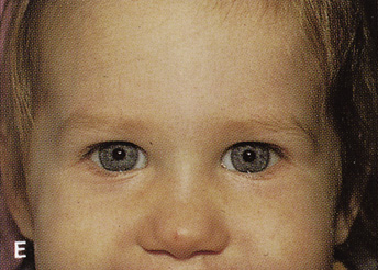
the procedure. E. Postoperative appearance of the patient. F. The postoperative CT scan shows complete removal of the lymphangioma. This large and diffuse lesion would have been difficult to remove with any other approach.

A burr hole is placed in the midline just above the orbital rim. This burr hole usually enters the frontal sinus. A second burr hole is placed anteriorly in the temporalis fossa at the junction of the cranium and orbit so that both compartments are exposed. Two or three additional holes are made in the frontal bone connecting the first two holes. The orbital rim is cut from the midline inferiorly, and the lateral orbital rim is cut from the temporalis fossa anteriorly. The dura is freed from the undersurface of the bone flap and is elevated superiorly, and the orbital roof is cracked off. The frontal bone, orbital roof, and supraorbital rim break off in one piece. The brain is retracted superiorly, and the remaining orbital roof is removed with bone rongeurs (see Fig. 14C).

After removal of the bony roof, the periorbita is visible. Typically, the periorbita is thin, and the levator rectus muscle and frontal nerve are visible beneath it. If exposure of the posterior optic nerve is desired, the dura can be elevated over the optic canal. The canal can be unroofed to decompress or explore the optic nerve, and the dura may be opened to view the intracranial optic nerve and the chiasm. At the orbital apex, the annulus may be cut to allow more anterior dissection and removal of the optic nerve in cases such as optic nerve glioma or meningioma. Because the superior orbital fissure and its contents lie lateral to the nerve, the intraconal space is entered on the medial side of the optic nerve. The orbital dissection can be carried out with a minimal amount of brain retraction after the en bloc removal of the frontal bone, supraorbital rim, and orbital roof.

After the dissection, the dura is closed and the frontal bone flap is plated or wired back into position (see Fig. 14D). The orbital roof is functionally restored with the replacement of the bone flap. The sinuses must be sealed off with muscle, pericranium, or other tissue. The coronal flap is closed in layers. The postoperative appearance is unchanged because the bone flap is replaced in one piece. Problems with globe ptosis, enophthalmos, pulsatile proptosis, or meningitis are rare. However, extensive mobilization of the temporalis may result in cosmetically significant temporal atrophy. Orbital apical dissection often results in extraocular motility dysfunction as a result of traction on the third, fourth, or sixth cranial nerves, but cranial nerve function usually recovers unless the nerves have been transected.

Indications

Transcranial orbitotomy provides access to the superior two-thirds of all the orbital compartments (Fig. 15). In some cases, the craniotomy is used only to provide access to the orbit that is otherwise not possible, such as biopsy of an orbital apex mass. Its primary use is for exploration of tumors involving the orbital apex, or large tumors extending above and medial to the optic nerve. The transcranial orbitotomy also is used as part of combined procedures for approaching tumors involving the orbit as well as the anterior and middle cranial fossae. Most commonly, this involves sphenoid wing meningioma resection.

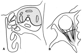
Fig. 15. Schematic of areas amenable to transcranial orbitotomy. Coronal (A) and axial (B) sections.

The performance of a transcranial orbitotomy requires that the orbital surgeon has a good working relationship with a neurosurgeon interested in diseases of the orbitocranial junction. Often, the surgeons working together on these procedures are part of a larger craniofacial team that uses combined expertise to approach other complicated tumor extirpations and reconstructions of the face and skull base.

The transcranial orbitotomy offers unsurpassed exposure of the superior orbit, orbital apex, and chiasm. Its relatively low morbidity makes this orbitotomy the procedure of choice when safe, wide exposure of the posterosuperior orbit is necessary. It is not indicated for orbital apex lesions lying inferior to the optic nerve, in which an extended lateral orbitotomy and temporal craniotomy may be required.

Back to Top
DEEP MEDIAL ORBITOTOMY

FRONTOETHMOIDAL ORBITOTOMY

Several spaces in and around the medial orbit may be entered by use of a frontoethmoidal orbitotomy. These spaces include the subperiosteal space, peripheral surgical space, and ethmoid and sphenoid sinuses. Although this approach is included here in the discussion of “deep” orbital approaches because it is often used in conjunction with removal of the medial orbital wall (anterior ethmoidectomy), it is also used without bone removal to drain pus or blood in the subperiosteal space. This approach has been largely replaced by the transcaruncular orbitotomy, described in the following.

Procedure

A frontoethmoidal, or Lynch, skin incision is marked halfway between the medial canthus and bridge of the nose. It extends superiorly and inferiorly in a curved fashion approximately 2 to 3 cm (Fig. 16). The exact limits of the incision depend on the location of the underlying lesion. Injection of local anesthetic with epinephrine provides vasoconstriction, which greatly enhances hemostasis. After the skin is incised, a unipolar cutting cautery is used to extend the incision deeply to the periosteum. Bleeding may be encountered, especially in the area of the angular artery and vein. Exposure is enhanced with 4-0 silk sutures passed into the orbicularis muscle and clamped to the drapes.

Fig. 16. A. Patient with right orbital cellulitis. B. Coronal CT scan demonstrating subperiosteal abscess formation from frontal and ethmoidal sinusitis. C. Frontoethmoidal orbitotomy incision marked for abscess drainage.

The periosteum is exposed and incised with a Freer elevator and then is reflected off the bone posteriorly. It is generally quite adherent to the curved contour of the medial canthal bones, especially at the medial canthal tendon. The anterior lacrimal crest is encountered inferiorly. Care should be taken not to damage the lacrimal sac with the elevator. The posterior lacrimal crest is visible behind the sac. Adequate mobilization of the periosteal connections to the anterior lacrimal crest gives a large area of exposure. The periosteal elevation is carried superiorly in the area of the trochlea. Elevation of the periosteum opens the subperiosteal space. Blood or pus caused by fracture or infection, if present in this space, is encountered at this point.

Elevation of the periorbita along the medial orbital wall posterior to the lacrimal sac progresses easily. Orbital fractures involving the thin ethmoid bone are seen at this point. The anterior ethmoidal artery is seen at the junction of the ethmoidal and frontal bones where the orbital roof meets the medial orbital wall (see Fig. 7). Usually it is found on a line extending posteriorly from the superior border of the medial canthal tendon. This artery either should be thoroughly cauterized with the bipolar cautery or clipped with a vascular clip before cutting. As the dissection in the subperiosteal space moves posteriorly, the orbit narrows. The posterior ethmoidal artery is identified. This is a reliable landmark for the optic foramen, which lies approximately 5 mm behind the ethmoidal artery.

Indications

The frontoethmoidal medial orbitotomy allows access for a variety of procedures in the subperiosteal and peripheral surgical spaces and sinuses (Fig. 17). Its main use is for processes involving both the frontal or ethmoid sinuses and the orbit. Entrance into the subperiosteal space is obtained easily. Drainage of subperiosteal blood or pus occurs as the space is entered.

Fig. 17. Schematic demonstration of areas amenable to frontoethmoidal orbitotomy. Coronal (A) and axial (B) views. This approach can be used for exposure of the medial orbit, ethmoid and sphenoid sinuses, and optic canal.

TRANSCARUNCULAR ORBITOTOMY

The peripheral extraconal and subperiosteal spaces of the medial orbit are explored more often with a transconjunctival incision placed just lateral to the caruncle, in the semilunar fold, and then extended superiorly and inferiorly into the medial fornices.22 This approach offers access to the medial subperiosteal space without requiring a cutaneous incision.

Procedure

A subconjunctival injection of Xylocaine with epinephrine or a drop of topical Neo-Synephrine 2.5% usually is used to aid vasoconstriction. If mild pupillary dilation is unwanted, this is omitted. The upper and lower eyelids are retracted with Desmarres vein retractors and Westcott scissors are used to cut the conjunctiva between the caruncle and plica vertically for a distance of 1.5 to 2 cm (Fig. 18A). The incision is limited superiorly by the medial levator aponeurosis, but inferiorly it can be extended laterally along the inferior border of the tarsus all the way across to the lateral orbital rim. Exploration of the floor and medial wall is enhanced by disinsertion of the inferior oblique muscle at its origin on the medial inferior rim. Dissection is carried posterior to the lacrimal sac using Steven's tenotomy scissors to spread soft tissues overlying the medial orbital wall just posterior to the posterior lacrimal crest. Once bone is encountered, sharp dissection through the periosteum is performed with cutting cautery to expose the medial orbital wall over the anterior ethmoid sinus. Periorbita is elevated posteriorly to identify and protect the anterior and posterior ethmoidal arteries (see Fig. 18B). This plane of dissection passes medial to the medial rectus muscle, but lateral and then posterior to the lacrimal sac. The exposure through the transcaruncular incision is similar to that afforded by the frontoethmoidal or “Lynch” incision but does not leave any visible cutaneous scar (see Fig. 18C). At the completion of the procedure, the conjunctiva can be reposited without need for suture approximation.

Fig. 18. A. Incision for transcaruncular medial orbitotomy. The incision is placed just lateral to the caruncle and medial to the plica semilunaris. B. Axial diagrammatic scheme of route of dissection for transcaruncular orbitotomy. The incision in the medial fornix allows dissection to remain lateral to the lacrimal sac but medial to the globe and medial rectus muscle. Posterior to the sac, dissection is carried to the medial bony wall, where periosteum then can be incised and elevated posteriorly. C. The globe and medial rectus are drawn laterally by a malleable retractor, and the upper and lower lids are distracted to expose the medial extraperiosteal orbital space.

Indications

The transcaruncular approach can be used to approach the medial peripheral or extraperiosteal space while avoiding the skin incision of the frontoethmoidal approach for a medial orbitotomy. Its most common indication is for the ethmoidectomy portion of a thyroid orbital decompression or repair of medial orbital fractures. When it is extended across the lower lid and combined with a lateral canthotomy, exposure of the medial, inferior, and lateral 270° of the orbit can be obtained.

Back to Top
DEEP INFERIOR ORBITOTOMY

TRANSANTRAL ORBITOTOMY

Lesions in the subperiosteal space that involve the orbital floor and roof of the maxillary sinus can be approached through an intraoral incision in the buccal sulcus above the upper canine incisors (Fig. 19). Dissection is carried through mucosa and soft tissue to the periosteum over the face of the maxilla. Dissection is carried superiorly in the subperiosteal plane to just below the inferior orbital rim. Care must be taken to identify the infraorbital nerve as it exits the foramen just below the rim. Osteotomies then are made in the face of the maxilla, and the underlying maxillary sinus is exposed. The sinus mucosa is excised and the roof of the sinus exposed in this fashion. Removal of the sinus roof (the orbital floor) allows exposure of the inferior periorbita. Care must be taken to avoid damage to the infraorbital nerve that runs within the bone of the floor. The inferior rectus muscle is encountered immediately above the periorbita, and it must be protected and retracted during deeper orbital dissection.

Fig. 19. Transantral inferior orbitotomy. The orbital floor (A) (roof of maxillary sinus) is exposed by an incision in the buccal sulcus and removal of the anterior face of the maxillary bone. The infraorbital nerve (B) traverses the floor of the orbit and exits on the face of the maxilla from the infraorbital foramen (C).

Indications

This approach often is used for orbital decompression in Graves' ophthalmopathy and cases of primary sinus pathology with secondary orbital involvement. It can be combined with a transconjunctival inferior orbitotomy approach to provide elevation and protection of orbital contents before removal of the orbital floor from below. It can be used to approach inferior peripheral lesions near the apex where an anterior-inferior orbital approach may not allow sufficiently deep access. However, better exposure may be afforded by a lateral orbitotomy in those cases.

Back to Top
ANTERIOR ORBITAL APPROACHES
Whenever possible, it is preferable to approach orbital lesions without the increased operative time and potential morbidity associated with osteotomies and bone removal. Anterior lesions and many encapsulated deeper lesions in the lateral, superior, medial, or inferior orbit often can be approached without the need for bone removal. Each of these locations is addressed separately.
Back to Top
SUPERIOR ANTERIOR ORBITOTOMIES

UPPER EYELID CREASE APPROACH

Anterior lesions in the anterior superior orbit can be approached with incisions placed in the eyelid crease, within or beneath the brow, or through a coronal flap approach. The upper eyelid crease approach offers excellent scar camouflage and direct access with minimal undermining or dissection; it is preferable whenever possible. Because of the extreme elasticity and distensibility of the upper eyelid soft tissues, lid crease incisions allow ready access to the superior orbital rim, the peripheral superior orbit, and more superficial lesions overlying the medial and lateral orbital rims as well. Incisions placed within the brow or just below the brow are rarely performed because the supraorbital rim usually can be approached as easily with a lid crease incision. The eyelid crease approach for deep orbital lesions without bone removal is described in the preceding.

Procedure

The lid crease incision is marked at a point approximately 2 mm below the pre-existing lid crease because of the tendency for postoperative scar retraction to cause superior migration of the crease. For superomedial lesions, the incision medial to the punctum is angled superonasally toward the medial brow (Fig. 20). For lateral lesions, the incision beyond the lateral canthus can be directed superolaterally toward the tail of the brow (Fig. 21).

Fig. 20. A. Axial CT scan of a superonasal orbital cyst. B. Nasal extension of the superior lid crease is directed up at a right angle to the crease, toward the nasal brow. C. Incision through skin and orbicularis and superior dissection to expose and incise septum allows removal of the anterior orbital cyst.

   
Fig. 21. A. Sagittal MR image demonstrating a large hematic cyst in the right superior orbit. B. Lid crease incision marked across the width of the right upper lid and extending laterally toward the tail of the brow. C. Dissection plane deep to skin and orbicularis and superficial to orbital septum allows ready exposure of the supraorbital rim. D,E. Periosteum over the supraorbital rim is incised with a No. 15 blade and dissection carried out to expose the full extent of the hematic cyst (E). F. After removal of the cyst, simple skin closure is accomplished with a running absorbable suture in the lid crease. A drain is brought out through the temporal edge of the incision. G. Postoperative appearance of the patient. Excellent scar camouflage is achieved by placing the incision within the eyelid crease.

A lid fixation suture is placed through the marginal tarsus and clamped to the drape below. This stabilizes the lid and makes identification of the appropriate dissection plane much easier. The skin is incised, and dissection is carried through the underlying orbicularis to the postorbicular fascial plane. This plane overlies the orbital septum and levator aponeurosis, and dissection superiorly in this plane keeps the orbital septum intact.

Dissection in the postorbicular plane allows ready access to the superior orbital rim and good exposure of both extraperiosteal and anterior peripheral intraorbital lesions. If a lesion is extraperiosteal, the periosteum overlying the orbital rim is incised and the extraperiosteal space is entered and the periorbita elevated (see Fig. 21D). Care must be taken to locate the supraorbital foramen or notch along the superomedial orbital rim to avoid damage to the neurovascular bundle. Lesions that lie within the peripheral orbital space require opening of the orbital septum and dissection superficial to the levator aponeurosis. Once the orbital dissection is completed, skin closure is carried out with a running cutaneous absorbable suture. The orbital septum should never be sutured closed because this might result in foreshortening of the septum with resultant lagophthalmos. There is no need to reform the lid crease by fixating subcutaneous tissues to the levator aponeurosis. The lid crease naturally reforms around the incision and dissection plane, and aponeurotic fixation may cause superior migration of the postoperative lid crease.

Indications

The upper eyelid crease approach allows excellent access to lesions in the superior orbit at or anterior to the equator of the globe. Encapsulated lesions deep within the orbit often can be removed through this anterior approach provided their superficial surface extends anterior to the equator of the globe (Fig. 22). The lid crease incision also allows simultaneous exposure and surgery on the underlying levator aponeurosis when necessary. This approach has also proved useful in approaching superficial dermoid cysts overlying the medial and lateral orbital rims.23,24

 
Fig. 22. A. Appearance of patient with a superonasal lesion in the peripheral orbital space. B. Axial MR image demonstrates encapsulated lesion extending deeply into the orbit. C,D. A lid crease incision is used to expose the anterior portion of the mass, which is then extracted with the aid of a cryoprobe (D). E. Postoperative appearance of patient with excellent scar camouflage of the eyelid crease incision.

The medial intraconal space also may be accessed through a medial upper eyelid crease incision. This requires exposure of the medial border of the levator aponeurosis and levator muscle and dissection through the intermuscular septum extending between the superior and medial rectus muscles. The superior oblique tendon must be identified and retracted superiorly, so that the medial intraconal space can be entered (Fig. 23).25

Fig. 23. A. The medial intraconal space also may be accessed through a medial upper eyelid crease incision. B. This requires exposure of the medial border of the levator aponeurosis and levator muscle and dissection through the intermuscular septum extending between the superior and medial rectus muscles. The superior oblique tendon must be identified and retracted superiorly, so that the medial intraconal space can be entered.

In the case of more deeply placed superior orbital lesions, it is possible to use an air drill to burr away the superior orbital rim to provide better exposure and allow deeper access in the superior orbit (Fig. 24). Because the contour of the brow is established by the supraorbital ridge, fairly extensive removal of the superior orbital rim can be carried out without cosmetic deficiency. However, care must be taken to identify the supraorbital nerve and avoid damage to it during this maneuver, or hypesthesia over the forehead and scalp can result. The amount of bone removal also is limited by the degree of pneumatization of the overlying frontal sinuses, although inadvertent entry into the sinus usually is not a problem as long as the nasofrontal duct is not compromised.

Fig. 24. A. Sagittal CT scan of a lesion in the peripheral superior orbit behind the equator of the globe. B. Lid crease incision and superior dissection are used to expose the superior orbital rim. C. The superior orbital rim then can be burred away to provide greater access to the more posterior orbital lesion.

VERTICAL-LID-SPLIT SUPERIOR ORBITOTOMY

The upper eyelid crease incision gives excellent access to the peripheral superior orbital space. However, lesions that lie intraconally are difficult to approach with a transverse lid crease incision as dissection must be performed medial to the medial horn of the levator muscle to avoid transecting and disinserting the levator aponeurosis and Müller's muscle. However, vertically splitting the upper lid allows a vertical separation of the levator aponeurosis and Müller's muscle and does not disinsert it from its normal insertions on the tarsal plate. This approach provides an excellent exposure of the superomedial orbit and is useful for approaching lesions that lie medial to the optic nerve.26 In this situation, it is often an attractive alternative to a transcranial superior orbitotomy, which might otherwise be required (Fig. 25).

Fig. 25. A,B. Coronal (A) and axial (B) CT scans demonstrating well-encapsulated mass posteriorly in the superonasal orbit. An excellent alternative to transcranial orbitotomy for this lesion is an anterior approach via a vertical-lid splitting incision.

Procedure

A vertical lid incision is made full-thickness through the lid margin transecting eyelid skin, orbicularis muscle, tarsus, distal levator aponeurosis, Müller's muscle, and palpebral conjunctiva with straight iris scissors. It is important to remain exactly perpendicular to the lid margin (Fig. 26). The incision is then extended posteriorly through the conjunctival fornix and is reflected through the fornix back down the bulbar conjunctiva to the limbus. After traversing fornix and bulbar conjunctiva, dissection proceeds posteriorly into the medial orbit. The superior oblique tendon can be identified crossing from the trochlea, medially, to the superior equator of the globe, laterally, and may be retracted. Ready access to the intraconal space then is afforded in the superonasal quadrant between the medial rectus and superior rectus muscles.

Fig. 26. A. Straight iris scissors are used to make a full-thickness vertical incision at the junction of the medial and central one-third of the left upper lid. B. The incision is carried through fornix conjunctiva and then can be reflected inferiorly through bulbar conjunctiva as necessary. C,D. Sewall retractors retract the superior oblique muscle above and the globe laterally, so that dissection can be carried to the peripheral and intraconal spaces where the lesion is identified and removed with the aid of a cryoprobe (D). E. Full-thickness lid incision is reapproximated. F. Postoperative appearance of the patient. Excellent scar camouflage is afforded by the vertical incision placed within the eyelid skin. Schematic of areas of orbit amenable to the vertical-lid split anterior approach. Coronal (G) and axial (H) views.

After removal of the lesion, repair is carried out in a fashion identical to that used for repair of a lid marginal laceration. The bulbar, fornix, and palpebral conjunctiva are reapproximated and the tarsal plate meticulously realigned at the lid margin with a vertical mattress 7-0 Vicryl suture. Additional sutures to realign the lash line are followed by closure of the orbicularis and skin.

Indications

This approach allows for excellent scar camouflage with maintenance of appropriate lid height and contour. It is useful for exposure of superior orbital lesions that lie medial to the optic nerve (see Fig. 26G, H). Well-encapsulated peripheral and intraconal lesions posterior to the globe may be reached fairly readily with this approach.

Back to Top
MEDIAL ANTERIOR ORBITOTOMIES

TRANSCONJUNCTIVAL MEDIAL ORBITOTOMY

The transconjunctival orbitotomy is used to enter Tenon's space or the medial intraconal space through an incision in the medial perilimbal bulbar conjunctiva. If the intraconal space is to be entered, disinsertion of the medial rectus muscle usually is required. Out-fracture or removal of the lateral rim through a lateral canthotomy approach can be performed in conjunction with a medial transconjunctival approach. This allows improved access by permitting the globe to be displaced further laterally (Fig. 27).

Fig. 27. A. Area of orbit accessible by transconjunctival medial orbitotomy. B. Exposure of medial orbitotomy is increased by combining it with lateral orbitotomy to allow lateral displacement of the globe.

Procedure

A drop of topical Neo-Synephrine 2.5% or a subconjunctival injection of lidocaine with epinephrine facilitates hemostasis. If mild pupillary dilation is unwanted, this should be omitted. A lid speculum is placed and a 90° to 180° medial conjunctival peritomy is performed centered on the 3 o'clock meridian. The peritomy can be enlarged if more exposure is necessary. Two relaxing incisions are made radially at the ends of the peritomy. Stevens' tenotomy scissors are used to bluntly spread open the two medial quadrants of anterior Tenon's space, exposing the sclera and medial rectus muscle. The medial rectus muscle can be disinserted if exposure deep into the intraconal space is necessary. Visualization of the anterior part of the optic nerve can be obtained without disinsertion of the muscle. If the muscle is removed, a standard von Pirquet suture should be used to reinsert the muscle. A suture can be passed through the muscle origin for retraction of the eye. Additional sutures or muscle hooks can be looped under the superior and inferior rectus muscles if additional retraction of the eye is needed.

Sewall and ribbon orbital retractors are used to retract the eye and Tenon's capsule (Fig. 28). The intraconal space may be entered by sharp or blunt dissection of Tenon's capsule. A Westcott scissors or Freer periosteal elevator may be used for this purpose. The orbital retractors can be repositioned over moist neurosurgical cottonoids to prevent orbital fat from prolapsing forward. An operating microscope is helpful for obtaining appropriate illumination and magnification when working in the medial intraconal space.

Fig. 28. A. Patient with a medial orbital mass resulting in lateral displacement of the left globe. B. Coronal CT scan reveals the mass to be in the medial peripheral surgical space. C. Transconjunctival orbitotomy approach with conjunctival incision and dissection medial to the globe. In this case, the medial rectus muscle was not disinserted because the lesion lay in the peripheral surgical space outside the muscle cone. D. Postoperative appearance of the patient. Note that the lateral displacement of the globe has resolved. The conjunctival incision leaves no visible scar.

Indications

The most common use of the transconjunctival orbitotomy is to expose the anterior portion of the peripheral extraocular muscle and intraconal spaces. In particular, the optic nerve can be biopsied or fenestrated by use of this approach. When exposure is too limited to safely biopsy deep intraconal masses, transconjunctival orbitotomy can be combined with a lateral orbitotomy with bone removal. This allows the eye to be shifted laterally, making more room to work in the medial orbit (see Fig. 27B). The transconjunctival medial orbitotomy can be extended vertically into the upper lid, as mentioned, to provide exposure of the superonasal quadrant

FRONTOETHMOIDAL MEDIAL ORBITOTOMY

A frontoethmoidal incision or transcaruncular incision can be used to enter the anteromedial extraperiosteal space without performing bone removal along the medial wall. These approaches are discussed under Deep Medial Orbitotomy.

Back to Top
LATERAL ANTERIOR ORBITOTOMY
The utility of the upper eyelid crease incision in exposing the lateral orbit while leaving the bony orbital rim intact is described in the preceding. A canthotomy incision without removal of the underlying lateral bony rim also allows access to the anterior retrobulbar space. This is a particularly effective approach for performing fenestration of the optic nerve sheath and can be done without disinsertion of the lateral rectus muscle.27

PROCEDURE

A 1- to 1.5-cm lateral canthotomy incision is made, and dissection with unipolar cautery is carried down to the lateral rim (Fig. 29A). Periosteum is incised along the long axis of the lateral rim from superior to inferior and elevated from the lateral orbital rim. The periorbita is then elevated from the inner aspect of the lateral orbital rim, and a longitudinal incision from anterior to posterior is made in the periorbita paralleling the underlying lateral rectus muscle.

Fig. 29. A. Lateral canthotomy incision is used to expose the lateral orbital rim (B). Dissection then is carried through periorbita, allowing ready entry into the intraconal space and exposure of the anterior optic nerve (C).

The lateral rim may be left in place and dissection carried out anterior to its posterior recess at the lateral canthus (see Fig. 29B). Because the lateral rim recesses posteriorly at the canthus, the posterior surface of the globe and anterior retrobulbar space usually are accessible. If exposure is limited, the lateral rim can be partially thinned with a bone burr until there is ready access to the retrobulbar space. Sharp dissection through the intermuscular septum allows entry into the retrobulbar intraconal space (see Fig. 29C). Closure is accomplished by simply reapproximating the superior and inferior crura of the lateral canthal tendon and closing the canthotomy skin incision.

INDICATIONS

Anterior peripheral and intraconal lesions and the lateral retrobulbar optic nerve can be readily approached with this technique. There is no need for disinsertion of the lateral rectus muscle because its belly can be retracted out of the field.

Back to Top
ANTERIOR INFERIOR ORBITOTOMIES
Access to the anterior inferior orbit can be obtained through cutaneous incisions placed in either the lower lid crease or subciliary area or through transconjunctival incisions placed in the inferior fornix or beneath the tarsal plate (Fig. 30). With any of these approaches, dissection can be carried inferiorly to expose the orbital septum and infraorbital rim, and exploration then can be carried posteriorly into the orbit. Although access to the anterior orbit historically was obtained by incisions placed directly over the infraorbital rim, this can result in objectionable scarring and lymphedema and is not recommended.

Fig. 30. Sagittal diagram of the lower lid. An incision through skin and orbicularis can be placed in the subciliary area (1) or in a lower lid crease (2). In either case, dissection is carried through orbicularis to enter the postorbicular fascia plane on the anterior surface of the orbital septum. An incision through conjunctiva at the inferior border of the tarsal plate also leads into the postorbicular fascia plane, but from a posterior approach (3). An incision directly through the fornix gains access to the retroseptal space immediately (4).

PERCUTANEOUS APPROACH

An incision can be marked out just beneath the lashes of the lower lid, or in the lower lid crease a few millimeters below. The lower lid crease incision may be preferable to a subciliary incision because a wider band of pretarsal orbicularis is left to oppose the tendency for postoperative contracture and ectropion. A traction suture through the marginal tarsus provides superior traction and helps with identification of the dissection plane. The incision is carried through skin and orbicularis into the postorbicular fascia plane. A flap of skin and underlying orbicularis muscle then is elevated inferiorly by blunt and sharp dissection while keeping the orbital septum intact. In this fashion, the orbital rim can be exposed. From this location, the periorbita of the floor can be incised and elevated to access extraperiosteal lesions. Lesions in the inferior peripheral space or intraconal space can be accessed by opening the orbital septum widely and dissecting posteriorly within the orbital fat (Fig. 31).

Fig. 31. A. Infant with inferior orbital mass causing supraplacement of the right globe. B. Coronal CT scan demonstrating a diffuse mass involving the entire orbital floor. C. Lower lid crease incision through skin and orbicularis allows dissection inferiorly to the orbital rim and then through orbital septum to expose the mass. D. Postoperative appearance of the patient after debulking of a capillary hemangioma and intralesional steroid injection. E. Same patient 1 year later. Note the excellent scar camouflage afforded by lower lid crease incision. F. Postoperative CT scan shows dramatic response of capillary hemangioma to intralesional steroid injection.

Care must be taken to identify and avoid damage to the inferior oblique muscle arising from the medial inferior orbital rim. The inferior oblique may be elevated from its origin on the medial inferior orbital rim to enhance exposure. It may be resutured or merely allowed to passively resettle at the completion of orbital dissection.

Upon completion of orbital dissection, closure is carried out by simple running suture of the skin. Usually it is not necessary to close periosteum at the rim except in selected cases, such as when placing a subperiosteal implant for fracture repair. The orbital septum should never be closed because this risks lower lid retraction and foreshortening.

Indications

This approach allows excellent access to anterior extraperiosteal peripheral or intraconal lesions. The main disadvantages of a percutaneous lower lid incision are the possibility of visible scar formation and contracture of the skin muscle flap, which may produce lower lid retraction, scleral show, or ectropion in up to 10% of cases.28

TRANSCONJUNCTIVAL APPROACH

A preferable approach to the inferior orbit is through a transconjunctival incision. This eliminates the percutaneous scar and risk of lower lid retraction. The transconjunctival incision can be made through conjunctiva and lower lid retractors at the inferior border of the tarsal plate, or the incision can be made directly through the inferior fornix. The authors' preference is to incise through conjunctiva and retractors just inferior to the tarsal plate so that dissection can be carried out in the postorbicular fascia plane while keeping the orbital septum intact (Fig. 32). Other surgeons prefer incision directly through the fornix. This minimizes dissection within the postorbicular fascia plane but results in prolapse of orbital fat into the operative field because dissection occurs posterior to the septum.

Fig. 32. A. Schematic diagram of subtarsal transconjunctival lower lid orbitotomy. Initial dissection allows the orbital septum to be kept intact to prevent orbital fat from prolapsing into the operative field. B. After the flap is retracted, the peripheral orbit may be entered by incising the orbital septum, or the subperiosteal space may be entered by incising and elevating periorbita at the inferior rim.

Procedure

The transconjunctival incisions usually are accompanied by lateral canthotomy and inferior cantholysis to allow the lower lid to swing away from the rim and provide better access to the inferior orbit.29 Straight iris scissors can be used to perform a lateral canthotomy; then they are rotated 90° to cut through the inferior crus of the lateral canthal tendon (Fig. 33). The incision then is carried across the lower lid at the inferior border of the tarsal plate, disinserting conjunctiva and lower lid retractors. A traction suture passed through the cut edge of the conjunctiva and retracted superiorly allows for ready identification of the dissection plane just superficial to the orbital septum. This plane is identical to the one that is approached through the lower lid crease incision. It differs only in that it is entered posteriorly rather than anteriorly. The surgeon's view while approaching the inferior orbital rim through the subtarsal incision is identical to that during percutaneous approaches, and dissection may proceed extraperiosteally along the floor or through orbital septum into the intraorbital tissues.

   
Fig. 33. A. Patient with left-sided proptosis caused by large, well-encapsulated intraconal mass (B). C. Canthotomy and inferior cantholysis allow disinsertion of the lower lid from the lateral orbital rim. D. A subtarsal incision with iris scissors is used to disinsert conjunctiva and lower lid retractors from the inferior border of the tarsal plate. E. Dissection is then carried through orbital septum to expose and remove the large cavernous hemangioma. F. Closure is by simply reapproximating the conjunctiva to the inferior border of the tarsal plate and reattaching the inferior crus to the superior crus of the lateral canthal tendon. G. Postoperative appearance shows excellent scar camouflage afforded by the transconjunctival approach.

After orbital dissection is completed, the conjunctiva and retractors are reapproximated to the inferior border of the tarsus with a continuous running absorbable suture. The inferior crus of the lateral canthal tendon then is sutured to the superior crus with an absorbable suture, and the canthotomy incision in the skin is closed with one or two interrupted skin sutures.

Indications

Extraperiosteal, peripheral, and anterior intraconal lesions can be approached through either percutaneous or transconjunctival incisions (Fig. 34). Because excellent exposure is afforded by either approach, the authors' preference is to avoid a cutaneous scar and the potential complication of lower eyelid retraction or ectropion by approaching the inferior orbit through a conjunctival incision.28 On rare occasions it can be combined with an osteotomy of the inferior orbital rim to provide wider access to the inferior orbit. In this case, care must be taken to avoid damage to the infraorbital neurovascular bundle where it exits the foramen just below the inferior rim.

Fig. 34. Schematic of orbital area accessible by anterior inferior orbitotomy. (A) Coronal and (B) axial views.

Back to Top
COMBINED APPROACHES
On occasion, more than one orbital approach may be required to completely expose and safely remove a lesion. A good orbital surgeon remains flexible throughout the surgical procedure and uses creative combinations of approaches as circumstances dictate. The surgeon also should recognize when specialists in other disciplines, such as otolaryngology or neurosurgery, may help in the definitive management of pathologic orbital processes and should not hesitate to obtain their assistance.

Combining one of the anterior approaches with a lateral canthotomy and removal of the lateral wall often is helpful to give access to deeper orbital structures. Lateral orbitotomy with out-fracture or removal of the lateral wall allows greater access to the medial orbit by permitting greater displacement of the globe laterally. Similarly, lateral orbitotomy may be combined with transconjunctival inferior orbitotomy (Fig. 35) or a superior orbitotomy to allow more extended access. Transconjunctival inferior orbitotomy can be extended into a transcaruncular medial orbitotomy to allow extensive exposure of the medial and inferior orbits. Lesions that encircle the optic nerve occasionally can be divided and removed through separate medial and lateral approaches that allow the separate portions of the lesion to be directly accessed without crossing the optic nerve.

 
Fig. 35. A. Patient with multiple cystic masses in the inferior and lateral orbit. These are causing supraplacement of the globe and expansion of the bony orbit as demonstrated on coronal CT scan (B). C. Approach is through a combined modified Berke lateral orbitotomy and transconjunctival inferior orbitotomy. After the lateral orbital rim is removed, wide exposure of the lateral and inferior orbits is afforded (D). E. Postoperative appearance of patient, revealing excellent scar camouflage afforded by the small lateral canthotomy incision and the hidden transconjunctival incision.
Back to Top
REFERENCES

1. Dortzbach RK, Kronish JW, Gentry LR: Magnetic resonance imaging of the orbit. Part I. Physical principles. Ophthal Plast Reconstr Surg 5:151, 1989

2. Dortzbach RK, Kronish JW, Gentry LR: Magnetic resonance imaging of the orbit. Part II. Clinical applications. Ophthal Plast Reconstr Surg 5:160, 1989

3. Kersten RC, Kersten JL, Kulwin DR, et al: Chronic hematic cysts of the orbit: Role of MRI in diagnosis. Ophthalmology 95:1549, 1988

4. Flaherty PM, Wolfgang EL, Sergott RC, et al: Color Doppler imaging. A new noninvasive technique to diagnose and monitor carotid cavernous sinus fistulas. Arch Ophthalmol 109:552, 1991

5. Reseh D, Ossoinig K, Nerad JA: Diagnosis and localization of deep orbital organic foreign bodies. Orbit 6:3, 1987

6. Lee AG, Brazis PW, Garrity JA, White M: Imaging for neuro-ophthalmic and orbital disease. Am J Ophthalmol 138:852, 2004

7. Custer PL, Trinkas KM: Hemorrhagic complications of oculoplastic surgery Ophthal Plast Reconstr Surg 18:409, 2002

8. Kronlein RU: Zur Pathologie und operativen Behandlung der Dermoidcysten der Orbita. Beitr Klin Chir 4:149, 1888

9. Maroon JC, Kennerdell JS: Surgical approaches to the orbit. Indications and techniques. J Neurosurg 60:1226, 1984

10. Jones BR: Surgical approaches to the orbit. Trans Ophthalmol Soc UK 90:269, 1970

11. Leone CR: Surgical approaches to the orbit. Ophthalmology 86:930, 1979

12. Kersten RC: Lateral canthotomy incision for lateral orbitotomy Ophthal Plast Reconstr Surg 15:19. 1999

13. Harris GJ, Logani SC: Eyelid crease incision for lateral orbitotomy Ophthal Plast Reconstr Surg 15:9. 1999

14. Stewart WB, Levin PS, Toth BA: The technique of coronal scalp flap approach to the lateral orbitotomy. Arch Ophthalmol 106:1724, 1988

15. Patrinely JR, Cech DA: Hemicoronal flap approach for lateral orbitotomy. Arch Ophthalmol 107:1421, 1989

16. Long JC, Ellis PP: Total visual loss following orbital surgery. Am J Ophthalmol 71:218, 1971

17. Berke RN: A modified Krönlein operation. Arch Ophthalmol 51:609, 1954

18. Dandy WE: Results following the transcranial operative attack on orbital tumors. Arch Ophthalmol 25:191, 1941

19. Jane JA, Park TS, Pobereskin LH, et al: The supraorbital approach: Technical note. Neurosurgery 11:537, 1982

20. Housepian EM: Microsurgical anatomy of the orbital apex and principles of transcranial orbital exploration. Clin Neurosurg 25:556, 1978

21. Rootman J, Stewart B., Goldberg RA eds. Orbital Surgery: A Conceptual Approach. New York: Lippincott Williams & Wilkins, 1995

22. Garcia GH, Gldberg RA, Shorr N: The transcaruncular approach in repair of orbital fractures: A retrospective study. J Craniomaxillofac Trauma 4(1):7;1998

23. Kersten RC: The eyelid crease approach to superficial lateral dermoid cysts. J Pediatr Ophthalmol Strabismus 25:48, 1988

24. Wolfley DE: The lid crease approach to the superomedial orbit. Ophthalmic Surg 16:652, 1985

25. Pelton RW, Patel BC: Superomedial lid crease approach to the medial intraconal space: A new technique for access to the optic nerve and central space. Ophthal Plast Reconstr Surg 17(4):241, 2001

26. Smith B: The anterior surgical approach to orbital tumors. Trans Am Acad Ophthalmol Otolaryngol 70:607, 1966

27. Kersten RC, Kulwin DR: Optic nerve sheath fenestration through a lateral canthotomy incision. Arch Ophthalmol III:870, 1993

28. Lacy MF, Pospisil OA: Lower blepharoplasty, post orbicularis approach to the orbit: A prospective study. Br J Oral Maxillofac Surg 25:398, 1987

29. McCord CD, Moses JL: Exposure of the inferior orbit with fornix incision and lateral canthotomy. Ophthalmic Surg 10:53, 1979

Back to Top