|
|
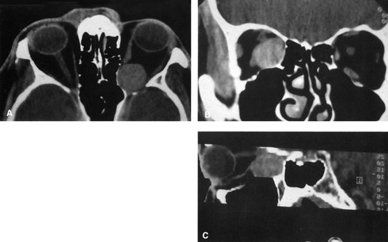
| Fig. 1. A. Axial CT scan demonstrating a large, well-encapsulated lesion in the orbital apex. Coronal (B) and sagittal (C) scans demonstrate that the mass lies inferior and medial to the optic nerve within the intraconal space. This information is useful in planning the surgical approach to the mass, which should avoid traversing the optic nerve. |