

|
|
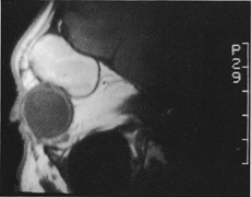
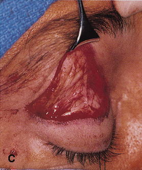
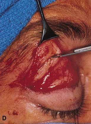
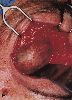
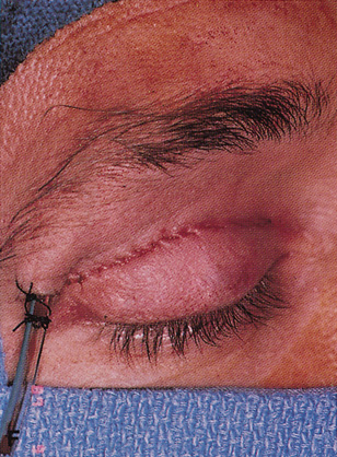
| Fig. 21. A. Sagittal MR image demonstrating a large hematic cyst in the right superior orbit. B. Lid crease incision marked across the width of the right upper lid and extending laterally toward the tail of the brow. C. Dissection plane deep to skin and orbicularis and superficial to orbital septum allows ready exposure of the supraorbital rim. D,E. Periosteum over the supraorbital rim is incised with a No. 15 blade and dissection carried out to expose the full extent of the hematic cyst (E). F. After removal of the cyst, simple skin closure is accomplished with a running absorbable suture in the lid crease. A drain is brought out through the temporal edge of the incision. G. Postoperative appearance of the patient. Excellent scar camouflage is achieved by placing the incision within the eyelid crease. |