

|
|
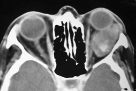
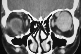
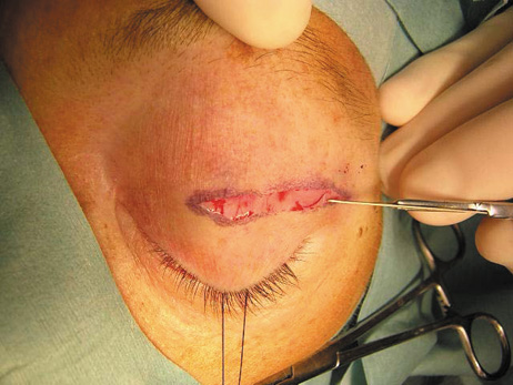
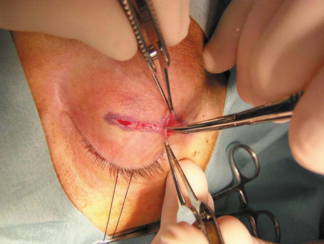
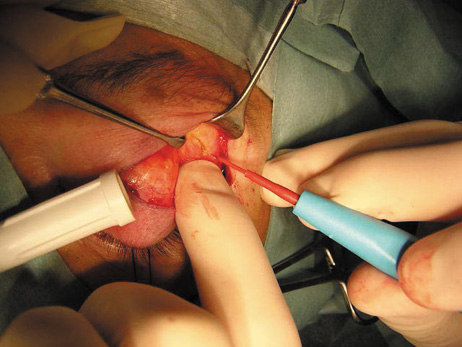
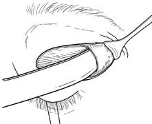
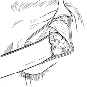
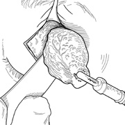
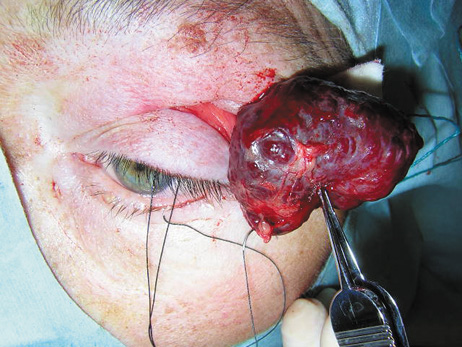
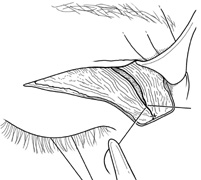
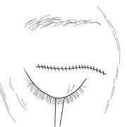
| Fig. 9. A,B. Coronal and axial CT images of a large intraconal neoplasm. C. Because it was felt to represent a well-encapsulated cavernous hemangioma, this lesion was a candidate for removal via an eyelid crease orbitotomy without bone removal. The eyelid crease incision marked. D. Incision made with scalpel. E. Orbicularis muscle is tented up and incised to expose the underlying septum. F. Dissection of a skin-muscle flap deep to orbicularis exposes the orbital septum and superior orbital bony rim. G. Cutting cautery is used to incise periosteum along the superior and lateral rims; finger palpation of the bone helps to direct this incision. H. Periorbita is elevated along the mesial surface of the lateral orbital rim in order to expose the deep orbital tissues. I. The cavernous hemangioma is visualized in the wound. Retraction is provided by one or more malleable retractors. J. Cryoprobe is affixed to the hemangioma to facilitate manipulation of the lesion. K. Large cavernous hemangioma after removal through the eyelid crease incision which was accomplished without bone removal. L. Periorbita is reattached over the lateral rim. M. The eyelid crease incision is closed with a running suture. |